This human anatomy ClipArt gallery offers 825 illustrations of the human skeletal system, including images of both the axial skeleton and the appendicular skeleton. The human axial skeleton includes 80 bones formed by the vertebral column (spine), the thoracic cage (e.g., ribs, sternum), and the skull. The axial skeleton is responsible for the upright position of the body. The human appendicular skeleton is composed of 126 bones formed by the pectoral girdles, the upper and lower limbs, and the pelvic girdle. These bones function in locomotion as well as protection of vital organs.
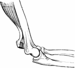
Fracture of the olecranon.

Fractured Olecranon

Fracture of the olecranon.

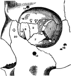
The orbit of the eye.

The Orbit of the Eye

The orbit of the eye.

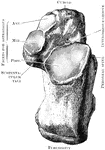
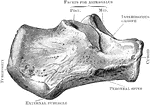
The right os calcis as seen from above.

Os Calcis Viewed from Above

The right os calcis as seen from above.

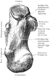
The right os calcis as seen from below.

Os Calcis Viewed from Below

The right os calcis as seen from below.

The os calcis as seen from the inner side.

Inner Side of Os Calcis

The os calcis as seen from the inner side.

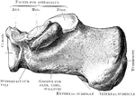
The os calcis as seen from the outer side.

Outer Side of Os Calcis

The os calcis as seen from the outer side.

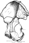
The external surface of the right os innominatum.

Os Innominatum

The external surface of the right os innominatum.

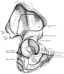
The internal surface of the right os innominatum.

Os Innominatum

The internal surface of the right os innominatum.

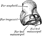
The left os magnum, the outer side seen from below.

Os Magnum

The left os magnum, the outer side seen from below.

The left os magnum seen from the internal posterior surface.

Os Magnum

The left os magnum seen from the internal posterior surface.

Ossifying cartilage showing loops of blood vessels.

Ossifying Cartilage

Ossifying cartilage showing loops of blood vessels.

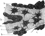
Osteoblasts from the parietal bone of a human embryo thirteen weeks old. Labels: a, bony septa with the cells of he lacunae; b, layers of osteoblasts; c, the latter in transition to bone corpuscles.

Osteoblasts from Embryo

Osteoblasts from the parietal bone of a human embryo thirteen weeks old. Labels: a, bony septa with…
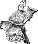
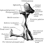
Right palate bone from behind.

Palate Bone from Behind

Right palate bone from behind.

Right palate bone as seen from behind.

Palate Bone Viewed from Behind

Right palate bone as seen from behind.

Palate bone. Palate bones form the back part of the roof of the mouth; part of the floor and outer wall of the nasal fossae, and a very small portion of the floor of the orbit.

Human Palate Bone

Palate bone. Palate bones form the back part of the roof of the mouth; part of the floor and outer wall…
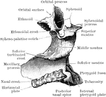
Right palate bone seen from inner side.

Inner Side of Palate Bone

Right palate bone seen from inner side.

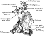
Outer aspect of palate bone.

Outer Aspect of Palate Bone

Outer aspect of palate bone.

Right palate bone seen from outer side.

Outer Side of Palate Bone

Right palate bone seen from outer side.

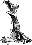
The palate bones.

The Palate Bones

The palate bones.

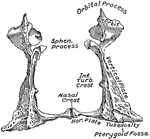
The two palate bones in their natural position.

Palate Bones

The two palate bones in their natural position.

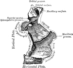
Left palate bone. Internal view.

Internal View of Palate

Left palate bone. Internal view.

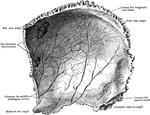
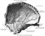
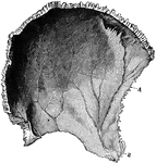
Parietal bone of the human skull, inner surface. The parietal bones form the greater part of the sides and roof of the skull. Labels: A, parietal depression; E, furrow for ramification of arteries.

Parietal Bone of the Human Skull

Parietal bone of the human skull, inner surface. The parietal bones form the greater part of the sides…
Right parietal bone, inner surface.

Inner Surface of the Parietal Bone

Right parietal bone, inner surface.

The inner surface of the right parietal bone.

Inner Surface of the Parietal Bone

The inner surface of the right parietal bone.

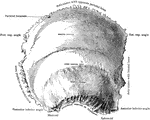
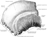
Right parietal bone, outer surface.

Outer Surface of the Parietal Bone

Right parietal bone, outer surface.

The outer side of the right parietal bone.

Outer Surface of the Parietal Bone

The outer side of the right parietal bone.

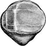
The right patella, dorsal surface.

Patella

The right patella, dorsal surface.

View of the posterior surface of the right patella, showing diagrammatically the areas of contact with the femur in different positions of the knee.

Patella

View of the posterior surface of the right patella, showing diagrammatically the areas of contact with…
Anterior surface (A) and posterior surface (B) of right patella.

Patela

Anterior surface (A) and posterior surface (B) of right patella.

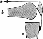
Diagram to show mechanism of fracture of the patella by muscular action. a, Line of action of quadriceps muscle; b, femur; c, tibia.

Mechanism of Fracture of the Patella by Muscular Action

Diagram to show mechanism of fracture of the patella by muscular action. a, Line of action of quadriceps…
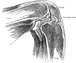
Showing position of patella in relation to condyles of femur with knee flexed at a right angle.

Position of Patella in Relation to Condyles of Femur with Knee Flexed

Showing position of patella in relation to condyles of femur with knee flexed at a right angle.

Showing position of patella in relation to condyles of femur with knee partially flexed.

Position of Patella in Relation to Condyles of Femur with Knee Partially Flexed

Showing position of patella in relation to condyles of femur with knee partially flexed.
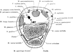
Section through the patella and one inch above the condyles.

Cross Section Through Patella

Section through the patella and one inch above the condyles.

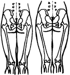
Diagram showing the position of the pelvic and thigh bones in back view in the male and female.

Position of the Pelvic and Thigh Bones in the Male and Female

Diagram showing the position of the pelvic and thigh bones in back view in the male and female.
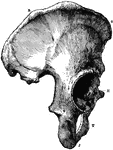
The Os Innominatum, or nameless bone, so called from bearing no resemblance to any known object, is a large irregular shaped bone, which, with its fellow of opposite side, forms the sides and front wall of the pelvic cavity. Labels: R, O, crest of ilium, just below O is seen the anterior superior spinous process; J, tuberosity of ischium; t, part of pubes, between J and T is seen the thyroid foramen: H, acetabulum, below H is seen end of ubic bone which, with its fellow of opposite side, forms the symphysis pubis.

Part of the Human Pelvic Bone

The Os Innominatum, or nameless bone, so called from bearing no resemblance to any known object, is…

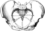
The diameters of the pelvic inlet in the female.

Pelvic Inlet

The diameters of the pelvic inlet in the female.

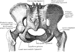
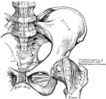
Pelvic joints as seen from behind.

Pelvic Joints Seen from Behind

Pelvic joints as seen from behind.

Pelvic joints as seen from the front.

Pelvic Joints Seen from Front

Pelvic joints as seen from the front.

The diameters of the pelvic outlet in the female.

Pelvic Outlet

The diameters of the pelvic outlet in the female.

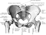
The anterior view of the articulations of the pelvis and hip.

Pelvis

The anterior view of the articulations of the pelvis and hip.

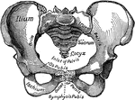
The pelvis of the human body. 1: Hip Bones; 2: Sacrum; 3: Coccyx.

Pelvis

The pelvis of the human body. 1: Hip Bones; 2: Sacrum; 3: Coccyx.

The Pelvis bone.

The Pelvis

The Pelvis bone.

The pelvis.

Pelvis

The pelvis.

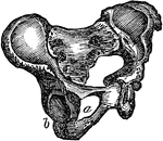
The pelvis. Labels: a, the sacrum; b, the right and the left innominatum.

The Pelvis

The pelvis. Labels: a, the sacrum; b, the right and the left innominatum.

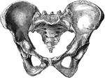
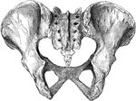
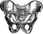
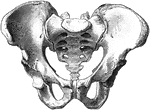
Female pelvis from before.

Female Pelvis from Before

Female pelvis from before.

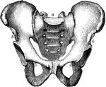
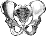
Male pelvis from before.

Male Pelvis from Before

Male pelvis from before.

The pelvis from behind.

Pelvis from Behind

The pelvis from behind.

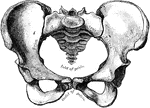
Female pelvis.

Female Pelvis

Female pelvis.

The female pelvis.

Female Pelvis

The female pelvis.

The female pelvis as seen from in front.

Female Pelvis

The female pelvis as seen from in front.

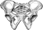
Male pelvis (top) and female pelvis (bottom). The pelvis is stronger and more massively constructed than either the cranial or thoracic cavity. It is composed of 4 bones, the ossa innominata, forming sides and front, and the sacrum and coccyx, completing it behind. The female pelvis differs from that of the male in making it better adapted for giving birth. The most notable differences are that the female pelvis is wider in every direction, giving more room for the child to pass and shallower, which lessens the distance through which the child has to be propelled; and lastly, the bones are thinner and smoother.

Human Pelvis, Male and Female

Male pelvis (top) and female pelvis (bottom). The pelvis is stronger and more massively constructed…
Anterior view of the male pelvis.

Male Pelvis

Anterior view of the male pelvis.

The male pelvis.

Male Pelvis

The male pelvis.

The male pelvis as seen from in front.

Male Pelvis

The male pelvis as seen from in front.

Side view of pelvis.

Side View of Pelvis

Side view of pelvis.

The phalanges of the fingers (palmar aspect).

Phalanges

The phalanges of the fingers (palmar aspect).

The phalanges of the toes (dorsal aspect).

Phalanges

The phalanges of the toes (dorsal aspect).

Phalanges of right second toe, plantar surface.

Phalanges of Second Toe

Phalanges of right second toe, plantar surface.

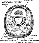
Horizontal section through the middle of the first phalanx.

Phalanx

Horizontal section through the middle of the first phalanx.

The pisiform may be known by its small size and by its presenting a single articular facet. It is situated on a plane anterior to the other bones of the carpus; it is spheroidal in form, with its long diameter directed vertically. Shown is the left pisiform, showing posterior and lateral surfaces.

Pisiform Bone

The pisiform may be known by its small size and by its presenting a single articular facet. It is situated…