This human anatomy ClipArt gallery offers 825 illustrations of the human skeletal system, including images of both the axial skeleton and the appendicular skeleton. The human axial skeleton includes 80 bones formed by the vertebral column (spine), the thoracic cage (e.g., ribs, sternum), and the skull. The axial skeleton is responsible for the upright position of the body. The human appendicular skeleton is composed of 126 bones formed by the pectoral girdles, the upper and lower limbs, and the pelvic girdle. These bones function in locomotion as well as protection of vital organs.
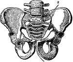
Pelvic Bone, Male
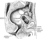
A bony structure located at the bottom of the spine. The human sacrum forms the back part of the pelvis,…
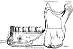
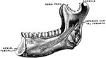
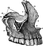
Mandible
The mandible is the largest and strongest bone of the face. It serves for the reception of the lower…
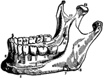
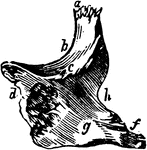
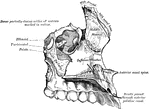
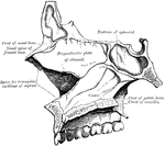
Maxilla
The largest bones of the face, excepting the mandible, and form, by their union, the whole of the upper…
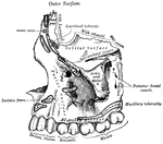
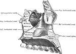
Maxilla
The largest bones of the face, excepting the mandible, and form, by their union, the whole of the upper…
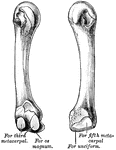
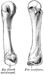
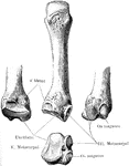
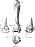
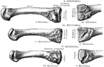
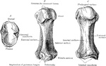
Metacarpal and Phalangeal Bones of the Fingers
The metacarpal and phalangeal bones of the fingers, with their tendons and ligaments. Labels: 1, metacarpal…
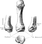
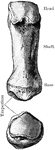
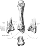

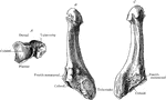
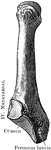
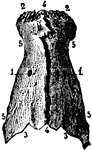
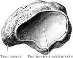
Metatarsal Bones
View of the bases and shafts of the second, third, and fourth metatarsal bones of the right foot.

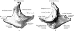
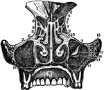
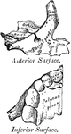
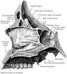
The Nasal Bones
The nasal fossae (bones), which together form the cavity of the nose and are separated from either other…
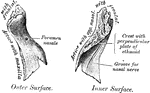
Human Nostril Bone
Inferior turbinated bone, convex surface. The inferior turbinated bones are situated on the outer wall…
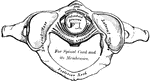
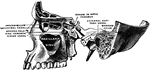
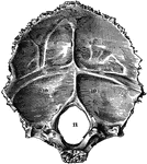
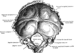
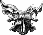
Occipital Bone and Cervical Vertebrae
Occipital bone and first three cervical vertebrae with ligaments, from in front.
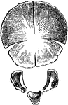
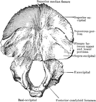
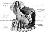
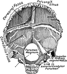
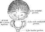
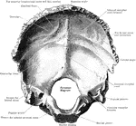
Occipital Bone of the Human Skull
Occipital bone of the human skull, inner surface. It is situated at the back and base of the skull.…
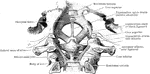
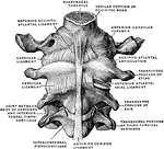
Ligaments of the Occipital Bone
Dissection from behind the ligaments connecting the occipital bone, the atlas, and the axis with each…
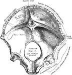
Occipito-atlantal Articulation
The occipito-atlantal articulation, which is a double condyloid formed by the condyles of the occipital…