This human anatomy ClipArt gallery offers 825 illustrations of the human skeletal system, including images of both the axial skeleton and the appendicular skeleton. The human axial skeleton includes 80 bones formed by the vertebral column (spine), the thoracic cage (e.g., ribs, sternum), and the skull. The axial skeleton is responsible for the upright position of the body. The human appendicular skeleton is composed of 126 bones formed by the pectoral girdles, the upper and lower limbs, and the pelvic girdle. These bones function in locomotion as well as protection of vital organs.
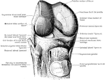
The knee joint, which consists of three articulations in one.

Knee Joint

The knee joint, which consists of three articulations in one.

The knee joint, which consists of three articulations in one.

Knee Joint

The knee joint, which consists of three articulations in one.

A knee joint cut open from behind to show the ligaments.

Knee Joint

A knee joint cut open from behind to show the ligaments.

The knee joint. Labels: h, the patella; f, the tendon.

The Knee Joint

The knee joint. Labels: h, the patella; f, the tendon.

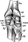
Dissection of knee joint from behind.

Dissection of Knee Joint From Behind

Dissection of knee joint from behind.

Dissection of knee joint from the front with patella thrown down.

Dissection of Knee Joint From Front

Dissection of knee joint from the front with patella thrown down.
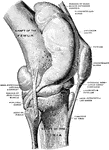
Right knee joint from the lateral surface. The joint cavity and several bursae have been injected with a stiffening medium and then dissected out.

Knee Joint from Lateral Surface

Right knee joint from the lateral surface. The joint cavity and several bursae have been injected with…
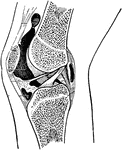
Right knee joint showing interior ligaments.

Knee Joint Showing Interior Ligaments

Right knee joint showing interior ligaments.

Section through right knee joint.

Cross Section Through Knee Joint

Section through right knee joint.

Epiphyseal lines in the neighborhood of the knee joint and their relationship to the synovial membrane.
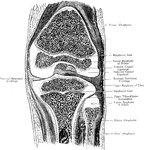
Epiphyseal lines in the Knee Joint

Epiphyseal lines in the neighborhood of the knee joint and their relationship to the synovial membrane.

A frontal section through the right knee joint of a boy. Seen from behind.

Frontal Section Through Knee Joint

A frontal section through the right knee joint of a boy. Seen from behind.
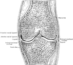
Frontal section through middle of right knee joint. Seen from behind.

Frontal Section Through Knee Joint

Frontal section through middle of right knee joint. Seen from behind.
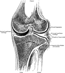
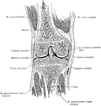
Frontal section through knee joint, showing articulating surfaces and epiphyseal lines.

Frontal Section Through Knee Joint

Frontal section through knee joint, showing articulating surfaces and epiphyseal lines.
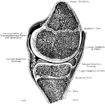
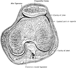
Horizontal section through right knee joint.

Horizontal Section Through Knee Joint

Horizontal section through right knee joint.

Right knee joint opened, and the knee flexed. Seen from before.

Opened and Flexed Knee Joint

Right knee joint opened, and the knee flexed. Seen from before.

A sagittal section through the right knee joint of a boy. Seen from the outer side.

Sagittal Section Through Knee Joint

A sagittal section through the right knee joint of a boy. Seen from the outer side.
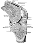
Right knee joint. Sagittal section through the external condyle of the femur. Mesal half of section, from lateral side. The knee is slightly flexed; the joint surfaces have been pulled a little apart.

Sagittal Section Through Knee Joint

Right knee joint. Sagittal section through the external condyle of the femur. Mesal half of section,…
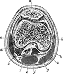
Transverse section of the knee joint through the center of the patella. Labels: a, Bursa patellae; b, internal lateral ligament and inner condyle; c, external lateral ligament and outer condyle; d, biceps; e, semimembranosus; f, semitendinosus; g, gracilis tendon; h, sartorius. 1, internal popliteal nerve; 2, external popliteal nerve; 3, internal saphenous vein.

Transverse Section of the Knee Joint

Transverse section of the knee joint through the center of the patella. Labels: a, Bursa patellae; b,…
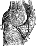
A vertical section of the knee joint. Labels: femur; 3, patella; 2, 4, ligaments of the patella; 5, tibia; 6, cartilage of the tibia; 12, cartilage of the membrane.

Vertical Section of the Knee Joint

A vertical section of the knee joint. Labels: femur; 3, patella; 2, 4, ligaments of the patella; 5,…
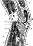
Vertical section of knee joint distended with fluid. Labels: a, Vastus externus; b, crureus; c, short head, and d, long head of biceps; e, plantaris; f, gastrocnemius; g, popliteus; h, soleus; i, tibialis posticus; j, bursa patellae; k, ligamentum patellae; l, ligamentum mucosum; m, anterior crucial ligament; n, external semilunar cartilage. 1, external popliteal nerve; 2, popliteal artery.

Vertical Section of the Knee Joint

Vertical section of knee joint distended with fluid. Labels: a, Vastus externus; b, crureus; c, short…
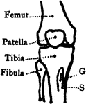
Bones of the knee also showing muscle. Labels: s, insertion of the sartorius; g, insertion of the gracilis.
Bones of the Knee

Bones of the knee also showing muscle. Labels: s, insertion of the sartorius; g, insertion of the gracilis.

Median frontal section of the right knee, viewed from in front.

Median Frontal Section Through Knee

Median frontal section of the right knee, viewed from in front.

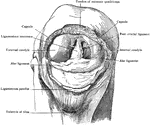
Patella removed from right knee, which is strongly flexed to show alar ligaments and ligamentum mucosum. A probe is passed beneath the latter.

Patella Removed from Knee

Patella removed from right knee, which is strongly flexed to show alar ligaments and ligamentum mucosum.…
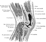
Sagittal section of the right knee, viewed from the outer side. The joint cavity proper lies to each side of the anterior crural ligament.

Sagittal Section Through Knee

Sagittal section of the right knee, viewed from the outer side. The joint cavity proper lies to each…

An anterior view of the right knee-joint.

Knee-joint

An anterior view of the right knee-joint.

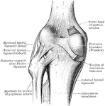
An posterior view of the right knee-joint.

Knee-joint

An posterior view of the right knee-joint.

The right knee-joint showing interior ligaments.

Knee-joint

The right knee-joint showing interior ligaments.

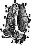
The inner surface of the right lachrymal bone.

Lachrymal Bone

The inner surface of the right lachrymal bone.

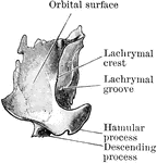
The orbital surface of the right lachrymal bone.

Lachrymal Bone

The orbital surface of the right lachrymal bone.

Right lachrymal bone, inner aspect. Upper part completes anterior ethmoidal cells, lower looks into middle nasal meatus.

Inner Aspect of Lachrymal Bone

Right lachrymal bone, inner aspect. Upper part completes anterior ethmoidal cells, lower looks into…

Lachrymal Bone. The lachrymal are the smallest and most fragile bones fo the face. They are situated at the front part of the inner wall of the orbit.

Human Lachrymal Facial Bone

Lachrymal Bone. The lachrymal are the smallest and most fragile bones fo the face. They are situated…
The lacrimal bone (part of the eye orbit).

The Lacrimal Bone

The lacrimal bone (part of the eye orbit).

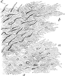
Lamellae torn off from a decalcified human parietal bone at some depth from the surface. Labels: a, lamellae, showing reticular fibers; b, darker part, where several lamellae are superposed; perforating fibers. Apertures through which perforating fibers has passed, are seen especially in the lower part of the figure.

Lamellae

Lamellae torn off from a decalcified human parietal bone at some depth from the surface. Labels: a,…
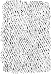
Shown is a thin layer peeled off from a soft bone, which is intended to represent the reticular structure of a lamella.

Reticular Structure of a Lamellae

Shown is a thin layer peeled off from a soft bone, which is intended to represent the reticular structure…

A view of the left scaphoid seen from behind.

Left Scaphoid

A view of the left scaphoid seen from behind.

A view of the left scaphoid seen from in front.

Left Scaphoid

A view of the left scaphoid seen from in front.

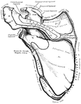
The anterior surface of the left scapula.

Left Scapula

The anterior surface of the left scapula.

The posterior surface or the left scapula.

Left Scapula

The posterior surface or the left scapula.

"Bones of the leg. a, femur; b, tibia; c, fibula; d, tarsal bones; e, metatarsal bones; f, phalanges; g, patella." —Martin, 1917

Leg Bones

"Bones of the leg. a, femur; b, tibia; c, fibula; d, tarsal bones; e, metatarsal bones; f, phalanges;…

Bones of the leg (tibia and fibula).

Leg Bones

Bones of the leg (tibia and fibula).

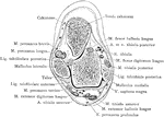
Section two and a half inches above right ankle joint.

Cross Section of Leg, Two and a Half Inches above Ankle

Section two and a half inches above right ankle joint.
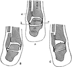
The right tibia and fibula in their normal relation, rear view.

Bones of the Lower Leg

The right tibia and fibula in their normal relation, rear view.

Section through the external malleolus.

Cross Section Through Malleolus of Leg

Section through the external malleolus.

Ligaments of the right leg, from in front.

Ligaments of the Leg

Ligaments of the right leg, from in front.

Transverse section through the lower third of the leg. Labels: a, tibialis anticus; b, extensor longus hallucis; c, extensor communis digitorum; d, peroneus brevis; e, peroneus longus; t, tibialis posticus; g, flexor longus digitorum; h, flexor longus hallucis; i, gastrocnemius and soleus; j, short saphenous nerve and vein; k, anterior tibial vessels and nerve; l, peroneal vessels; m, posterior tibial vessels and nerve; n, musculo-cutaneous nerve.

Transverse Section Through the Lower Leg

Transverse section through the lower third of the leg. Labels: a, tibialis anticus; b, extensor longus…

"Little openings (Haversian canals) are seen, and around them are arranged rings of bone with little dark spaces (<em>lacunae</em>), from which branch out fine dark lines (<em>analiculi</em>)." &mdash; Blaisedell, 1904

Cross-section from a shaft of a long bone

"Little openings (Haversian canals) are seen, and around them are arranged rings of bone with little…

The longitudinal section of a long bone.

Structure of a Long Bone

The longitudinal section of a long bone.

The lower extremity of the human body. 1: Head of femur; 2: Femur; 3: Patella; 4: Tibia; 5: Fibula; 6: Tarsus; 7: Metatarsus; 8: Phalanges.

Lower Extremity

The lower extremity of the human body. 1: Head of femur; 2: Femur; 3: Patella; 4: Tibia; 5: Fibula;…
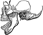
Dislocation of the lower jaw.

Dislocation of the Lower Jaw

Dislocation of the lower jaw.

The last lumbar vertebra and the sacrum seen from the ventral side.

Lumbar and Sacrum of Spine

The last lumbar vertebra and the sacrum seen from the ventral side.

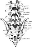
Diagram of the lumbar interlaminar spaces, showing the position of the fourth lumbar spine.

Lumbar Showing Position of Fourth Lumbar Spine

Diagram of the lumbar interlaminar spaces, showing the position of the fourth lumbar spine.
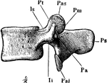
A lumbar vertebra, seen from the left side. Labels: Ps, spinous process; Pas, anterior articular process; Pai, posterior articular process; Pt, transverse process.

A Lumbar Vertebra

A lumbar vertebra, seen from the left side. Labels: Ps, spinous process; Pas, anterior articular process;…
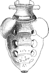
The last lumbar vertebra and the sacrum seen from the ventral side.

The Last Lumbar Verebra and the Sacrum

The last lumbar vertebra and the sacrum seen from the ventral side.
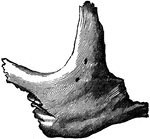
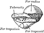
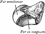
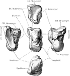
The right magnum bone.

Magnum Bone

The right magnum bone.

Malar (cheek) bone. The malar bones form the prominence of the cheek, and part of the outer wall and floor of the orbit.

Human Malar (Cheek) Bone

Malar (cheek) bone. The malar bones form the prominence of the cheek, and part of the outer wall and…

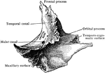
Right malar bone.

Malar Bone

Right malar bone.

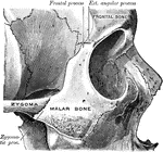
Right malar bone in situ.

Malar Bone

Right malar bone in situ.

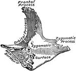
The outer side (A) and inner side (B) of the right malar bone.

Malar Bone

The outer side (A) and inner side (B) of the right malar bone.

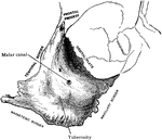
Right malar bone, inner aspect.

Inner Aspect of Malar Bone

Right malar bone, inner aspect.

Right malar bone, outer aspect.

Outer Aspect of Malar Bone

Right malar bone, outer aspect.