This human anatomy ClipArt gallery offers 825 illustrations of the human skeletal system, including images of both the axial skeleton and the appendicular skeleton. The human axial skeleton includes 80 bones formed by the vertebral column (spine), the thoracic cage (e.g., ribs, sternum), and the skull. The axial skeleton is responsible for the upright position of the body. The human appendicular skeleton is composed of 126 bones formed by the pectoral girdles, the upper and lower limbs, and the pelvic girdle. These bones function in locomotion as well as protection of vital organs.

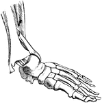
Ligaments on outer aspect of ankle and on dorsum and outer aspects of foot.

Outer Aspect of Foot Ligaments

Ligaments on outer aspect of ankle and on dorsum and outer aspects of foot.

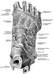
Ligaments on plantar aspect of foot.

Plantar Aspect of Foot Ligaments

Ligaments on plantar aspect of foot.

The internal border of the skeleton of the foot.

Foot Skeleton

The internal border of the skeleton of the foot.

The external border of the skeleton of the foot.

Foot Skeleton

The external border of the skeleton of the foot.

Section through the bones and ligaments of the foot. The parts of the joints are well shown.

Bones and Ligaments of the Foot

Section through the bones and ligaments of the foot. The parts of the joints are well shown.

The bones of the foot. Labels: Ca, Calcaneum, or heel bone; Ta, articular surface for tibia on the astragalus; Cb, the cuboid bone

Bones of the Foot

The bones of the foot. Labels: Ca, Calcaneum, or heel bone; Ta, articular surface for tibia on the astragalus;…

Bones of the foot (tarsus, metatarsus, and phalanges).

Bones of the Foot

Bones of the foot (tarsus, metatarsus, and phalanges).

Bones of the foot. At <em>e d f g h</em> are the 7 bones of the tarsus; at <em>a</em> are the 5 bones of the metatarsus; and at <em>b c</em> are the 14 bones of the toes (26 in all).

Bones of the Foot

Bones of the foot. At e d f g h are the 7 bones of the tarsus; at a are the 5 bones…

The bones of the foot. Labels: Ca, calcaneum, or os calcis; Ta, articular surface for tibia on the astragalus; N, scaphoid bone; CI, CII, first and second cuneiform bones; Cb, cuboid bone; M1, metatarsal bone of great toe.

Bones of the Foot

The bones of the foot. Labels: Ca, calcaneum, or os calcis; Ta, articular surface for tibia on the astragalus;…

"Bones of Human Foot, or Pes, the third principal segment of the hind limb, consisting of tarsus, metatarsus, and phalanges. a, astralagus; ca, calcaneum; n, navicular, or scaphoid; co, cuboid; c1, c2, c3, entocuneiform, mesocuneiform, and ectocuneiform, or inner, middle, and outer cuneiform bones. The foregoing seven bones constitute the tarsus, and m1 to m5, first to the fifth metatarsal, constitue the metatarsus. The remaining fourteen bones are the phalanges, three to each digit excepting the great toe; h, distal phalanx of the hallux or great toe." -Whitney, 1911

Bones of Human Foot

"Bones of Human Foot, or Pes, the third principal segment of the hind limb, consisting of tarsus, metatarsus,…

The upper surface of the bones of the foot. Labels: 1, the surface of the astragulus or ankle bones, where it unites with the tibia. 2, The body of the astragulus. 3, Calcis or heel-bone. 4, The scaphoid. 5,6,7, The cuneiform. 8, The cuboid. 9,The metatarsal bones. 10,11, The phalanges of the great toe. 12, 13, 14, The phalanges of the other toes.

Bones of the Foot

The upper surface of the bones of the foot. Labels: 1, the surface of the astragulus or ankle bones,…

The bones of the right foot, viewed from above.

Bones of the Foot

The bones of the right foot, viewed from above.

Bones of right foot, dorsal aspect. A, outer series; B; inner series.

Bones of the Foot

Bones of right foot, dorsal aspect. A, outer series; B; inner series.

Oblique section of the foot showing six synovial membranes.

Bones of the Foot

Oblique section of the foot showing six synovial membranes.

Section through the right tarsometatarsal joint. Upper surface.

Cross Section Through Tarso-Metatarsal Joint of Foot

Section through the right tarsometatarsal joint. Upper surface.

Joints of the right foot, from the back of the foot.

Joints of the Foot

Joints of the right foot, from the back of the foot.

Longitudinal section through right foot in axis of first metatarsal bone.

Longitudinal Section Through Foot

Longitudinal section through right foot in axis of first metatarsal bone.

Bones of the upper surface of the left foot. Labels: 1, astragalus; 2, its anterior face; 3, os calcis; 4, navicular, or scaphoid; , internal cuneiform; 6, middle cuneiform; 7, external cuneiform; 8, cuboid bone; 9, 9, metatarsal bones; 10, first phalanx of the big toe; 11, second phalanx of the big toe; 12, the first, 13, second and 14, third phalanges of the other toes.

Upper Surface of the Left Foot

Bones of the upper surface of the left foot. Labels: 1, astragalus; 2, its anterior face; 3, os calcis;…

Transverse section through the bones of the forearm (radius and ulna), taken at about the middle of their length (schematized).

Forearm Bones

Transverse section through the bones of the forearm (radius and ulna), taken at about the middle of…
Bones of the forearm.

Bones of the Forearm

Bones of the forearm.

Front view of the bones of the forearm

Back View of the Bones of the Forearm

Front view of the bones of the forearm

Bones of the right forearm,with their ligaments; volar aspect.

Bones and Ligaments of Forearm

Bones of the right forearm,with their ligaments; volar aspect.

The bones of the right forearm, dorsal view.

Dorsal View of the Bones of the Forearm

The bones of the right forearm, dorsal view.

Front view of the bones of the forearm

Front View of the Bones of the Forearm

Front view of the bones of the forearm

The bones of the right forearm, ventral view.

Ventral View of the Bones of the Forearm

The bones of the right forearm, ventral view.

A fracture of the fibula, with dislocation of the foot outward.

Fractured Fibula

A fracture of the fibula, with dislocation of the foot outward.

An oblique fracture of the shaft of the tibia.

Fractured Tibia

An oblique fracture of the shaft of the tibia.

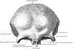
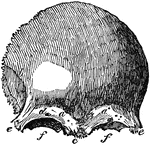
A front view of the frontal bone; a,a, frontal sinuses; b, the temporal arch, beneath which lies the temporal muscle, which closes the lower jaw; d, the supra-orbitary hole for a passage of a branch of the fifth pair of nerves.

Frontal Bone

A front view of the frontal bone; a,a, frontal sinuses; b, the temporal arch, beneath which lies the…

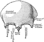
The frontal bone, seen from in front.

Frontal Bone

The frontal bone, seen from in front.

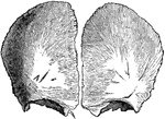
The frontal bone at birth. Developed from two lateral halves.

Frontal Bone at Birth

The frontal bone at birth. Developed from two lateral halves.

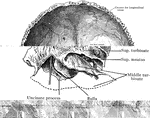
The frontal bone from behind.

Frontal Bone from Behind

The frontal bone from behind.

Frontal bone of the human skull, outer surface. The frontal bone forms the forehead, roof of the orbital cavities, and the nasal cavity. Labels: 1, frontal eminence; 7, roof of orbital cavitiy; 10, orbital arch.

Frontal Bone of the Human Skull

Frontal bone of the human skull, outer surface. The frontal bone forms the forehead, roof of the orbital…

The frontal bone viewed from below.

Frontal Bone Viewed From Below

The frontal bone viewed from below.

Anterior view of the frontal bone.

Anterior View of Frontal Bone

Anterior view of the frontal bone.

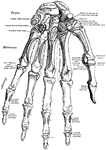
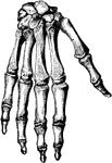
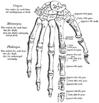
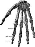
The bones of palmar surface of the left hand.

Hand

The bones of palmar surface of the left hand.

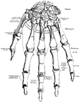
The bones of dorsal surface of the left hand.

Hand

The bones of dorsal surface of the left hand.

Bones of the hand.

Bones of the Hand

Bones of the hand.

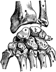
Bones and ligaments of the hand. There are 27 bones in all, including 8 small bones called the carpal bones (d). These are tightly packed together, and lie next to the bones of the forearm. The metacarpal bones (e) make the framework of the flat part of the hand and are very much like the first row of the bones of the fingers (f) but they are firmly bound together by ligaments. Ligaments (d d) bind the metacarpal bones together at their beginning and other ligaments bind them at their ends (e e) where the bones of the fingers are jointed with them. Other ligaments bind the carpal bones together (a,b,c,g).

Bones and Ligaments of the Hand

Bones and ligaments of the hand. There are 27 bones in all, including 8 small bones called the carpal…
A plan of the development of the bones of the hand.

Hand Development

A plan of the development of the bones of the hand.

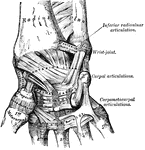
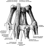
A anterior view of the ligaments of the wrist and hand.

Hand Ligaments

A anterior view of the ligaments of the wrist and hand.

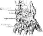
A posterior view of the ligaments of the wrist and hand.

Hand Ligaments

A posterior view of the ligaments of the wrist and hand.

The bones of the hand (carpus, metacarpus, and phalanges).

Bones of the Hand

The bones of the hand (carpus, metacarpus, and phalanges).

Bones of the hand.

Bones of the Hand

Bones of the hand.

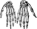
The bones of the right hand.

Bones of the Hand

The bones of the right hand.

Metacarpal bones and first phalanges of the second to the fifth of the right hand, with ligaments, from the volar surface.

Bones of the Hand

Metacarpal bones and first phalanges of the second to the fifth of the right hand, with ligaments, from…

The bones of the hand.

Bones of the Hand

The bones of the hand.

Section through the distal end of the right metacarpal bones.

Cross Section Through Distal End of the Metacarpal Bones of the Hand

Section through the distal end of the right metacarpal bones.

Section through the middle of the right metacarpal bones.

Cross Section Through Middle Metacarpal Bones of the Hand

Section through the middle of the right metacarpal bones.
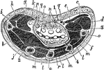
Horizontal section of the hand through the middle of the thenar and hypothenar eminences. Labels: a, metacarpal bone; b, first dorsal interosseous; c, palmaris brevis; d, abductor quinti digiti; e, flexor brevis min. dig.; f, opponens min. dig.; g, flexor brevis poll.; h, abductor brevis poll.; i, opponens poll.; j, adductor poll.; k, flexor long. poll.; l, dorsal interossei; m, palmar interossei; n, flexor sublimis; o, flexor profundus; p, superfic. volae; q, median nerve, and (on inner side) ulnar artery nerve; r, deep volar arch; 1, palmar fascia; 2, outer septum; 3, inner septum; 4, deep fascia of palm.

Horizontal Section of the Hand

Horizontal section of the hand through the middle of the thenar and hypothenar eminences. Labels: a,…
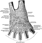
Joints of the right hand, from the back of the hand.

Joints of the Hand

Joints of the right hand, from the back of the hand.

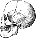
Bones of the head.

Bones of the Head

Bones of the head.

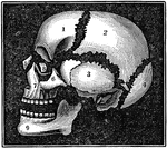
A diagram of the bones of the head. Label: 1, frontal lobe; 2, parietal bone; 3, temporal bone; 4, occipital bone; 5, nasal bone; 6, malar bone; 7, upper jaw; 8, os unguis; 9, lower jaw.

Bones of the Head

A diagram of the bones of the head. Label: 1, frontal lobe; 2, parietal bone; 3, temporal bone; 4, occipital…
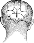
The rear view of a human face.

Human Head

The rear view of a human face.

Line of pressure between hip and knee.

Line of Pressure Between Hip and Knee

Line of pressure between hip and knee.

Ligaments and bones of the hip joint and pelvis. Labels: 1, posterior sacro-iliac ligament; 2, greater sacro sciatic ligament; 3, lesser sacro-sciatic ligament; 4, greater sacro-sciatic notch; 5, lesser sacro-sciatic notch; 6, cotyloid ligament around the acetabulum; 7, ligamentum teres; 8, line of attachment of the capsular ligament of the hip-joint, posteriorly; 9, obturator ligament.

Bones and Ligaments of the Hip and Pelvis

Ligaments and bones of the hip joint and pelvis. Labels: 1, posterior sacro-iliac ligament; 2, greater…
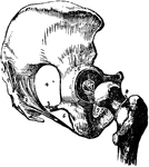
Dislocation of the hip upon the dorsum ilii.

Hip Dislocation upon the Dorsum Illi

Dislocation of the hip upon the dorsum ilii.

Hip dislocation upon the pubes.

Hip Dislocation upon the Pubes

Hip dislocation upon the pubes.

The hip joint. 1 and 2: Capsular ligament.

Hip Joint

The hip joint. 1 and 2: Capsular ligament.

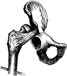
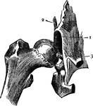
Hip joint with capsular ligament cut away. 1: Margin of socket; 2: Portion of capsular ligament; 3: Round ligament.

Hip Joint

Hip joint with capsular ligament cut away. 1: Margin of socket; 2: Portion of capsular ligament; 3:…

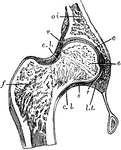
Section through the hip joint.

Hip Joint

Section through the hip joint.