This human anatomy ClipArt gallery offers 825 illustrations of the human skeletal system, including images of both the axial skeleton and the appendicular skeleton. The human axial skeleton includes 80 bones formed by the vertebral column (spine), the thoracic cage (e.g., ribs, sternum), and the skull. The axial skeleton is responsible for the upright position of the body. The human appendicular skeleton is composed of 126 bones formed by the pectoral girdles, the upper and lower limbs, and the pelvic girdle. These bones function in locomotion as well as protection of vital organs.
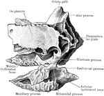
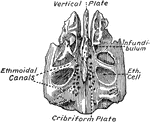
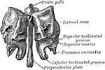
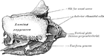
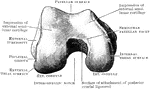
Ethmoid Bone of the Human Skull
Ethmoid bone, posterior surface. The ethmoid bone is an exceedingly light, spongy bone, placed between…
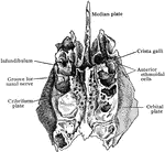
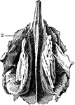
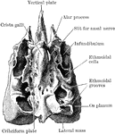
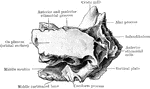
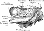
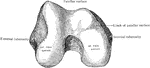
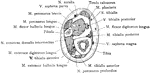
Inner Aspect of the Ethmoid Bone
The ethmoid bone, inner aspect from the left side, part of the middle turbinate having been removed.
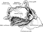
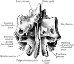
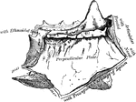
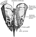
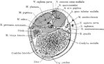
Perpendicular Plate of Ethmoid
Perpendicular plate of ethmoid, shown by removing the right lateral mass.
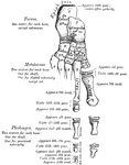
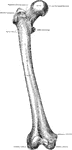
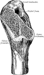
Femur
Section of the femur. 1: External view; 2: Cellular portion at end; 3: Hollow in middle; 4: Thick shell…
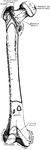
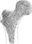
Femur
Posterior view of the femur (bone of the leg). Labels: 1, depression for round ligament; 2, the head;…
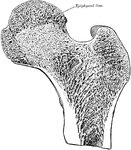
Femur
The thigh bone cut through the middle. Labels: b, hard bone; h and d, spongy bone; ma, marrow.

Ossification of the Femur and the Condition of Coxa Vara
Illustrating the ossification of the upper end of the femur and the condition of coxa vara. Labels:…
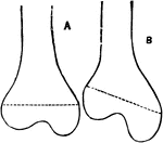
Femur in Knock-Knee
A, Normal Femur. B, Femur in an advanced state of knock-knee, showing the enlargement of the internal…
Calcar Femorale and its Relationship to Fractures of the Femur
The calcar femorale and its relationship to impacted fractures of the neck of the femur.
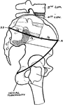
Displacement of the Femur
Diagram showing the line used by Nelaton to test upward displacement of the femur, and another which…
Femoral Spur of the Femur
In the midst of the cancellous tissue the femoral spur, which commences at the point where the neck…
Frontal Section Through Upper End of Femur
Frontal section through upper end of femur, showing arrangement of pressure and tension lamellae.
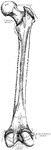
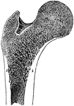
Human Femur Bone
The Femur (upper leg bone) is the longest, largest, and strongest bone in the skeleton. Labels: b, rounded…
Longitudinal Section of the Femur
The longitudinal section of the extremity of the femur, exhibiting the arrangement of the spongy substance.…
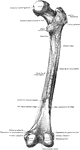
Anterior View of Human Right Femur
"Anterior View of Human Right Femur. ec, external condyle; etu, external tuberosity; ic, internal condyle;…
Youth Femur
"Right Femur of a Youth. E, E, epiphyses; gtr, ltr, greater and lesser trochanter; h, head; et, it,…
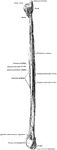
Fibula
The fibula (bone of leg). Labels: 1, head; 2, articular face; 3, insertion of external ligament; 4,…
Cross Section Four Inches Above the Lower End of the Fibula
Section four inches above the lower end of the fibula.
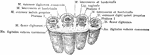
Metacarpal and Phalanges of Finger
Metacarpal bones and first phalanges of the third finger of the right hand, with ligaments, from the…

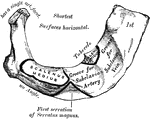
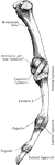
First Rib
The first rib is the shortest and the most curved of all the ribs; it is broad and flat, its surfaces…
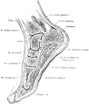
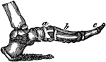
Sagittal Section of Foot and Ankle
Sagittal section of the foot and ankle passing through the great toe.
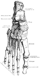
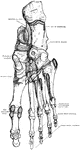
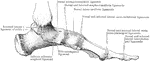
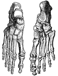
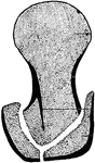
Foot Bones
"The foot. a, calcaneum; b, astragalus; c, cuboid; d, metatarsal, of which there are five; e, phalanges,…
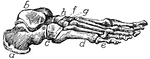
Side View of the Bone of the Foot
Bones of the foot, side view. In this figure the bones of the tarsus extend from the heel to a;…