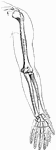
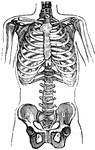
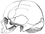
This human anatomy ClipArt gallery offers 825 illustrations of the human skeletal system, including images of both the axial skeleton and the appendicular skeleton. The human axial skeleton includes 80 bones formed by the vertebral column (spine), the thoracic cage (e.g., ribs, sternum), and the skull. The axial skeleton is responsible for the upright position of the body. The human appendicular skeleton is composed of 126 bones formed by the pectoral girdles, the upper and lower limbs, and the pelvic girdle. These bones function in locomotion as well as protection of vital organs.
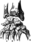
Transverse section of compact tissue of bone.

Transverse Section of Compact Bone

Transverse section of compact tissue of bone.

Bones of the arm

Arm Bones

Bones of the arm

Bones of the arm

Arm Bones

Bones of the arm

Bones of the leg

Leg Bones

Bones of the leg

A Brachycephalic cranium from above.

Brachycephalic Cranium

A Brachycephalic cranium from above.

A Brachycephalic cranium from the side.

Brachycephalic Cranium

A Brachycephalic cranium from the side.

Right calcaneum from above.

Calcaneum

Right calcaneum from above.

The left calcaneus. A. Posterior external view. B. Anterior internal view.

Calcaneus

The left calcaneus. A. Posterior external view. B. Anterior internal view.

Articulations of bones of the carpus (wrist area). Labels: 1, ulna; 2, radius; 3, inter-articular fibro-cartilage; 4, metacarpal of thumb; 5, metacarpal of first finger; 6, metacarpal of second finger; 7, metacarpal of third finger; 8, metacarpal of fourth finger; S, scaphoid; L, lunar; C, cuneiform; P, pisiform; T, T, trapezium and trapezoid; M, magnum; U, unciform.

Bones of the Carpus

Articulations of bones of the carpus (wrist area). Labels: 1, ulna; 2, radius; 3, inter-articular fibro-cartilage;…

"Cartilage or gristle is fibrous tissue glued together by a substance containing chondrine." — Richardson, 1906

Cartilage

"Cartilage or gristle is fibrous tissue glued together by a substance containing chondrine." —…
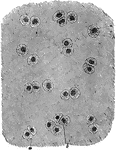
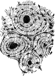
Portions of cartilage seen in section.

Cartilage Portion in Section

Portions of cartilage seen in section.

A thin slice of cartilage highly magnified, showing the cartilage cells (a,b) scattered through an almost structureless material.

Cartilage Tissue

A thin slice of cartilage highly magnified, showing the cartilage cells (a,b) scattered through an almost…
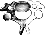
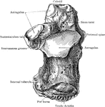
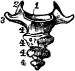
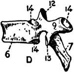
A cervical vertebra. Labels: Frt, vertebral foramen; Pai, anterior articular process; R, rudimentary rib.

A Cervical Vertebra

A cervical vertebra. Labels: Frt, vertebral foramen; Pai, anterior articular process; R, rudimentary…

A cervical vertebra of the spine, inferior surface. Labels: 1, spinous process, slightly bifid; 4, transverse process; 5, articular process, inferior surface. Below the arch, or hollow portion, is seen the solid portion, or body.

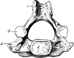
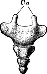
Human Cervical Vertebra Bone

A cervical vertebra of the spine, inferior surface. Labels: 1, spinous process, slightly bifid; 4, transverse…

B, the first cervical vertebra, the atlas. A, the atlas, and the second cervical vertebra, the axis; e, the odontoid process; c. the foramen.

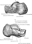
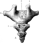
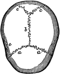
The First and Second Cervical Vertebrae

B, the first cervical vertebra, the atlas. A, the atlas, and the second cervical vertebra, the axis;…

The chest or ribcage of the human body.

Chest

The chest or ribcage of the human body.

The chest and pelvis. 1: The hip-bones.

Chest and Pelvis

The chest and pelvis. 1: The hip-bones.

The clavicle (collar bone).

Clavicle

The clavicle (collar bone).

The right clavicle, upper surface.

Clavicle

The right clavicle, upper surface.

Right clavicle and scapula with ligament, from without and somewhat in front.

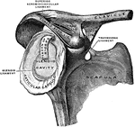
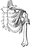
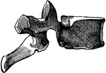
Clavicle and Scapula with Ligament

Right clavicle and scapula with ligament, from without and somewhat in front.

Fracture of the middle clavicle.

Fractured Clavical

Fracture of the middle clavicle.

The human clavicle bone (anterior portion of the shoulder girdle).

The Human Clavicle

The human clavicle bone (anterior portion of the shoulder girdle).

The vertebrae of the coccyx.

The Coccyx

The vertebrae of the coccyx.

The coccyx.

The Coccyx

The coccyx.

Anterior surface of the coccyx. Labels:5, sacrum; 6, cornu; 7, transverse process; 8, transverse process.

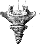
Coccyx, Anterior Surface of the

Anterior surface of the coccyx. Labels:5, sacrum; 6, cornu; 7, transverse process; 8, transverse process.

Posterior surface of the coccyx. Labels: 1, transverse process; 2, transverse process; 3, sacrum; 4, cornu.

Coccyx, Posterior Surface of the

Posterior surface of the coccyx. Labels: 1, transverse process; 2, transverse process; 3, sacrum; 4,…
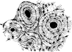
"Compact bone consists of a series of concentric layers of bone disposed around a canal called the Haversian canal which affords passage for the blood-vessels." — Richardson, 1906

Compact Bone

"Compact bone consists of a series of concentric layers of bone disposed around a canal called the Haversian…
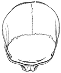
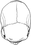
The bones of the top of the head are fastened together by what are called sutures which are locked together by little teeth of bone that shoot by each other, as shown in the figure. <em>b</em> is the suture at the top of the head between the two parietal bones; <em>a a</em> is that between the two parietal bones and the frontal bone in front; and <em>c c </em> is that between the parietal bones and the bone at the back part of the cranium. These bones are not completely formed in the newborn infant.

Cranial Sutures

The bones of the top of the head are fastened together by what are called sutures which are locked together…
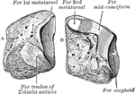
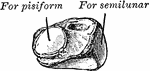
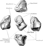
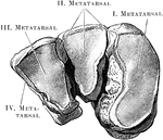
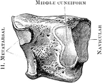
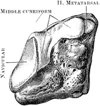
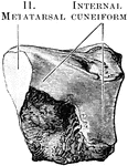
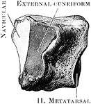
Right cuboid. A, inner aspect; B, posterior aspect, outer, and inferior surfaces.

Cuboid

Right cuboid. A, inner aspect; B, posterior aspect, outer, and inferior surfaces.

The left cuboid. A. Anterior internal view. B. Posterior external view.

Cuboid

The left cuboid. A. Anterior internal view. B. Posterior external view.

The right cuboid bone (outer side).

Cuboid

The right cuboid bone (outer side).

The right cuboid bone (inner side).

Cuboid

The right cuboid bone (inner side).

The left internal cuneiform. A. Anterior internal view. B. Posterior external view.

Cuneiform

The left internal cuneiform. A. Anterior internal view. B. Posterior external view.

The cuneiform may be distinguished by its pyramidal shape, and by its having an oval, isolated facet for articulation with pisiform bone. It is situated at the upper and inner side of the carpus. Shown is the left cuneiform, showing palmar and lateral surfaces.

Cuneiform Bone

The cuneiform may be distinguished by its pyramidal shape, and by its having an oval, isolated facet…
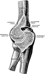
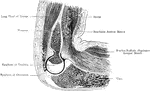
The right cuneiform bone.

Cuneiform Bone

The right cuneiform bone.

Anterior view of the three cuneiform bones of the right foot.

Cuneiform Bones

Anterior view of the three cuneiform bones of the right foot.

The right external cuneiform (inner side).

External Cuneiform

The right external cuneiform (inner side).

The right external cuneiform (outer side).

External Cuneiform

The right external cuneiform (outer side).

The right internal cuneiform (inner side).

Internal Cuneiform

The right internal cuneiform (inner side).

The right internal cuneiform (outer side).

Internal Cuneiform

The right internal cuneiform (outer side).

The right middle cuneiform (inner side).

Middle Cuneiform

The right middle cuneiform (inner side).

The right middle cuneiform (outer side).

Middle Cuneiform

The right middle cuneiform (outer side).

Right external cuneiform. A, inner aspect; B, outer aspect.

Right External Cuneiform

Right external cuneiform. A, inner aspect; B, outer aspect.

A deformed chest.

Deformed Chest

A deformed chest.

A deformed thorax and spinal column.

Deformed Thorax

A deformed thorax and spinal column.

A Dolichocephalic cranium from above.

Dolichocephalic Cranium

A Dolichocephalic cranium from above.

A Dolichocephalic cranium from the side.

Dolichocephalic Cranium

A Dolichocephalic cranium from the side.

Side view of the dorsal vertebra.

Dorsal vertebra

Side view of the dorsal vertebra.

A dorsal vertebra, side view.

Side View of a Dorsal Vertebra

A dorsal vertebra, side view.

The elbow joint (articulatio cubiti).

Elbow Joint

The elbow joint (articulatio cubiti).

Right elbow joint, cut through at right angles to the axis of trochlea humeri, from the ulnar side.

Elbow Joint

Right elbow joint, cut through at right angles to the axis of trochlea humeri, from the ulnar side.

Anterior view of the elbow joint.

Anterior View of the Elbow Joint

Anterior view of the elbow joint.

A view from behind of a frontal section through the right elbow joint.

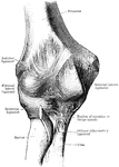
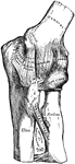
Frontal Section Through Elbow Joint

A view from behind of a frontal section through the right elbow joint.

Inner aspect of the elbow joint.

Inner Aspect of the Elbow Joint

Inner aspect of the elbow joint.

A sagittal section through the left elbow joint of a child. View from the inner side.

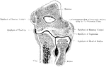
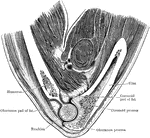
Sagittal Section Through Elbow Joint

A sagittal section through the left elbow joint of a child. View from the inner side.

Vertical section through the trochlear part of the elbow joint.

Vertical Section Through Elbow Joint

Vertical section through the trochlear part of the elbow joint.

Left elbow joint, showing posterior and external ligaments.

Elbow Showing External Ligaments

Left elbow joint, showing posterior and external ligaments.

Left elbow joint, showing anterior and internal ligaments.

Elbow Showing Internal Ligaments

Left elbow joint, showing anterior and internal ligaments.

Posterior view of elbow, showing relative position of condyles and olecranon. A, in extension; B, in flexion.

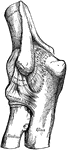
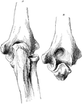
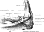
Posterior View of Elbow

Posterior view of elbow, showing relative position of condyles and olecranon. A, in extension; B, in…
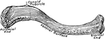
The eleventh and twelfth ribs have each a single articular facet on the head, which is of rather large size; they have no neck or tuberosity, and are pointed at the extremity.

Eleventh Rib

The eleventh and twelfth ribs have each a single articular facet on the head, which is of rather large…