This human anatomy ClipArt gallery offers 825 illustrations of the human skeletal system, including images of both the axial skeleton and the appendicular skeleton. The human axial skeleton includes 80 bones formed by the vertebral column (spine), the thoracic cage (e.g., ribs, sternum), and the skull. The axial skeleton is responsible for the upright position of the body. The human appendicular skeleton is composed of 126 bones formed by the pectoral girdles, the upper and lower limbs, and the pelvic girdle. These bones function in locomotion as well as protection of vital organs.
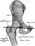
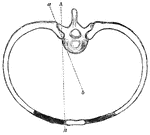
Pubic Spine
Diagram showing that the pubic spine (tubercle) and the tip of the great trochanter are on the same…
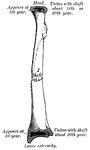
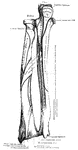
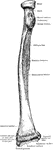
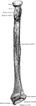
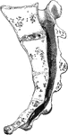
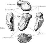
Radius
Anterior view of radius (bone of the arm) of the right side. Labels: 1, cylindrical head; 2, surface…
Carpal Articular Surface of the Radius
Carpal articular surface of the radius, and triangular fibro cartilage of the wrist.
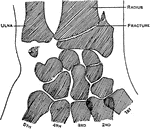
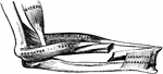
Colle's Fraction of the Radius
Showing the situation of Colle's fracture of the radius, with fracture of the styloid of the ulna. The…
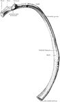
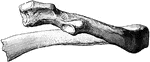
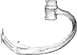
Fourth Rib
Fourth rib. Labels: a, vertebral extremity, called the head, which is connected with the bodies of the…
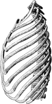
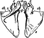
The Ribs
The ribs of the left side, with the dorsal and two lumbar vertebrae, the rib cartilages, and the sternum.
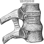
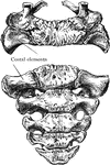
Ribs and Corresponding Vertebral Bodies
Ribs and corresponding vertebral bodies in their ligaments, viewed from the right.
Movement of Ribs in Inspiration
The elevation of the ribs is accompanied by a slight opening out of the angle which the bony part forms…
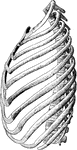
Ribs of the Left Side
The ribs of the left side, with the dorsal and two lumbar vertebrae, the rib-cartilages and the sternum.
Movement of Ribs
The axes of rotation of rib movement is two; one corresponding with a line drawn through two articulations…
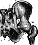
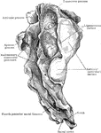
Sacro-ischiatic Articulation
The sacro-ischiatic articulation (joint), which is an amphiarthrosis between the sacrum and the ischium.
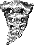
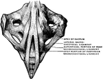
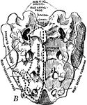
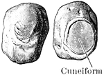
Human Sacrum
A large triangular bone as the base of the spine. Resides in between the two hip bones.
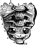
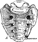
Superior and Anterior Surface of Sacrum
Superior and anterior view of young sacrum of about five years.
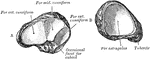
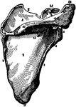
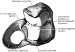
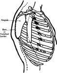
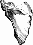
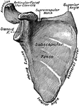
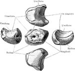
Scapula
Venter of scapula. Labels: 1, 1, 1, oblique ridges; 2, 2, fossa for subscapularis muscle; 3, superior…
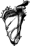
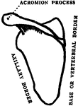
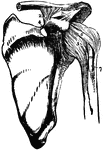
Scapula
Scapula. Labels: a, superior angle; d, the glenoid cavity, or socket for the round head of the arm bone;…
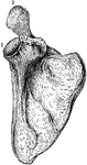
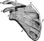
The Human Scapula
The human scapula bone (shoulder blade). Labels: 1, glenoid cavity; 2, end of the spine of scapula.
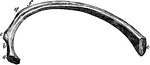
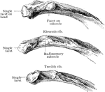
Second Rib
The second rib is much longer than the first, but bears a very considerable resemblance to it in the…
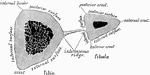
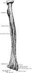
Shank Bones
A section of the bones of the crus (shank of the leg) taken at about the middle of their length (schematized)…
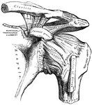
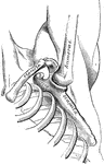
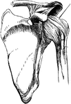
Shoulder
The left shoulder-joint, scapuloclavicular articulations, and proper ligaments of the scapula.
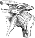
Bones and Ligaments of the Shoulder Articulation
Ligaments of the acromio-clavicular and scapulo-humeral articulations (joints of the shoulder). Labels:…
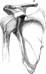
Shoulder blade
"The Scapula, or shoulder blade, is one of the two bones, the other being the clavicle, which form the…
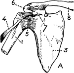
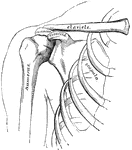
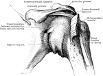
Shoulder bones and ligaments
"Shoulder bones and ligaments. 1. Humerus, 3. Scapula, 4. Tendon of biceps 5. Capsular ligament 6. Acromion…
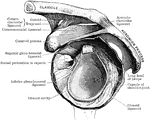
Capsular Ligament of the Shoulder Joint
Capsular ligament of the shoulder joint cut across and humerus removed.
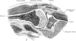
Frontal Section Through Shoulder Joint
The frontal section through the right shoulder joint of a boy. Arm abducted to a right angle.