This human anatomy ClipArt gallery offers 265 illustrations of the central nervous system, including external and dissected views of the brain and spinal cord.
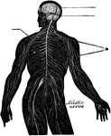
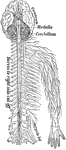
The Nervous System
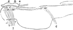
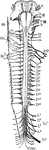
View of the nervous system of man, showing the nerve centers (brain and spinal cord) giving off nerves…
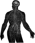
Nervous System
View of the nervous system in man, showing the nervous centers (the brain and the spinal near row) where…
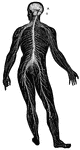
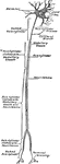
Nervous System
A representation of the brain, spinal cord and spinal nerve. Labels: 1, The cerebrum. 2. The cerebellum.…
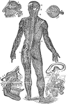
Nervous System
The human nervous system includes the brain, spinal cord, and the nerves. Labels: A, cerebrum; B, cerebellum.
Nervous System Diagram
Diagram of nervous system. Labels: a, a, cortex of cerebral hemispheres; b, b, cell body and dendrites…
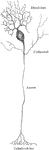
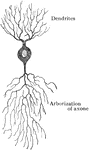
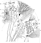
Neuron from the Cerebral Cortex
A neuron from the cerebral cortex. The axis-cylinder process, dendrites and collaterals are marked 1,…
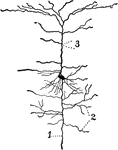
Neuron of Spinal Cord
Nerve cells of human spinal cord stained to show Nissl bodies. Labels: D, dendrites; A, axons; C, implantation…
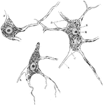
Variety of the Cell Bodies of Neurons
Showing some varieties of cell bodies of neurons. A, Unipolar (amacrine) cell from retina; B, Bipolar…
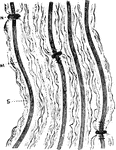
Behavior of the Nodes of Ranvier
Several fibers of a bundle of medullated nerve fibers acted upon by silver nitrate to show peculiar…
Development of the Opercula
Diagram to illustrate the development of the opercula which cover the insula. A, Sylian fossa before…
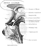
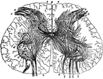
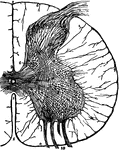
Optic Thalami
The optic thalamus is the principle object in the forebrain. It is a large ovoid mass of gray matter,…
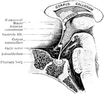
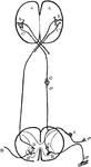
Pituitary Region in a Child
Mesial section through the pituitary region in a child of twelve months old.
Pons
Diagram of a longitudinal section through the pons, showing the relation of the nuclei for the ocular…
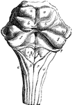
Anterior Surface of the Pons Varolii and Medulla Oblongata
Ventral or anterior surface of the pons Varolii, and medulla oblongata. Labels: a, anterior pyramids;…
Dorsal Surface of the Pons Varolii
Dorsal or posterior surface of the pons Varolii, corpus quadrigemina, and medulla oblongata. The peduncles…
Section Through Pons Varolii
Section through the lower part of the human pons varolii immediately above the medulla.
Section across the Pons
Section across the pons, about the middle of fourth ventricle. py., pyramidal bundles; po., transverse…

Section of the Pons
The pons is composed chiefly of transverse fibers arranged in coarse bundles, longitudinal fibers gathered…
Section of the Pons
The pons is composed chiefly of transverse fibers arranged in coarse bundles, longitudinal fibers gathered…
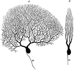
Purkinje Cell
Two purkinje cells from silver preparation of cerebellar cortex; A, side view; B, cell in profile; a,…
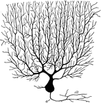
Purkinjean Cell from Cerebellum
Purkinjean cell from human cerebellum, as seen in a plane transverse to the long axis of a cerebellar…
Sagittal Section Through Cerebellar Folium
Transverse section through a cerebellar folium. Labels: A, axon of cell Purkinje; F, moss fibers; K…
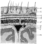
Layers of the Scalp and Membrane of the Brain
Diagram showing the layers of the scalp and membranes of the brain in section. Labels: a, skin; b, subcutaneous…
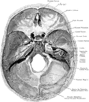
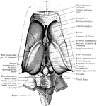
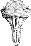
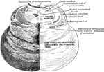
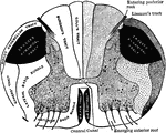
Base of the Skull
The base of the skull viewed from above. Three Fossae are recognized -the Anterior, Middle, and Posterior…
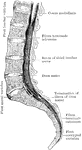
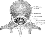
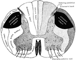
Sacral Region of Spinal Canal
The conus and medullaris and the filum terminale exposed within the spinal canal.
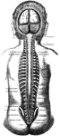
The Spinal Column and Brain
A section of the brain and spinal column. Labels: 1, The cerebrum (large brain). 2, The cerebellum (small…
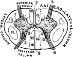
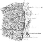
Spinal Cord
"Magnified view of transverse section of the spinal cord through the middle of the Lumbad Enlargement:…
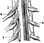
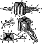
Spinal Cord
Portion of the spinal cord. 1: Body of cord; 2: A spinal nerve from left side of cord; 3: Anterior roots…
Spinal Cord
Diagrammatic view from before of the spinal cord and medulla oblongata, including the roots of the spinal…
Spinal Cord
"Diagram of a cross-section of the spinal cord through the roots of spinal nerves. c, central canal;…
Spinal Cord
The spinal cord is a double cord, the two halves being joined by a narrow portion, but the outside sheath…
The Spinal Cord and Medulla Oblongata
The spinal cord and medulla oblongata. Labels: A, from the ventral, and B, from the dorsal aspect; C…
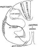
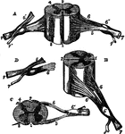
Spinal Cord and Nerve Roots
Diagrams of spinal cord and nerve roots. Labels: A, a small portion of the cord seen from the ventral…
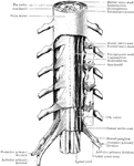
The Position of the Spinal Cord and Spinal Nerves in the Spinal Canal
The skull and spinal canal of a child from behind with the Dura Mater slit open and ribs with the transverse…
Spinal Cord and Spinal Nerves
Scheme of the arrangement of the membranes of the spinal cord and the roots of the spinal nerves.
Spinal Cord Section
A thin transverse section of half the spinal cord magnified about 10 diameters. Labels: 1, anterior…
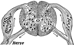
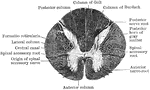
Section of the Spinal Cord
Diagrammatic transverse section of the spinal cord showing the conduction paths.
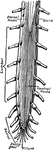
Different View of the Spinal Cord
Different views of a portion of the spinal cord from the cervical region, with roots of the nerves slightly…
Lower End of the Spinal Cord
Lowered end of the spinal cord and the cauda equina, dorsal aspect. The dorsal roots of the right side…
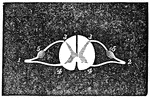
Section of Spinal Cord
"Diagram of a slice across the spinal cord, showing the roots of a spinal nerve to the arm on the left.…
Section of the Spinal Cord
Section of a spinal cord, one half of which shows the tracts of the white matter, and the other half…
Section Through Spinal Cord
Section through the conus medullaris and the cauda equina as they lie in the spinal canal.
Section Through Upper Part of Spinal Cord
Transverse section through upper part of the cervical region of the cord.
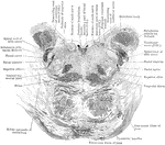
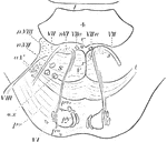
Spinal Cord, Spinal Nerves, and Base of Brain
Base of brain, spinal cord, and spinal nerves. Labels: V, 5th nerve; VI, 6th nerve; VII, a, facial nerve,…
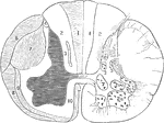
Transverse Section of Spinal Cord
Peripheral part of transverse section of spinal cord, showing nerve fibers subdivided into groups by…
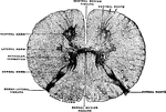
Transverse Section of the Spinal Cord
Transverse section of the spinal cord. Labels: 1, 2, Spinal nerves. 3, Origin of posterior root. 4,…
Transverse Section of the Spinal Cord
Transverse section of the spinal cord at the middle of the thoracic region. The neuroglia septum has…
Transverse Section Through Spinal Cord
Diagrammatic representation of a transverse section through the spinal cord. The nerve tracts in the…
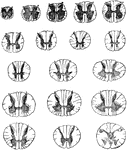
Transverse Sections of Spinal Cord
Transverse sections of the spinal cord at different levels, twice the natural size.