Leaves - compound (hand-shaped; leaflets, five); opposite; edge toothed. Outline - of leaflet, oval or long oval. Apex - taper-pointed. Base - pointed. Leaflets - three to seven inches long; one and a half to three inches wide. Bark - with a disagreeable odor. Flowers - small, yellowish-white. June. Fruit - about three fourths of an inch in diameter. Husk - prickly when young. Nut - smooth. Found - along the western slopes of the Alleghany Mountains - Pennsylvania to Northern Alabama and westward. General Information - A small, ill-scented tree (eighteen to thirty-five feet high). Its wood is light and hard to split. With the other species of the same genus it is preferred, above any other American wood, for the making of artificial limbs.

Genus Aesculus, L. (Buckeye, Horse Chestnut)

Leaves - compound (hand-shaped; leaflets, five); opposite; edge toothed. Outline - of leaflet, oval…

The wood is cut away so that about 1/4 or 1/2 inch of lead projects. The lead can then be sharpened.
Dull Edge Pencil

The wood is cut away so that about 1/4 or 1/2 inch of lead projects. The lead can then be sharpened.

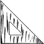
Triangles are made of various substances such as wood, rubber, celluloid, and steel.

45 Degree Triangle

Triangles are made of various substances such as wood, rubber, celluloid, and steel.

Triangles are made of various substances such as wood, rubber, celluloid, and steel.

30-60 Degree Triangle

Triangles are made of various substances such as wood, rubber, celluloid, and steel.

A piece of wood or metal to stamp a letter or character. "(1) the body, (2) the face, (3) the shoulder, (4) the nick and (5) the groove." -Foster, 1921

Type

A piece of wood or metal to stamp a letter or character. "(1) the body, (2) the face, (3) the shoulder,…
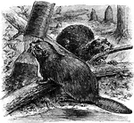
Beavers chewing on branches.

Beavers

Beavers chewing on branches.

The Union Label for wood workers.

Wood Workers Union Label

The Union Label for wood workers.

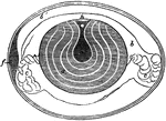
"Section of a Hen's Egg before Incubation. a, yolk, showing concentric layers; a', its semi-fluid center; b, inner dense part of the albumen; b', outer thinner part; c, twisted cords of albumen; h, the white spot, or germ cell." -Cooper, 1887

Egg Parts

"Section of a Hen's Egg before Incubation. a, yolk, showing concentric layers; a', its semi-fluid center;…

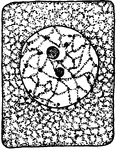
"A, embryonic cells from onion root tip; d, plasmatic membrane; c, cytoplasm; a, nuclear membrane enclosing the thread-like nuclear reticulum; b, nucleolus; e, plastids (black dots scattered about)." -Stevens, 1916

Onion Cells

"A, embryonic cells from onion root tip; d, plasmatic membrane; c, cytoplasm; a, nuclear membrane enclosing…

"B, older (onion) cells farther back from the root tip. The cytoplasm is becoming vacuolate; f, vacuole." -Stevens, 1916

Onion Cells

"B, older (onion) cells farther back from the root tip. The cytoplasm is becoming vacuolate; f, vacuole."…

In onion cells: "C, a cell from the epidermis of the mid-rib of Tradescantia zebrina, in its natural condition on the right, and plasmolyzed by salt solution on the left; g, space left by the recedence of the cytoplasm from the wall; the plasma membrane can now be seen as a delicate membrane bounding the shrunken protoplast." -Stevens, 1916

T. Zebrina Cell

In onion cells: "C, a cell from the epidermis of the mid-rib of Tradescantia zebrina, in its natural…

"A, cell from the epidermis of the upper side of the calyx of Tropaeolum majus with crystalline chromoplasts." -Stevens, 1916

T. Majus Cell

"A, cell from the epidermis of the upper side of the calyx of Tropaeolum majus with crystalline chromoplasts."…
"B, cells from the petal of Lupinus luteus with yellow chromoplasts." -Stevens, 1916

L. Luteus Cell

"B, cells from the petal of Lupinus luteus with yellow chromoplasts." -Stevens, 1916

"C, cell showing numerous chloroplasts scattered through the cytoplasm." -Stevens, 1916

Plant Cell

"C, cell showing numerous chloroplasts scattered through the cytoplasm." -Stevens, 1916

First stage in plant cell division: Protophase 1; "Resting cell ready to begin division." -Stevens, 1916

Plant Cell Division 1

First stage in plant cell division: Protophase 1; "Resting cell ready to begin division." -Stevens,…
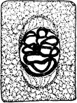
Second stage in plant cell division: Protophase 2; "the nuclear reticulum is assuming the form of a thickened thread, and the cytoplasm at opposite poles is becoming thread-like to form the spindle fibers." -Stevens, 1916

Plant Cell Division 2

Second stage in plant cell division: Protophase 2; "the nuclear reticulum is assuming the form of a…
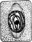
Third stage in plant cell division: Protophase 3; "The nuclear thread has divided longitudinally throughout the middle, and the spindle fibers have become more definite." -Stevens, 1916

Plant Cell Division 3

Third stage in plant cell division: Protophase 3; "The nuclear thread has divided longitudinally throughout…
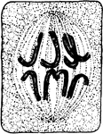
Fourth stage in plant cell division: Protophase 4; "The nuclear membrane and the nucleolus have disappeared, and the nuclear thread has become segmented into chromosomes which are assembling at the equator of the cell." -Stevens, 1916

Plant Cell Division 4

Fourth stage in plant cell division: Protophase 4; "The nuclear membrane and the nucleolus have disappeared,…
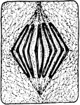
Fifth stage in plant cell division: Metaphase; "The metaphase, where the longitudinal halves of the chromosomes are being drawn apart preparatory to their journey toward opposite poles." -Stevens, 1916

Plant Cell Division 5

Fifth stage in plant cell division: Metaphase; "The metaphase, where the longitudinal halves of the…
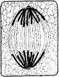
Sixth stage in plant cell division: Anaphase; "The anaphase, or movement of the chromosomes toward the poles, is about completed, connecting fibers extend from pole to pole." -Stevens, 1916

Plant Cell Division 6

Sixth stage in plant cell division: Anaphase; "The anaphase, or movement of the chromosomes toward the…
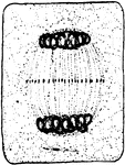
Seventh stage in plant cell division: Telophase; "Telophase where the chromosomes have begun to spin out in the form of a nuclear reticulum. The connecting fibers have begun to thicken in the equatorial plane." -Stevens, 1916

Plant Cell Division 7

Seventh stage in plant cell division: Telophase; "Telophase where the chromosomes have begun to spin…
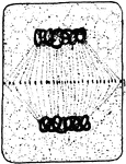
Eighth stage in plant cell division: "The connecting fibers have spread out and come into contact with the wall of the mother cell in the equatorial plane, and the thickening of the fibers throughout this plane has made a complete cell plate within which the dividing wall will be produced." -Stevens, 1916

Plant Cell Division 8

Eighth stage in plant cell division: "The connecting fibers have spread out and come into contact with…
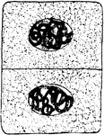
Ninth and final stage in plant cell division: "A nuclear membrane has been formed about each daughter nucleus, and the dividing cell-wall is complete." -Stevens, 1916

Plant Cell Division 9

Ninth and final stage in plant cell division: "A nuclear membrane has been formed about each daughter…
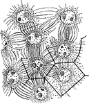
"Formation of endosperm in the embryo-sac of Agrimonia Eupatorium. Cell-walls are being formed between the nuclei." -Stevens, 1916

A. Eupatorium Cell

"Formation of endosperm in the embryo-sac of Agrimonia Eupatorium. Cell-walls are being formed between…
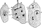
"Free cell formation of spores in the ascus of Erysiphe communis. A, ascus with single nucleus; C, cytoplasm; N, nucleus; NL, nucleolus; B, successive stages in nuclear division with the ascus; at X, early anaphase, nuclear membrane, NM, still persisting; R, kinoplasmic radiations from the poles; at Y, telophase, new nuclear membrane not yet formed; Z, a later stage where the nuclear membranes demark the daughter nuclei." -Stevens, 1916

E. Communis Cell

"Free cell formation of spores in the ascus of Erysiphe communis. A, ascus with single nucleus; C, cytoplasm;…

"Various stages of cell multiplication by budding of Saccharomyces cerevisiae." -Stevens, 1916

S. Cerevisiae Cell Multiplication

"Various stages of cell multiplication by budding of Saccharomyces cerevisiae." -Stevens, 1916

Developmental stages of collenchyma: "1 and 2, cross and longitudinal section of a collenchyma cell in its primary meristem condition." -Stevens, 1916

Collenchyma 1 and 2

Developmental stages of collenchyma: "1 and 2, cross and longitudinal section of a collenchyma cell…

Developmental stages of collenchyma: "3 and 4, longitudinal and cross sections of the same cell at a later stage, the walls in 4 have commenced to thicken at the angles." -Stevens, 1916

Collenchyma 3 and 4

Developmental stages of collenchyma: "3 and 4, longitudinal and cross sections of the same cell at a…

Developmental stages of collenchyma: "5 and 6, longitudinal and cross sections of a mature cell. The arrows indicate the planes through which the longitudinal sections were cut. The stippling inside the walls indicates the protoplasts." -Stevens, 1916

Collenchyma 5 and 6

Developmental stages of collenchyma: "5 and 6, longitudinal and cross sections of a mature cell. The…
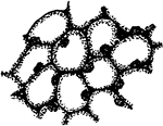
First developmental stage of stone cells: "1, in the primary meristem condition." -Stevens, 1916

Stone Cells 1

First developmental stage of stone cells: "1, in the primary meristem condition." -Stevens, 1916
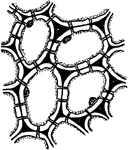
Second developmental stage of stone cells: "2, the cells have enlarged and the walls have begun to thicken and become pitted." -Stevens, 1916

Stone Cells 2

Second developmental stage of stone cells: "2, the cells have enlarged and the walls have begun to thicken…
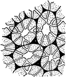
Third and final developmental stage of stone cells: "3, The walls are completed. The primary wall is black, cellulose additions white, and the lignified walls in 3 are stippled. Notice that the protoplasts have disappeared in 3, and the pits in some instances are branched." -Stevens, 1916

Stone Cells 3

Third and final developmental stage of stone cells: "3, The walls are completed. The primary wall is…

Developmental stages of bast fibers: "1 and 2, cross and longitudinal sections of primary meristem cells that are to become bast fibers." -Stevens, 1916

Bast Fibers 1 and 2

Developmental stages of bast fibers: "1 and 2, cross and longitudinal sections of primary meristem cells…

Developmental stages of bast fibers: 3 and 4, cross and longitudinal sections of meristem cells becoming bast fibers.

Bast Fibers 3 and 4

Developmental stages of bast fibers: 3 and 4, cross and longitudinal sections of meristem cells becoming…
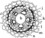
"Portion of a cross section of a root of Allium ascalonicum. h, large central, tracheal tube; i, xylem, and l, phloem portion of the vascular bundle; n, cortex cells; 1, endodermis with thin-walled cell at k to admit passage of materials; m, pericycle or pericambium." -Stevens, 1916

A. Ascalonicum Root

"Portion of a cross section of a root of Allium ascalonicum. h, large central, tracheal tube; i, xylem,…

First developmental stage of sieve tubes, companion cells, and phloem parenchyma. "A, a and b, two rows of procambial cells; in c and d, a has divided longitudinal and c is to become companion cells; d, a sieve tube, and b, phloem parenchyma." -Stevens, 1916

Plant Cell Development 1

First developmental stage of sieve tubes, companion cells, and phloem parenchyma. "A, a and b, two rows…

Second developmental stage of sieve tubes, companion cells, and phloem parenchyma. "B, c, companion cells, and d, a beginning sieve tube from c and d, respectively in A. The cross-walls in d are pitted; b, phloem parenchyma grown larger." -Stevens, 1916

Plant Cell Development 2

Second developmental stage of sieve tubes, companion cells, and phloem parenchyma. "B, c, companion…

Third developmental stage of sieve tubes, companion cells, and phloem parenchyma. "The pits in the cross-walls of the sieve tubes become perforations, and the nuclei gone from the cells composing the tube." -Stevens, 1916

Plant Cell Development 3

Third developmental stage of sieve tubes, companion cells, and phloem parenchyma. "The pits in the cross-walls…

"Stages in the development of the elements of the xylem. A, progressive steps in the development of a tracheal tube. 1, row of procambial or cambial cells that are to take part in the formation of a tube; 2, the same at a later stage enlarged in all dimensions; 3, the cells in 2 have grown larger, their cross-walls have been dissolved out, and the wall has become thickened and pitted; 4, the walls in 3 have become more thickened, the pits have an overhanging border, the walls have become lignified as indicated by the stippling, and finally the protoplasts have disappeared, and the tube is mature and dead." -Stevens, 1916

Xylem Development 1

"Stages in the development of the elements of the xylem. A, progressive steps in the development of…

Stages in the development of the elements of the xylem. "B, stages in the formation of tracheids from procambial or cambial cells. The steps are the same as in A, excepting that the cross-walls remain and become pitted." -Stevens, 1916

Xylem Development 2

Stages in the development of the elements of the xylem. "B, stages in the formation of tracheids from…

Stages in the development of the elements of the xylem. "C, steps in the development of wood fibers from cambial cells; 1, cambial cells; 2, the same grown larger in all dimensions with cells shoving past each other as they elongate; 3, a later stage with cells longer and more pointed and walls becoming thickened and pitted; 4, complete wood fibers with walls more thickened than in the previous stage and lignified, as shown by the stippling. The protoplasts in this last stage have disappeared and the fibers are dead." -Stevens, 1916

Xylem Development 3

Stages in the development of the elements of the xylem. "C, steps in the development of wood fibers…

Stages in the development of the elements of the xylem. "D, steps in the formation of wood parenchyma from cambial or procambial cells. 1, group of cambial or procambial cells; 2, the same enlarged in all dimensions; 3, the same with walls thickened and pitted; 4 and 5 show the same stages as 2 and 3, but here the cells have enlarged radially or tangentially more than they have vertically. The walls of these cells are apt to become lignified, but the cells are longer lived than the wood fibers." -Stevens, 1916

Xylem Development 4

Stages in the development of the elements of the xylem. "D, steps in the formation of wood parenchyma…

"Diagram showing the relation of this year's leaves to the wood of the current year." -Stevens, 1916
Plant Tissues

"Diagram showing the relation of this year's leaves to the wood of the current year." -Stevens, 1916

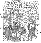
"Portion of a cross section throughout the stem of Dracaena marginata. P, parenchyma of cortex. V, meristematic zone of the pericycle by the activity of which the stem increases in diameter, with the addition of new vascular bundles. M, mature vascular bundle. N, nearly mature vascular bundle. O, newly formed procambium strand from which a vascular bundle is to arise. B, beginning of a procambium strand by the division of cells in the meristematic zone. F, parenchyma of the fundamental tissue." -Stevens, 1916

D. Marginata Stem

"Portion of a cross section throughout the stem of Dracaena marginata. P, parenchyma of cortex. V, meristematic…

"Multiple epidermis of leaf of mangrove in cross section. This serves as a water reservoir, and the relatively thick walls of the inner cells reenforce the protective power of the outer layer." -Stevens, 1916

Multiple Epidermis

"Multiple epidermis of leaf of mangrove in cross section. This serves as a water reservoir, and the…
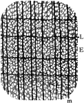
"Cross section of yellow poplar wood. E, early; L, late growth; m, medullary ray." -Stevens, 1916

Yellow Poplar Wood

"Cross section of yellow poplar wood. E, early; L, late growth; m, medullary ray." -Stevens, 1916
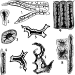
"Stone cells from different sources. 1, from coffee; 2, 3, and 4, from stem of clove; 5 and 6, from tea leaf; 7, 8, and 9, from powdered star anise seed." -Stevens, 1916

Stone Cells

"Stone cells from different sources. 1, from coffee; 2, 3, and 4, from stem of clove; 5 and 6, from…

"Camera-lucida outline of portion of cross section of cornstalk, showing at g bast fiber zone beneath the epidermis and surrounding the outermost vascular bundles." -Stevens, 1916

Cornstalk Bast Fibers

"Camera-lucida outline of portion of cross section of cornstalk, showing at g bast fiber zone beneath…

"Cross section of a portion of palm stem. e, xylem; f, phloem portions of vascular bundle; g, sclerenchyma tissue about vascular bundle; d, fundamental or ground tissue; c, larger tracheal tubes in vascular bundle." -Stevens, 1916

Palm Stem

"Cross section of a portion of palm stem. e, xylem; f, phloem portions of vascular bundle; g, sclerenchyma…

"A single root hair on a large scale, showing that it is an outgrowth of an epidermal cell, and the fact that it possesses a living protoplast and large vacuole filled with cell-sap and traversed by cytoplasmic strands. The nucleus is near the apex of the hair." -Stevens, 1916

Plant Root Hair

"A single root hair on a large scale, showing that it is an outgrowth of an epidermal cell, and the…

"Cross section through a water-absorbing scale of Tillandsia usneoides; a, a, water-absorbing cells partially filled with water." -Stevens, 1916

T. Usneoides Cell

"Cross section through a water-absorbing scale of Tillandsia usneoides; a, a, water-absorbing cells…
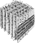
"Diagrammatic representation of a block of pine wood highly magnified. a, Early growth; b, late growth; c, intercellular space; d, bordered pit in tangential wall of late growth; m, f and e, bordered pit in radial wall of early growth from different points of view; h, row of medullary cells for carrying food; g, row of medullary ray cells for carrying water; k, thin place in radial wall of ray cells that carry food." -Stevens, 1916

Magnified Pine

"Diagrammatic representation of a block of pine wood highly magnified. a, Early growth; b, late growth;…
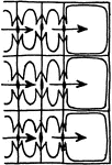
"Diagram to show the radial flow of water in pine wood, from the tracheids of the late growth of one year into those of early growth of the succeeding year." -Stevens, 1916

Water Flow in Pine

"Diagram to show the radial flow of water in pine wood, from the tracheids of the late growth of one…

"Diagram indicating by arrows how the water in the tracheids of pine passes longitudinally from one tracheid to another." -Stevens, 1916

Water Flow in Pine

"Diagram indicating by arrows how the water in the tracheids of pine passes longitudinally from one…
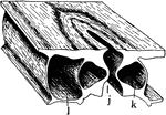
"A typical stoma in cross section and surface view combined. k, guard cell; j, the gap or stoma between the guard cells; l, epidermal cell bordering a guard cell." -Stevens, 1916

Stoma

"A typical stoma in cross section and surface view combined. k, guard cell; j, the gap or stoma between…

"A, diagram showing relative position of the guard cells in cross section in the open and closed positions; the heavier line indicates the open position." -Stevens, 1916

Stoma Guard Cells

"A, diagram showing relative position of the guard cells in cross section in the open and closed positions;…
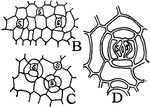
"B and C, early stages in the formation of stomata; at s, mother cells of guard cells are shown. D, s, s, two guard cells formed by the division of a mother cell." -Stevens, 1916

Stomata Formation

"B and C, early stages in the formation of stomata; at s, mother cells of guard cells are shown. D,…

"Diagram of apparatus showing how the guard cells draw apart; j, j, position of the rubber tubing when the water pressure is turned on." -Stevens, 1916

Guard Cell Experiment

"Diagram of apparatus showing how the guard cells draw apart; j, j, position of the rubber tubing when…

"Diagrammatic representation of a single palisade cell, with chloroplasts lining the walls." -Stevens, 1916

Palisade Cell

"Diagrammatic representation of a single palisade cell, with chloroplasts lining the walls." -Stevens,…
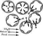
"Diagram to show the intake of carbon dioxide by the palisade cells from the intercellular spaces, the absorption by the chloroplasts of water from the cell-sap, and the passage of food from the chloroplasts into the cell-sap." -Stevens, 1916

Palisade Cell Intake

"Diagram to show the intake of carbon dioxide by the palisade cells from the intercellular spaces, the…