"Eudorina. The development of reproductive bodies within the colony from the ordinary vegetative cells (v). f, a mass of female cells; m, a mass of male or motile cells; f', a single female cell surrounded by male cells (m'); q, the boundary of the original colony." -Galloway, 1915

Eudorina

"Eudorina. The development of reproductive bodies within the colony from the ordinary vegetative cells…

"Diagram of flame cell, the internal terminus of the excretory tubules. c, cilia lining tubule; f, special cilia constituting the flame; n, nucleus of flame cell; p, cell processes; v, vacuole or cavity in cell communicating with the capillary tubules (t)." -Galloway, 1915

Flame Cell

"Diagram of flame cell, the internal terminus of the excretory tubules. c, cilia lining tubule; f, special…
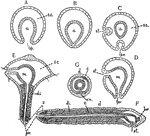
"Diagrams of stages in the metamorphosis of Polygordius, a primitive annelid. Ectoderm throughout is represented as nucleated without cell boundaries; the entoderm has the cell-boundaries shown, and the mesoderm is diagonally shaded. A, gastrula; B, same with blastopore closed; C and D represent formation of stomodaeum and proctodaeum from ectoderm; E, Trochosphere stage showing formation of segments in the posterior portion; F, adult (sagittal); G, adult (transverse). a, archenteron; bp., blostopore; br, brain; c, coelom; d, dorsal; di, dissepiments; m, mesenteron; pr., proctodaeum; s.c., segmentation cavity; st, stomodaeum; v.n., ventral nerve chain; z, zone of formation of nerve segments." -Galloway, 1915

Polygordius

"Diagrams of stages in the metamorphosis of Polygordius, a primitive annelid. Ectoderm throughout is…
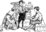
A group of boys moving wooden boxes and barrels.

Boys with Boxes

A group of boys moving wooden boxes and barrels.

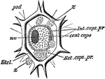
"Liteocircus annularis. cent. caps, central capsule; ext. caps. pr, extra-capsular protoplasm; int. cap. pr, intra-capsular protoplasm; nu, nucleus; psd, pseudopods; skel, skeleton; z, cells of zooxanthella." -Parker, 1900

L. Annularis

"Liteocircus annularis. cent. caps, central capsule; ext. caps. pr, extra-capsular protoplasm; int.…
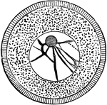
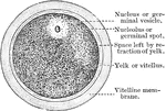
"Ovum of a Sea-Urchin, showing the radially striated cell-membrane, the protoplasm, containing yolk-granules, the large nucleus (germinal vescicle), with its network of chromatin and a large nucleolus (germinal spot)." -Parker, 1900

Sea Urchin Ovum

"Ovum of a Sea-Urchin, showing the radially striated cell-membrane, the protoplasm, containing yolk-granules,…
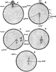
"Diagram illustrating the maturation and fertilization of the ovum. A, formation of first polar globule; B, beginning of fertilisation, sperms approaching the micropyle or aperture in the enclosing membrane of the ovum through which the sperm enters; C, formation of the male pronucleus; D, approximation of the male and female pronuclei; E, formation of segmentation-nucleus; female centrosome; male centrosome (the centrosomes are cell-structures not further referred to in this work); mem, egg-membrane; microp, micropyle; pol, polar bodies; female pronucleus; male pronucleus; seg. nucle, segmentation nucleus." -Parker, 1900

Ovum Fertilization

"Diagram illustrating the maturation and fertilization of the ovum. A, formation of first polar globule;…
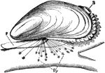
"Mytilus edulis, attached by byssus (By) to a piece of wood. F, foot; S, exhalant siphon." -Parker, 1900

Blue Mussel

"Mytilus edulis, attached by byssus (By) to a piece of wood. F, foot; S, exhalant siphon." -Parker,…

"Teredo navalis, in a piece of timber. P, pallets; SS, siphons; T, tube; V, valve; of shell." -Parker, 1900

Shipworm in Wood

"Teredo navalis, in a piece of timber. P, pallets; SS, siphons; T, tube; V, valve; of shell." -Parker,…

Of the Figwort family (Scrophulariaceae), the lousewort (Pedicularis canadensis).

Lousewort

Of the Figwort family (Scrophulariaceae), the lousewort (Pedicularis canadensis).

Of the Composite family (Compositae), the sharp-leaved wood aster (Aster acuminatus).

Sharp-Leaved Wood Aster

Of the Composite family (Compositae), the sharp-leaved wood aster (Aster acuminatus).
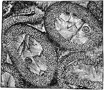
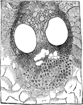
"Microscopical section of Fossil Wood, from clay iron-stone nodules; Oldham." -Taylor, 1904

Fossil Wood

"Microscopical section of Fossil Wood, from clay iron-stone nodules; Oldham." -Taylor, 1904

"Microscopic sections of fossil wood, fron Scarborough)." -Taylor, 1904

Wood Fossil

"Microscopic sections of fossil wood, fron Scarborough)." -Taylor, 1904

"Development of an oxalis leaf. A, full-grown leaf; B, rudimentary leaf, the leaflets not yet evident; C, more advanced stage, the leaflets appearing; D, a still more advanced stage." -Bergen, 1896

Oxalis Leaf Development

"Development of an oxalis leaf. A, full-grown leaf; B, rudimentary leaf, the leaflets not yet evident;…
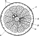
"Cross-section of an orange. a, axis of fruit with dots showing cut-off ends of fibro-vascular bundles; p, partition between cells of ovary; S, seed; c, cell of ovary filled with a pulp composed of irregular tubes full of juice; o, oil reservoirs near outer surface of rind; e, corky layer of epidermis." -Bergen, 1896

Orange

"Cross-section of an orange. a, axis of fruit with dots showing cut-off ends of fibro-vascular bundles;…
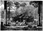
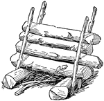
One way to build a campfire.

Campfire

One way to build a campfire.

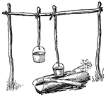
One way to build a campfire.

Campfire

One way to build a campfire.

One way to build a campfire.

Campfire

One way to build a campfire.

Tar is made from the turpentine by the smothered burning of the wood. When burning the wood, it is placed in a pile resembling a cone with the point cut off.

Manufacturing of Turpentine

Tar is made from the turpentine by the smothered burning of the wood. When burning the wood, it is placed…

"It is a very large tree, the trunk being sometimes three feet in diameter, although it is of little is as a timber tree, the wood being spongy." -Lupton

The Gutta-percha tree

"It is a very large tree, the trunk being sometimes three feet in diameter, although it is of little…

A simple hip roof.

Hip Style Roof Type

A simple hip roof.

A simple conical roof.

Conical Style Roof Type

A simple conical roof.

A simple m-roof.

Style M-Roof Type

A simple m-roof.

A simple king-post roof.

King-post Roof

A simple king-post roof.

A simple queen-post roof.

Queen-post Roof

A simple queen-post roof.

"A cord of wood is a solidly built pile of 8 feet long, 4 feet wide and 4 feet high. 1. How many cords, stove-wood measure, each stick 12 inches long, could be made from a full-sized cord of wood? 2. If the full cord as pictured above is bought for $3.00 and sawed into stove lengths as in problem (1) and sold for $1.25 a cord, what is the profit? 3. If 6 hours' time was required to saw this wood, and labor cost 25 cents per hour, was there anything gained on the transaction?" -Foster, 1921

Cord of Wood

"A cord of wood is a solidly built pile of 8 feet long, 4 feet wide and 4 feet high. 1. How many cords,…
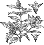
A tree belonging to the East Indies and the Malayan and Polynesian islands, remarkable for its fragrance. Its wood is used as a perfume, and is manufactured into glove-boxes and other light articles.

Sandal Wood

A tree belonging to the East Indies and the Malayan and Polynesian islands, remarkable for its fragrance.…
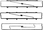
A particular method of uniting two pieces of timber together by the extremities, the end of one being cut or notched so as to fit into the other, making the part where the junction takes place of the same thickness as the rest of the pieces of timber.

Scarfing Design

A particular method of uniting two pieces of timber together by the extremities, the end of one being…

Diagram to show the relationship of nerve cell (NC) to Muscle (M) through its nerve fiber.

The Relation of Nerve Cells to Muscle

Diagram to show the relationship of nerve cell (NC) to Muscle (M) through its nerve fiber.

The development of muscular fibers from cells. Labels: a, simple cell. b, a pair of cells fused together. c, Three cells fused and their contents assuming the striated character. d, A muscular fiber exhibiting its original composition of cells.

The Development of Muscular Fibers from Cells

The development of muscular fibers from cells. Labels: a, simple cell. b, a pair of cells fused together.…

"Lizard's tail (Saururus cernuus). Portions of leaf-epidermis; U, upper epidermis; L, lower epidermis; ep, epidermal cell; st, guard-cells of the stomata." -Gager, 1916

Leaf Epidermis

"Lizard's tail (Saururus cernuus). Portions of leaf-epidermis; U, upper epidermis; L, lower epidermis;…
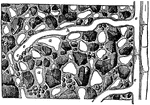
"Diagram to illustrate a root-hair (h) in the soil, and its relation to the soil-particles, the capillary film of water (w), and the air spaces (a); e, epidermal cell of the root, of which the root-hair is an out-growth, or branch." -Gager, 1916

Roots in Soil

"Diagram to illustrate a root-hair (h) in the soil, and its relation to the soil-particles, the capillary…
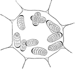
"Cell of Pellionia Daveauana, showing starch-grains. The black, crescent-shaped body on the end of each grain is the amyloplast." -Gager, 1916

Pellionia Plant Cell

"Cell of Pellionia Daveauana, showing starch-grains. The black, crescent-shaped body on the end of each…

A vegetable cell.

Vegetable Cell

A vegetable cell.

The diagram of a human ovum, showing the parts of an animal cell.

A Diagram of the Human Ovum

The diagram of a human ovum, showing the parts of an animal cell.

A cell with its reticulum disposed radically; from the intestinal epithelium in a worm.

Cell with its Reticulum Disposed

A cell with its reticulum disposed radically; from the intestinal epithelium in a worm.

The intracellular network of colorless and colored blood corpuscles. Labels: A, The colorless blood corpuscles showing the intracellular network, and two nuclei with intranuclear network. B. Colored blood corpuscle showing the intracellular network of fibrils.

Intracellular Network of Blood Corpuscles

The intracellular network of colorless and colored blood corpuscles. Labels: A, The colorless blood…

The nucleus when in a condition of rest is bounded by a distinct membrane, possibly derived from the spongioplasm of the cell. Shown is a diagram of a resting nucleus.

Nucleus at Rest

The nucleus when in a condition of rest is bounded by a distinct membrane, possibly derived from the…
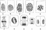
Karyokinesis. Labels: a, ordinary nucleus of a columnar epithelial cell; B, C, the same nucleus in the stage of convolution; D, the wreath or rosette from; E, the aster, or single star; F, a nuclear spindle the Descemet's endothelium of the frog's cornea; G, H, I, diaster; K, two daughter nuclei.

Karyokinesis

Karyokinesis. Labels: a, ordinary nucleus of a columnar epithelial cell; B, C, the same nucleus in the…

The early stages of karyokinesis. Labels: A, The thicker primary fibers remain and the achromatic spindle appears. B, The thick fibers split into two and the achromatic spindle becomes longitudinal.

Early Stages of Karyokinesis

The early stages of karyokinesis. Labels: A, The thicker primary fibers remain and the achromatic spindle…

The monaster stage of karyokinesis.

Monaster Stages of Karyokinesis

The monaster stage of karyokinesis.

Metakinesis- chromatic figure, spindle. Labels: A, early stage; B, later stage; C, latest stage-formation of disaster.

Metakinesis

Metakinesis- chromatic figure, spindle. Labels: A, early stage; B, later stage; C, latest stage-formation…

The final stage of karyokinesis. In the lower figure the changes are still more advanced than in the upper.

Final Stage of Karyokinesis

The final stage of karyokinesis. In the lower figure the changes are still more advanced than in the…

A, A young vegetable cell, showing cell cavity entirely filled with granular protoplasm enclosing a large oval nucleus with one or more nucleoli. B, An older cells from the same plant showing distinct cellulose wall and vacuolation of protoplasm.

Vegetable Cell

A, A young vegetable cell, showing cell cavity entirely filled with granular protoplasm enclosing a…

Squamous epithelium is found arranged as a single layer of flattened cells as the pigmentary layer of the retina. Shown is the pigment cells from the retina. A, cells still cohering, seen on their surface; a, nucleus indistinctly seen. In the other cells the nucleus is concealed by the pigment granules. B, two cells seen in profile; a, the outer or posterior part containing scarcely any pigment.

Pigment Cells from the Retina

Squamous epithelium is found arranged as a single layer of flattened cells as the pigmentary layer of…
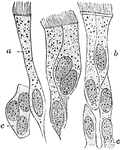
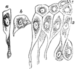
Ciliated epithelium from the human trachea. Large fully formed cell. Labels: b, shorter cell; c, developing cells with more than one nucleus.

Ciliated Epithelium

Ciliated epithelium from the human trachea. Large fully formed cell. Labels: b, shorter cell; c, developing…
Epithelium of the bladder. Labels: a, one of the cells of the first row; b, a cell of the second row; c, cells in situ, of first, second, and deepest layer.

Epithelium of the Bladder

Epithelium of the bladder. Labels: a, one of the cells of the first row; b, a cell of the second row;…
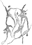
Ramified pigment cells, from the tissue of the choroid coat of the eye. Labels: a, cell with pigment; b, colorless fusiform cells.

Pigment Cells

Ramified pigment cells, from the tissue of the choroid coat of the eye. Labels: a, cell with pigment;…
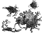
Flat, pigmented, branched connective tissue cells from the sheath of a large blood vessel of a frog's mesentery. The pigment is not distributed uniformly throughout the substance of the larger cell, consequently some parts of it look blacker than others (uncontracted state). In the two smaller cells most of the pigment is withdrawn into the cell body, so that they appear smaller, blacker, and less branched.

Connective Tissue Cells from a Frog

Flat, pigmented, branched connective tissue cells from the sheath of a large blood vessel of a frog's…
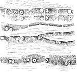
The cells in the tendons are arranged in long chains in the ground substance separating the bundle of fibers, and are more or less regularly quadrilateral with large round nuclei containing nucleoli, which are generally places so as to be contiguous in two cells. Show is the caudal tendon of young rat. Note arrangement, form, and structure of tendon cells.

Structure of Tendon Cells

The cells in the tendons are arranged in long chains in the ground substance separating the bundle of…

The branched character of the cells is seen. Shown is a transverse section from a cross section of the tail of a rabbit, showing sheath, fibrous septa, and branched connective tissue corpuscles. The spaces left white in the drawing represent the tendinous fibers in transverse section.

Branched Tendon Cells

The branched character of the cells is seen. Shown is a transverse section from a cross section of the…
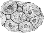
The matrix of hyaline cartilage has a dimly granular appearance like that of ground glass, and in man and higher mammals has no apparent structure. In some cartilages of the frog, however, even when examined in the fresh state, is is seem to be mapped out in polygonal blocks or cell-territories, each containing a cell in the center, and representing what is generally called the capsule of the cartilage cells. Shown is the fresh cartilage from a Triton.

Hyaline Cartilage Cells

The matrix of hyaline cartilage has a dimly granular appearance like that of ground glass, and in man…

Red marrow occupies the spaces in the cancellous tissue; it is highly vascular, and this maintains the nutrition of the spongy bone, the interstices of which it fills. Shown are the cells of the red marrow of the the guinea pig. Labels; a, a large cell, the nucleus of which appears to be partly divided into three by constrictions; b, a cell, the nucleus of which shows an appearance of being constricted into a number of smaller nuclei; c, so called giant cell, or myeloplaxe, with many nuclei; d, a smaller myeloplaxe, with three nuclei; e-i, proper cells of the marrow.

Cells of the Red Marrow

Red marrow occupies the spaces in the cancellous tissue; it is highly vascular, and this maintains the…
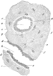
Transverse section of femur of a human embryo about eleven weeks old. Labels: a, rudimentary Haversian canal in cross sections; b, in longitudinal section; c, osteoblast; d, newly formed osseous substance of a lighter color; e, that of greater age; f, lacunae with their cells; g, a cell still united to an osteoblast.

Formation of Compact Bone in a Human

Transverse section of femur of a human embryo about eleven weeks old. Labels: a, rudimentary Haversian…

Unstriped muscle, or plain muscle, forms the proper muscular coats. Shown is unstriped muscle cells from the mesentery of a newt. The sheath exhibits transverse markings. B, from a similar preparation, showing that each muscle cell consists of a central bundle of fibrils, F, (contractile part), connected with the intranuclear network, N, and a sheath with annular thickenings, St. The cells show varicosities due to local contraction, and on these the annular thickenings are most marked.

Unstriped Muscle of a Newt

Unstriped muscle, or plain muscle, forms the proper muscular coats. Shown is unstriped muscle cells…

Nerve cells, also known as nerve corpuscles, comprise the second principle element of the nervous tissue. The are not generally present in the nerve trunk, but are found in collections of nervous tissue called ganglia. Shown are the ganglion nerve corpuscles of different shapes.

Ganglion Nerve Corpuscles

Nerve cells, also known as nerve corpuscles, comprise the second principle element of the nervous tissue.…
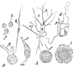
Different forms of ganglion cells. A, a, round ball-shaped unipolar cell from the human Gasserian ganglion. Two cells only show the process f; b, spindle shaped; c, multipolar ganglion cell from he spinal cord of the ox. d, D, Purkinjee ganglion cells from human cerebellum; ax, axis cylinder process; p, protoplasmic process; h, h, two cells surrounded with a nucleated sheath.

Different Forms of Ganglion Cells

Different forms of ganglion cells. A, a, round ball-shaped unipolar cell from the human Gasserian ganglion.…
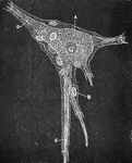
An isolated ganglion cell of a human, showing sheath with nucleated cell lining, B. A Ganglion cell, with nucleus and nucleolus. C, Branched process. D. Unbranched process.

Ganglion Cell, Structure of

An isolated ganglion cell of a human, showing sheath with nucleated cell lining, B. A Ganglion cell,…
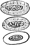
Acetic acid (dilute) causes the nucleus of the red blood cells in the frog to become more clearly defined; if the action is prolonged, the nucleus becomes strongly granulated, and all the coloring matter seems to be concentrated in it, the surrounding cell substance and outline of the cell becoming almost invisible; after a time the cells lose their color altogether. The cells shown represent the successive stages of change. A similar loss of color occurs in the red cells of human blood, which , however, from the absence of nuclei, seem to disappear entirely.

Effect of Acetic Acid on Red Blood Cells

Acetic acid (dilute) causes the nucleus of the red blood cells in the frog to become more clearly defined;…

A 2 percent solution of boric acid applied to nucleated red blood cells of a frog will cause the concentration of all he coloring matter in the nucleus; the colored body thus formed gradually quits its central position , and comes to be partly, sometime entirely, protruded from the surface of the now colorless cell. When applied to non-nucleated mammalian corpuscle its effect merely resembles that of other dilute acids.

Effect of Boric Acid on Red Blood Cells

A 2 percent solution of boric acid applied to nucleated red blood cells of a frog will cause the concentration…