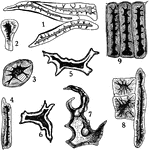
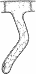
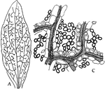
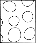
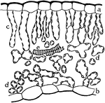
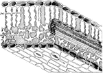
Stone Cells
"Stone cells from different sources. 1, from coffee; 2, 3, and 4, from stem of clove; 5 and 6, from…
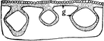
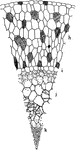
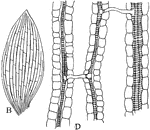
Cornstalk Bast Fibers
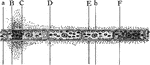
"Camera-lucida outline of portion of cross section of cornstalk, showing at g bast fiber zone beneath…
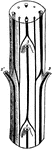
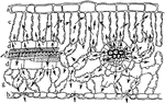
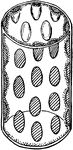
Palm Stem
"Cross section of a portion of palm stem. e, xylem; f, phloem portions of vascular bundle; g, sclerenchyma…
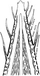
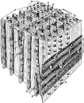
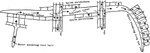
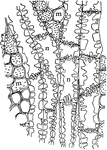
Plant Skeletal Tissues
"Diagram showing the progressive development of the skeletal tissues from the apex toward the base of…

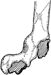
Plant Root Hair
"A single root hair on a large scale, showing that it is an outgrowth of an epidermal cell, and the…
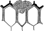
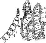
S. Oculata Aerial Root
"Portion of cross section through an aerial root of Stanhopea oculata. h, the velamen; i, exodermis;…
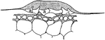
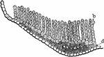
S. Macrantha Aerial Root
"Showing at f the felty body covering the passage way through the exodermis of the aerial root of Sobralia…
T. Usneoides Cell
"Cross section through a water-absorbing scale of Tillandsia usneoides; a, a, water-absorbing cells…
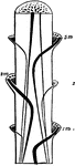
Palm Stem
"A, diagram of course of vascular bundles in a palm stem; 1m, 2m, 3m, bundles from the median portions…
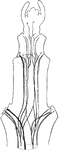
Cerastium Stem
"B, diagram of vascular bundles in external view and in cross section of the stem of Cerastium. The…
C. Viticella Stem
"Diagram showing the course of the vascular bundles in a stem of Clematis viticella. Median bundles…
Pit Development
"Different stages in the development of a bordered pit. b, The original, thin, primary wall; a, the…
Magnified Pine
"Diagrammatic representation of a block of pine wood highly magnified. a, Early growth; b, late growth;…
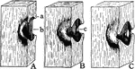
Water Flow in Plants
"Diagram to show the tangential flow of water from the side where there is less demand to the side where…
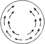
Water Flow in Plants
"Diagram showing by the arrows how the water can flow tangentially around a plant without traversing…
Water Flow in Pine
"Diagram to show the radial flow of water in pine wood, from the tracheids of the late growth of one…
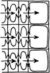
Water Flow in Pine
"Diagram indicating by arrows how the water in the tracheids of pine passes longitudinally from one…
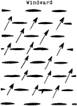
Water Flow in Plants
"Diagram showing the relation of the water-carrying tissues of the leaves to those of the stem, and…
Dicotyledon Leaf
A: "Camera-lucida drawing of a bleached leaf of a Dicotyledon, showing the course of the vascular bundles,…
Monocotyledon Leaf
B: "Camera-lucida drawing of a bleached leaf of... a Monocotyledon, showing the anastomosis of the parallel…
Leaf Water Flow
"Semi-diagrammatic cross section of a leaf showing by arrows how the water passes from the tracheal…
Leaf Water Flow
"Diagram to show the path of the water as it rises to, and escapes from, the leaves." -Stevens, 1916
Yellow Poplar Stem Tissue
The cross section of the stem of yellow poplar, which carries large amounts of water.
Water Cress Stem Tissue
The cross section of the stem of water cress, which carries very little water.
P. Galapageium Stem Tissue
The cross section of the stem of Pisdium Galapageium, which carries very little water.
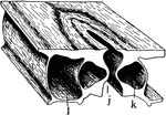
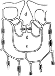
Stoma
"A typical stoma in cross section and surface view combined. k, guard cell; j, the gap or stoma between…
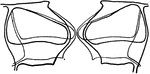
Stoma Guard Cells
"A, diagram showing relative position of the guard cells in cross section in the open and closed positions;…
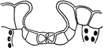
Stomata Formation
"B and C, early stages in the formation of stomata; at s, mother cells of guard cells are shown. D,…
Guard Cell Experiment
"Diagram of apparatus showing how the guard cells draw apart; j, j, position of the rubber tubing when…
Depressed Stoma
"A, depressed stoma of the under side of a leaf of Amherstia nobilis." -Stevens, 1916
Depressed Stoma
"B, depressed stoma of Hakea suaveolens. g stands beneath the guard cells; d, outer, and e inner, cavities."…
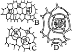
Intercellular Spaces of a Plant
"Diagram suggestive of the distribution of intercellular spaces throughout a plant. The heavy horizontal…
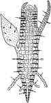
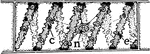
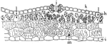
Juncus Stem
"A, cross section of stem of Juncus...the black lines traversing the section are really chains of cells…
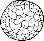
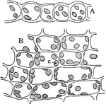
Leaf Epidermis
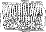
"Cross section of a portion of the blade of a leaf, showing upper epidermis at a, lower epidermis at…
Palisade Cell
"Diagrammatic representation of a single palisade cell, with chloroplasts lining the walls." -Stevens,…
Euphorbia Palisade Cells
"Starch grains from the palisade cells of a Euphorbia leaf." -Stevens, 1916

Palisade Cell Intake
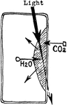
"Diagram to show the intake of carbon dioxide by the palisade cells from the intercellular spaces, the…
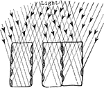
Plant Light Intake
"Diagram showing how the position of the chlorplasts against the vertical walls of the palisade cells…
Palisade Cell
"Diagram to show the activities going on in a palisade cell. The arrows from the chloroplasts into the…
Photosynthesis
"Diagram to show the effect of different portions of the spectrum on photosynthesis. a to F, different…
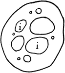
Intercellular Spaces of Leaf
"Showing intercellular spaces: f, between the palisade cells; e, in a leaf; g, border parenchyma; h,…
Leaf Architecture
"Diagram to show the architecture of a typical leaf in the region of one of the lateral veins. The shaded…
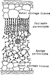
Rubber Leaf Cells
"Cross section through a portion of rubber leaf, showing the large percentage of water-storage tissues…
Indian Corn Leaf Cells
"Cross section of a portion of a leaf of Indian corn. a, upper and b, lower epidermis; c, c, palisade…
Codonanthe Leaf Tissues
"Cross section of a portion of leaf of Codonanthe, showing the water-storage tissue at f, and the chlorophyll-bearing…
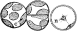
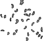
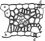
Spirogyra Chloroplast
"B, cell of Spirogyra, with spiral chloroplast at c, nucleus at n, and pyrenoid at e." -Stevens, 1916
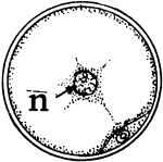
Spirogyra Chloroplast
"C, cross section of Spirogyra cell, with nucleus at n, and section of chloroplast and pyrenoid below."…
Marchantia Thallus
"Cross section through the thallus of Marchantia. j, stoma leading into a relatively large air-chamber…
Sphagnum Leaf
"Portion of leaf of Sphagnum, in cross section on the left, and surface view on the right. h, hole through…
P. Commune Leaf
"Cross section through a portion of leaf of Polytrichum commune. b, chains of chlorophyll-bearing cells;…
Moss Leaf Chloroplasts
"Cross section, A, and surface view, B, of a leaf of common moss, showing chloroplasts, c." -Stevens,…
Stored Food
"Diagram to show path of stored food upward through the tracheal tubes, and through the phloem portion…
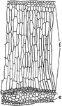
Yellow Poplar Wood
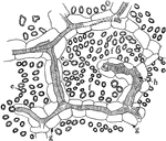
"Showing pitted connections between medullary rays and xylem parenchyma, and between contiguous xylem…