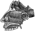
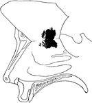
Mouth
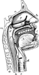
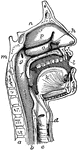
"The mouth, nose and pharynx, with the commencement of the gullet and larynx, as exposed by a section,…
Ethmoid Bone of the Human Skull
Ethmoid bone, posterior surface. The ethmoid bone is an exceedingly light, spongy bone, placed between…
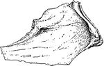
Human Vomer Nasal Bone
Vomer bone, a single bone placed at the back part of the nasal cavity, and forms part of the septum…
The Mouth, Nose, and Pharynx
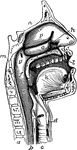
The mouth, nose, and pharynx, with the commencement of gullet (esophagus) and larynx, as exposed by…
The Mouth, Nose, and Pharynx
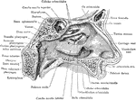
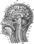
The mouth, nose, and pharynx, with the larynx and commencement of gullet (esophagus), seen in section.…
The Mouth, Nose, and Pharynx
The mouth, nose, and pharynx, with the commencement of the gullet (esophagus) and larynx, as exposed…
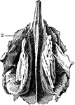
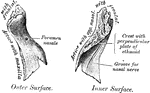
Inferior Turbinal Bones
Interior turbinal bones (or conchae nasales inferiores), which are situated in the nasal fossae.
The Nasal Bones
The nasal fossae (bones), which together form the cavity of the nose and are separated from either other…
Cartilage Tissue
Fibrous cartilage connective tissue from the symphysis pubis, magnified. Cartilage is a structure without…
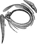
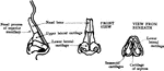
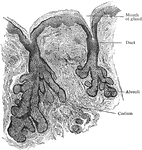
Tear Gland
The tear gland of the eye. The tears are carried from this factory b little ducts (b) and are poured…
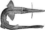
Torpedo
A device propelled through water with a ship as its target. "(a) Head; (b) air cylinder; (c) after body;…
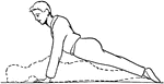
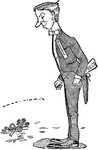
Exercise
"Place the hands on the floor, the body outstretched, face downward. Raise and lower the body from the…
Eyelids, Viewed from the Front
The eyelids viewed from before; a,a, the lachrymal canals; b, the lachrymal sack. The lachrymal sac…
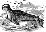
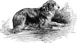
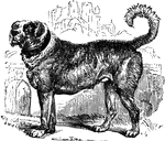
Hooded Seal
The genus Cystophora includes the large bladder-nose, hooded or crested seal of the Greenland seas,…
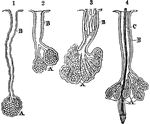
Oil Glands and Ducts
Oil-glands and ducts. 1, A, Oil-gland from the scalp; B, Its duct. 2, A, Two glands from the skin of…
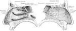
A Side View of the Passage of the Nostrils
A side view of the passage of the nostrils. 4, The distribution of the first olfactory pair of nerves.…
Olfactory System
The olfactory system. Labels: a, b, c, d, interior of the nose, which is lined by a mucous membrane;…
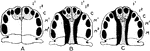
Incisor Relation to Palatal Cleft
Illustrating the relationship of the lateral incisor tooth to the palatal cleft. A, Normal hard palate.…
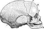
Side View of Fetal Skull
Side view of a fetal skull. The coronal suture extends from the top of the head downwards on either…
Nasal Cavity
The lateral wall of the left nasal cavity. The greater part of the middle turbinated bone has been excised…
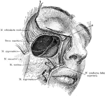
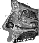
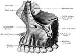
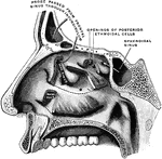
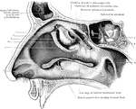
Dissection of the Maxillary Sinus
Exposure of the right maxillary sinus, after removal of facial muscles. The (*) indicates the opening…
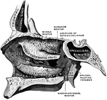
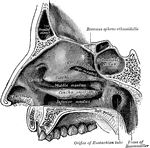
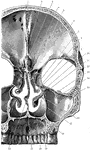
External Wall of Nasal Cavity
External wall of right nasal fossa, parts of the turbinates having been cut away to show the orifices…
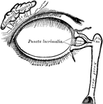
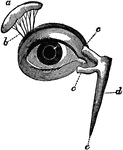
Lacrimal Apparatus
The lacrimal apparatus consists of the lacrimal gland, which secretes the tears, and its excretory ducts,…
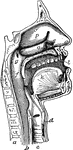
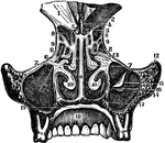
Section of the Head and Neck
Section of head and neck from front to back. Labels: 1, windpipe; 2, larynx; 3, spinal marrow; 4, pharynx;…
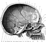
Skull Seen From Side
Shown is the inner aspect of the left half of the skull sagittally divided. Labels: 1, suture between…
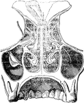
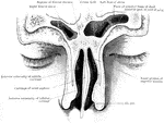
Coronal Section of Skull
Shown is a coronal section passing inferiorly through interval between between the first and second…
Outer Wall of Nose
View of the outer wall of the nose. The turbinated bones having been removed. Labels: 1, vestibule,…
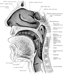
Section of the Head and Neck
Sagittal section through mouth, tongue, larynx, pharynx, and nasal cavity. The section was slightly…
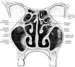
Section Through Nose and Frontal Sinuses
Vertical coronal section through the nose and frontal sinuses.
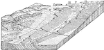
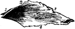
Hog-back Diagram
Hog-backs (RR) changing into cuestas (CC) and these into steps (HH) by progressive flattening of strata;…
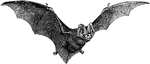
Pug-Nosed Bat
The pug-nosed bat. "Bat, one of the group of wing-handed, flying mammals, having the fore-limb peculiarly…

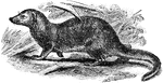
Common Kusimanse
The Common Kusimanse (Crossarchus obscurus), also known as the Long-nosed Kusimanse, is a small, diurnal…
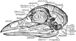
Skull of a Common Fowl
"Fig. 62 Skull of common fowl, enlarged. from nature by Dr. R.W. Shufeldt, U.S.A. The names of bones…
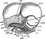
Chick Head
"Fig 66 - Head of a chick, second stage, after five days of incubation, section in profile; x6 diameters.…