Diagrammatic longitudinal section of the body. Labels: a, the neural tube, with its upper enlargement in the skull cavity at a'; N, the spinal cord; N', the brain; ee, vertebrae forming the solid partition between the dorsal and ventral cavities; b, the pleural, and c, the abdominal division of the ventral cavity, separated from one another by the diaphragm, d; i, the nasal, and o, the mouth chamber, opening behind into the pharynx, from which one tube leads to the lungs, l, and another to the stomach, f; h, the heart; k, a kidney; s, the sympathetic nervous chain. From the stomach, f, the intestinal tube leads through the abdominal cavity to the posterior opening of the alimentary canal.

Longitudinal Section of Body

Diagrammatic longitudinal section of the body. Labels: a, the neural tube, with its upper enlargement…

Diagram showing the position of the thoracic and abdominal organs. labels: 1, lower border of right lung; 2, the same of the left lung; 3, liver, right lobe; 4, liver, left lobe; 5, suspensory ligament of the liver; 6, fundus of gall-bladder; 7, cardia of stomach; 8, fundus of stomach; 9, lower border of stomach; 10, position of pylorus; 11, caecum; 12, vermiform appendix; 13, ascending colon; 14, right flexure of colon; 15, transverse colon, concealed by, 19, convolutions of the small intestine; 20, termination of ileum, ascending from right to left; 21, bladder, distended, partly covered by peritoneum; 22, the part of the bladder which is not covered by peritoneum.

Section Across the Forearm

Diagram showing the position of the thoracic and abdominal organs. labels: 1, lower border of right…
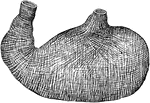
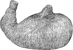
The muscular coat (lining) of the stomach, a type of involuntary muscle involved in the contraction of the stomach.

Muscle Lining of Stomach

The muscular coat (lining) of the stomach, a type of involuntary muscle involved in the contraction…
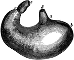
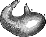
The stomach. Labels: d, lower end of the gullet; a, position of the cardiac aperture; b, the fundus; c, the pylorus; e, the first part of the small intestine; along a,b,c, the great curvature; between the pylorus and d, the lesser curvature.

The Stomach

The stomach. Labels: d, lower end of the gullet; a, position of the cardiac aperture; b, the fundus;…
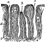
A thin section through the gastric mucous membrane which lines the stomach, perpendicular to its surface, magnified about 25 diameters. a, a simple peptic gland; b, a compound peptic gland; c, a mucous gland.

Section through the Gastric Mucous Membrane

A thin section through the gastric mucous membrane which lines the stomach, perpendicular to its surface,…

Wall of human stomach. Labels: E, epithelium; G, glands; Mm, muscularis mucosae.

Stomach Wall

Wall of human stomach. Labels: E, epithelium; G, glands; Mm, muscularis mucosae.

Diagram of the abdominal part of the alimentary canal (digestive system). Labels: C, the cardiac, and P, the pyloric end of the stomach; D, the duodenum; J, I, the convolutions of the small intestine; CC, the caecum with the vermiform appendix; AC, ascending, TC, transverse, and DC, descending colon; R, the rectum.

Alimentary Canal

Diagram of the abdominal part of the alimentary canal (digestive system). Labels: C, the cardiac, and…
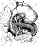
The stomach, pancreas, liver, and duodenum, with part of the rest of the small intestine and the mesentery; the stomach and liver have been turned up so as to expose the pancreas. Labels: V, stomach; D, D',D'', duodenum; L, spleen; P, pancreas; R, right kidney; T, jejunum; Vf, gall bladder; h, hepatic duct; c, cystic duct; ch, common bile duct; 1, aorta; 2, an artery (left coronary) of the stomach; 3, hepatic artery; 4, splenic artery; 5, superior mesenteric artery; 6, superior mesenteric vein; 7, splenic vein; Vp, portal vein.

The Stomach, Pancreas, Liver, and Duodenum

The stomach, pancreas, liver, and duodenum, with part of the rest of the small intestine and the mesentery;…

The portal vein and its branches. Labels: l, liver, under surface; gb, gall bladder; st, stomach; sp, spleen; p, pancreas; du, duodenum; ac, ascending colon; cd, descending colon; a,b,c,d,e, the portal vein and its branches. Portions of the duodenum and colon have been removed.

The Portal Vein and its Branches

The portal vein and its branches. Labels: l, liver, under surface; gb, gall bladder; st, stomach; sp,…

"The stomach pump injects liquid into a poisoned person's stomach and then withdraws the liquid and poison mixture. Bulbs C and D contain valves that open and close depending if B is upright or inverted." —Quackenbos 1859

Stomach Pump

"The stomach pump injects liquid into a poisoned person's stomach and then withdraws the liquid and…
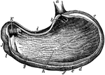
The stomach, the principal organ of digestion. It is a dilated part of the alimentary canal, situated between the termination of the esophagus and the beginning of the small intestine.

The Stomach

The stomach, the principal organ of digestion. It is a dilated part of the alimentary canal, situated…

The pancreas, a compound racemose gland about 5.5 inches long and 1.5 inches wide, situated transversely across the posterior wall of the abdomen behind the stomach and in front of the first lumbar vertebra.

The Pancreas

The pancreas, a compound racemose gland about 5.5 inches long and 1.5 inches wide, situated transversely…

The spleen, a soft, brittle, highly vascular organ, of dark purplish color, in size about 5 x 3 x 1.5 inches, about 7 ounces, situated deeply in the left hypochondrium, between the fundus of the stomach in front and internally, and the diaphragm behind and externally. It is connected with the stomach, diaphragm, and the left kidney through folds of the peritoneum.

The Spleen

The spleen, a soft, brittle, highly vascular organ, of dark purplish color, in size about 5 x 3 x 1.5…

Columnar epithelium lining a gland. It consists of conical cells laid side by side, their ends forming the surface of the membrane and is found in the stomach, intestines, and elsewhere.

Columnar Epithelial Tissue

Columnar epithelium lining a gland. It consists of conical cells laid side by side, their ends forming…

Diagram of the peritoneum, a serous membrane covering all the contents of the abdominal cavity. Labels: D, diaphragm; L, liver; S, stomach; C, transverse colon; D, transverse duodenum; P, pancreas; I, small intestines; R, rectum; B, bladder.

The Peritoneum

Diagram of the peritoneum, a serous membrane covering all the contents of the abdominal cavity. Labels:…
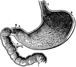
The inside of the stomach with the beginning of the intestines. At 3 is the left end and at 4 is the right end. At 1 is the opening of the gullet into the stomach. At 5 is a valve (the pylorus) which is sometimes shut, so as to prevent anything from passing from the stomach into the intestines. 8 indicates the inside lining of the stomach which produces gastric juices. 6 shows the duct from the liver.

The Stomach

The inside of the stomach with the beginning of the intestines. At 3 is the left end and at 4 is the…
Organs of the digestive system.

Organs of the Digestive System

Organs of the digestive system.

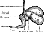
The four stomachs of the sheep, a grass-eating animal. The beginning of the intestines are also shown, which reach in their full length to 28 times the size of the sheep's body.

Sheep, Stomachs of

The four stomachs of the sheep, a grass-eating animal. The beginning of the intestines are also shown,…
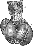
The stomach of a grain-eating bird, which has a gizzard that functions to crush the seeds to pieces since birds have no teeth to crush their food. It has on its inside two hard surfaces which rub and press against each other by muscles to grind food. At <em>b</em> is the gizzard cut open, showing two hard grinding surfaces, and at <em> a </em> above is the part from which oozes the gastric juice.

Stomach of a Bird

The stomach of a grain-eating bird, which has a gizzard that functions to crush the seeds to pieces…
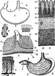
Views of the stomach. Labels: A. stomach (human). B. Same, anterior wall removed. C. Portion of stomach, pylorus and duodenum. D. Section through coats of stomach. E. Fundus of a cardiac gland. F. Epithelium from surface of stomach. G. Arterie sand veins of mucous membrane. w. Esophagus. 2. Duodenum. 3. Pylorus. $. Cardia. 5. Lesser curvature. 6. Greater curvature. 7. Anterior surface. 8. Pyloric valve. 9. Mucous membrane, with rugae. 10. Inner surface of gastric mucous membrane. 11. Mucous membrane of duodenum with villi. 12. Pyloric gastric glands. 13. Circular or deep muscular fibers. 17. Mucous membrane. 18. Muscularis mucosae. 19. Submucous coat. 20. Circular muscular layer. 21. Longitudinal muscular layer. 22. Serous coat. 23. Orifice of gland. 24. Fundus of gland.

Views of the Stomach

Views of the stomach. Labels: A. stomach (human). B. Same, anterior wall removed. C. Portion of stomach,…
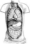
Diagram of the Body opened from the front to show the contents of the ventral cavity. Labels: d, diaphragm; h, heart; lu, lungs; st, stomach; li, liver; si, small intestines; c, large intestine

Ventral Cavity of the Body

Diagram of the Body opened from the front to show the contents of the ventral cavity. Labels: d, diaphragm;…

Diagrammatic longitudinal section of the Body. Labels: a, the neural tube, with its upper enlargement in the skull cavity at a'; N, the spinal cord; N', the brain; ee, vertebrae forming the solid partition between the dorsal and ventral cavities; b, the pleural, and c, the abdominal division of the ventral cavity, separated from one another by the diaphragm, d; i, the nasal, and o, the mouth chamber, opening behind into the pharynx, from which one tube leads to the lungs, l, and the heart; k, a kidney; s, the sympathetic nervous chain. From the stomach, f, the intestinal tube leads through the abdominal cavity to the posterior opening of the alimentary canal.

Longitudinal Section of the Body

Diagrammatic longitudinal section of the Body. Labels: a, the neural tube, with its upper enlargement…
The muscular coat of the stomach.

Muscular Coat of the Stomach

The muscular coat of the stomach.

The stomach. Labels: d, lower end of the gullet; a, position of the cardiac aperture; b, the fundus; c, the pylorus; e, the commencement of the small intestine; along a, b, c, the great curvature; between the pylorus and d, the lesser curvature.

The Stomach

The stomach. Labels: d, lower end of the gullet; a, position of the cardiac aperture; b, the fundus;…
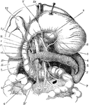
The stomach, pancreas, liver, and duodenum, with part of the rest of the small intestine and the mesentery; the stomach and liver have been turned up so as to expose the pancreas. Labels: V, stomach; D, D', D', duodenum; L, spleen P, pancreas; R, right kidney; T, jejunum; Vf, gallbladder; h, hepatic duct; c, cystic duct; ch, common bile-duct; 1, aorta, 2, an artery (left coronary) of the stomach; 3, hepatic artery; splenic artery; , superior mesenteric artery; 6, superior mesenteric vein; 7, splenic vein; Vp, portal vein.

Digestive Organs

The stomach, pancreas, liver, and duodenum, with part of the rest of the small intestine and the mesentery;…

Diagram of the abdominal part of the alimentary canal. Labels: C, the cardiac, and P, the pyloric end of the stomach, A; D, the duodenum; J, I, the convolutions of the small intestine; CC, the caecum with the vermiform appendix; AC, ascending, TC, transverse, and DC, descending colon; SF, sigmoid flexure; R, the rectum.

Alimentary Canal

Diagram of the abdominal part of the alimentary canal. Labels: C, the cardiac, and P, the pyloric end…
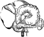
Arteries of the abdominal organs. Labels: 1, the liver; 2, the stomach; 3, upper gut; 4, pancreas; 6, great mesenteric artery; 7, gastric branch; 8, spleen; 9, pyloric branch; 10, pancreatic branch;    11, hepatic artery; 12, duodinal branch; 13, cystic artery; 14, branches to the stomach; 15, 16, spleenic arteries; 17, gastro-epiploic artery; 18, descending aorta; 19, great mesenteric artery.

Arteries of the Abdominal Organs

Arteries of the abdominal organs. Labels: 1, the liver; 2, the stomach; 3, upper gut; 4, pancreas; 6,…
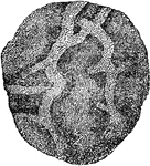
A portion of the stomach, showing its internal surface or mucous coat. Mucous membranes line various cavities of the body, which are exposed to the air.

Mucous Membrane

A portion of the stomach, showing its internal surface or mucous coat. Mucous membranes line various…
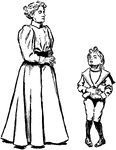
A boy complaining of a stomach ache. The mother looks down at him as if she knows he is pretending.
Stomach Ache

A boy complaining of a stomach ache. The mother looks down at him as if she knows he is pretending.

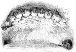
"The 'replete' workers, with their social stomachs distended with the sweet exudations of oak galls, hang from the vaulted roof of the chamber in a cluster and function as so many food reservoirs." -National Geographic, 1912

Replete Ants

"The 'replete' workers, with their social stomachs distended with the sweet exudations of oak galls,…

"The family stomach or repletes of the honey ant of the garden of the gods (Myrmecocystus hortideorum)." -National Geographic, 1912

Honey Ant Replete

"The family stomach or repletes of the honey ant of the garden of the gods (Myrmecocystus hortideorum)."…

"The family stomach or repletes of the honey ant of the garden of the gods (Myrmecocystus hortideorum)." -National Geographic, 1912

Honey Ant Replete

"The family stomach or repletes of the honey ant of the garden of the gods (Myrmecocystus hortideorum)."…
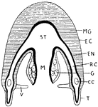
"Structure of a Medusoid. ST., Stomach; M., manubrium; V., velum; T., tentacle; C.C., circumference canal; G., gonad; R.C., radial canal; EN., endoderm; EC., ectoderm; MG, mesogloea." -Thomson, 1916

Medusoid Structure

"Structure of a Medusoid. ST., Stomach; M., manubrium; V., velum; T., tentacle; C.C., circumference…

"Vertical section of Aurelia. m., Mouth; st., stomach; r.c., radial canal; R., reproductive organs; g.f., gastric filaments; g.p., sub-genital cavity; l., marginal tentacle; s., sense organ; the shaded part is mesogloea." -Thomson, 1916

Aurelia

"Vertical section of Aurelia. m., Mouth; st., stomach; r.c., radial canal; R., reproductive organs;…
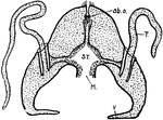
"Hydroctena. A medusoid with suggestion of Ctenophore structure, but a true medusoid nonetheless. ab.o. Aboral sensory organ; T., retractile tentacle; v., velum; M., mouth; ST., stomach" -Thomson, 1916

Hydroctena

"Hydroctena. A medusoid with suggestion of Ctenophore structure, but a true medusoid nonetheless. ab.o.…

"Dissection of lob-worm from dorsal surface. m., Opening of retracted buccal cavity; i., gullet; gl., diverticula on first diaphragm; gl''., oesophageal glands; d., dorsal blood vessels; ef1., first efferent branchial; g., stomach intestine; n6., sixth nephridium; ef 13., thirteenth efferent branchial; af13., thirteenth afferent branchial; a., anus; af1., first afferent branchial; h., heart of left side." -Thomson, 1916

Lob Worm

"Dissection of lob-worm from dorsal surface. m., Opening of retracted buccal cavity; i., gullet; gl.,…
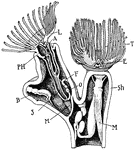
"Diagram of an Ectoproctous Polyzoon (Plumatella). L., Lophophore; PH., pharynx; A., anus; S., stomach; M., retractor muscle; F., funiculus, a cord of mesodermic tissue; O., cells that form "statoblast" buds; B., an ordinary bud; E., epistome over the mouth; T., tentacles; Sh., outer wall of zooecium." -Thomson, 1916

Plumatella

"Diagram of an Ectoproctous Polyzoon (Plumatella). L., Lophophore; PH., pharynx; A., anus; S., stomach;…
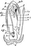
"Diagram of the structure of Sepia. a., Eight short arms around mouth; l.a., one of the two long arms; b., beak of the mouth; c.g., cerebral ganglia, with commissures to the others; E., eye; g., gullet; d.g., digestive gland (the "salivary glands" are not represented); st., stomach; a., anus; sh., shell-sac with sepiostaire; k., kidney; R., reproductive organ; br.h., branchial heart; g., a gill; i.b., ink-bag; m.c., mantle cavity; f., funnel." -Thomson, 1916

Cuttlefish Structure

"Diagram of the structure of Sepia. a., Eight short arms around mouth; l.a., one of the two long arms;…

"'Nurse' of Doliolum mulleri. I., Inhalant, E., exhalant aperture; C., ciliated band round pharynx (P.); En., endostyle; O., "otocyst"; N., nerve-ganglion; H., heart; (E., oesophageal opening; D., stomach; A., anus; Cl., cloaca; DO., dorsal organ; M., muscle bands." -Thomson, 1916

Doliolum Mulleri

"'Nurse' of Doliolum mulleri. I., Inhalant, E., exhalant aperture; C., ciliated band round pharynx (P.);…

"Sexual individual of Doliolum mulleri. G., gonads; B., gill-slits; I., Inhalant, E., exhalant aperture; C., ciliated band round pharynx (P.); En., endostyle; O., "otocyst"; N., nerve-ganglion; H., heart; (E., oesophageal opening; D., stomach; A., anus; Cl., cloaca; DO., dorsal organ; M., muscle bands." -Thomson, 1916

Doliolum Mulleri

"Sexual individual of Doliolum mulleri. G., gonads; B., gill-slits; I., Inhalant, E., exhalant aperture;…
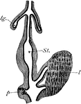
"Origin of lungs, liver, and pancreas in the chick. The mesoderm is shaded; the endoderm dark. lg., One of the lungs; St., stomach; l., liver; p., pancreas." -Thomson, 1916

Chick Development

"Origin of lungs, liver, and pancreas in the chick. The mesoderm is shaded; the endoderm dark. lg.,…
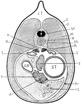
"Section through a young newt. c.t., Connective tissue; E., epidermis; D., dermis; S.C., spinal cord; M., muscle; N., notochord; Sh., mesodermic sheath of notochord; K., kidney; l., lung; S., spleen; ST., stomach; Pe., peritoneum; L., liver; d., duct of the pancreas (P); G.B., gall-bladder; V., dorsal aorta." -Thomson, 1916

Newt Anatomy

"Section through a young newt. c.t., Connective tissue; E., epidermis; D., dermis; S.C., spinal cord;…
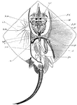
Skeleton of skate. nc, nasal capsules; pq, palato-pterygo-quadrate cartilage; M, Meckel's cartilage; hm, hyo-mandibular; h.br., fifth hypobranchial; v.pl., vertebral plate; st, sensory tubes; sc, scapular region; c, coracoid region; p.pt., anterior basal cartilage or pro-pterygium; m.pt., meso pterygium; mt.pt., meta-pterygium; pu., pubic bar; st, stomach; sv, spiral valve of intestine. -Thomson, 1916

Skate Skeleton

Skeleton of skate. nc, nasal capsules; pq, palato-pterygo-quadrate cartilage; M, Meckel's cartilage;…

"Duodenum of rabbit. P., Pyloric end of stomach; g.b., gall-bladder with bile duct and hepatic ducts; p.d., pancreatic duct." -Thomson, 1916

Rabbit Duodenum

"Duodenum of rabbit. P., Pyloric end of stomach; g.b., gall-bladder with bile duct and hepatic ducts;…

"Stomach of sheep. a, OEsophagus; c, rumen or paunch; d, reticulum or honeycomb-bag; e, psalterium or many-plies; f, abomasum or reed; b, beginning of duodenum." -Thomson, 1916

Sheep Stomach

"Stomach of sheep. a, OEsophagus; c, rumen or paunch; d, reticulum or honeycomb-bag; e, psalterium or…
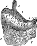
The human stomach. Labels: a, the esophagus or gullet; b, the cardiac portion; c, the left extremity; d, the small extremity; e, the pylorus tied; g,g, the omentum or caul, which is attached to the outside of the stomach, and falls over the intestines like a curtain.

Stomach

The human stomach. Labels: a, the esophagus or gullet; b, the cardiac portion; c, the left extremity;…

The digestive system. This figure represents the whole tract of the intestinal canal, not exactly in its natural position, but spread out so as to show the relative proportions; f, the esophagus; g, cardia; h, pylorus; i, i, the duodenum, about twelve fingers; breadth in length; k, hepatic duct; l, gall bladder; m, cystic duct; n, ductus communis, formed by the union of both; o, the opening of this duct into the duodenum; p, pancreatic duct; q, its opening into the duodenum; r, jejunum; s, ilium; these constitute the small intestines, and are about 26 feet in length, or five times the length of the body; t, termination of ilium in the coecum; , u, superior fold of valve of colon; v, inferior do. ; w, coecum; x, vermiform process; y, y, colon; z, rectum. The coecum, colon, and rectum, form the large intestines, and are about 6 feet in length; the coecum being about 4 inches long, and the same in diameter. The arrows show the direction which the food takes in digestion.

Stomach

The digestive system. This figure represents the whole tract of the intestinal canal, not exactly in…
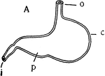
"Stomach of Dog (A)...c, cardiac portion; p, pyloric portion; o, esophagus; i, intestine." -Galloway, 1915

Dog Stomach

"Stomach of Dog (A)...c, cardiac portion; p, pyloric portion; o, esophagus; i, intestine." -Galloway,…
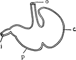
"Stomach of Rat (B)...c, cardiac portion; p, pyloric portion; o, esophagus; i, intestine." -Galloway, 1915

Rat Stomach

"Stomach of Rat (B)...c, cardiac portion; p, pyloric portion; o, esophagus; i, intestine." -Galloway,…
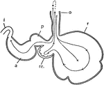
"Diagram of the stomach of a ruminant. o, esophagus; r, rumen or paunch; re., reticulum, or honeycomb; p, psalterium or manyplies; a, abomasum or rennet; i, intestine." -Galloway, 1915

Ruminant Stomach

"Diagram of the stomach of a ruminant. o, esophagus; r, rumen or paunch; re., reticulum, or honeycomb;…
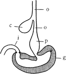
"Diagram of the stomach and esophagus of the fowl. o, esophagus; c, crop; p, proventriculus or glandular stomach; g, gizzard or grinding stomach; i, intestine." -Galloway, 1915

Fowl Stomach

"Diagram of the stomach and esophagus of the fowl. o, esophagus; c, crop; p, proventriculus or glandular…

"General view of the viscera of a male frog, from the right side. a, stomach; b, urinary bladder; c, small intestine; cl, cloacal aperture; d, large intestine; e, liver; f, bile duct; g, gall bladder; h, spleen; i, lung; k, larynx; l, fat body; m, testis; n, ureter; o, kidney; p, pancreas; s, cerebral hemisphere; sp, spinal cord; t, tongue; u, auricle; ur, urostyle; v, ventricle; v.s, vesicula seminalis; w, optic lobe; x, cerebellum; y, Eustachian recess; z, nasal sac." -Parker, 1900

Frog Viscera

"General view of the viscera of a male frog, from the right side. a, stomach; b, urinary bladder; c,…
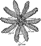
"Asterias rubens. Digestive system. an, anus; card. st, cardiac division of the stomach; int. caec, intestinal caeca; madr, madreporite; ply. caec, pyloric caeca; pyl. st, pyloric division of the stomach." -Parker, 1900

Common Starfish

"Asterias rubens. Digestive system. an, anus; card. st, cardiac division of the stomach; int. caec,…
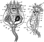
"Brachionus rubens. A, from the dorsal aspect; B, from the right side. a, anus; br, brain; d. f. dorsal feeler; c. gl, cement gland; cl, cloaca; c. l, ciliary lobes; c. v, contractile vesicle; e, eye-spot; int, intestine; lr, lorica; l. f, lateral feeler; m, muscular bands; nph, nephridial tubes; ov, ovary; ph, pharynx; st, stomach; t, tail; tr, d, trochal disc; vt, vitellarium." -Parker, 1900

Brachionus Rubens

"Brachionus rubens. A, from the dorsal aspect; B, from the right side. a, anus; br, brain; d. f. dorsal…

"Sepia officinalis, enteric canal. a, anus; b. d, one of the bile ducts; b. m, buccal mass; c, caecum; i, ink-sac; i. d, ink-duct; j, jaws; l, l, liver lobes; oe, oesophagus; p, pancreatic appendages1; r, rectum; s. g, salivary glands; st, stomach." -Parker, 1900

Cuttlefish Enteric Canal

"Sepia officinalis, enteric canal. a, anus; b. d, one of the bile ducts; b. m, buccal mass; c, caecum;…

"Lacerta agilis. General view of the viscera in their naturaal relations. Bl, urinary bladder; Ci, post-caval vein; ED, rectum; GB, gall-bladder; H, heart; Lg, Lg', the lungs; M, stomach; MD, small intestine; Oe, oesophagus; Pn, pancreas; Tr, trachea." -Parker, 1900

Sand Lizard Viscera

"Lacerta agilis. General view of the viscera in their naturaal relations. Bl, urinary bladder; Ci, post-caval…
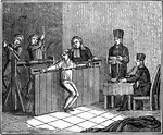
The torture of a free mason. Caption below illustration: "They next set my back against a thick board, at each extremity of which was pulley; through which a rope ran, that was fastened at the end of the chain at my wrist. The tormentors then, stretching these ropes by means of a roller, pressed and bruised my stomach in proportion as the ropes were drawn tighter.

Torture of a Free Mason

The torture of a free mason. Caption below illustration: "They next set my back against a thick board,…
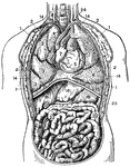
"Thorax and abdomen. 1, 1, 1, 1. Muscles of the chest. 2, 2, 2, 2. Ribs. 3, 3, 3. Upper, middle and lower lobes of the right lung. 4, 4. Lobes of the left lung. 5, Right ventricle of the heart. 6. Left ventricle. 7. Right auricle. 8. Left auricle. 9. Pulmonary artery. 10. Aorta. 11. Descending vena cava. 12. Trachea. 13. Oesophagus. 14, 14, 14, 14. Pleura. 15, 15. Diaphragm. 16, 16. Right and left lobes of the liver. 17. Gall cyst. 18. Stomach. 19. Duodenum. 20. Ascending colon. 21. Transverse colon. 22. Descending colon. 23, 23. Small intestine. 24. Thoracic duct opening into the left subclavian vein. 25. Spleen." -Foster, 1921

Thorax and Abdomen

"Thorax and abdomen. 1, 1, 1, 1. Muscles of the chest. 2, 2, 2, 2. Ribs. 3, 3, 3. Upper, middle and…

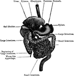
Labels: O, esophagus; S, stomach; SI, small intestine; LI, large intestine, Sp spleen; L, liver (raised up); G, gall bladder; Pa, pancreas.

General View of the Alimentary Canal

Labels: O, esophagus; S, stomach; SI, small intestine; LI, large intestine, Sp spleen; L, liver (raised…
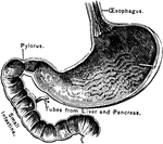
The opening part of the stomach where the esophagus joins it is called the cardiac opening; the one at the opposite end, where the small intestine joins it, is called the pylorus.

A Section of the Stomach

The opening part of the stomach where the esophagus joins it is called the cardiac opening; the one…