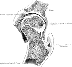
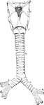
Frontal Section Through Hip Joint
A frontal section through the left hip joint of a boy. Front view.
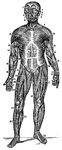
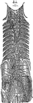
Front View of the Superficial Muscles of the Body
A front view of the superficial muscles of the body. Labels: 1, The frontal swells of the occipito-frontalis.…
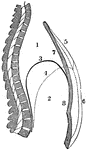
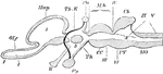
A Side View of the Lacteals and Thoracic Duct
A side view of the lacteal and thoracic duct. Labels: 1, Small intestine. 2, Lacteals. 3, Thoracic duct.…
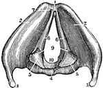
View of the Great Lymphatic Trunks
View of the great lymphatic trunks. Labels: 1, 2 Thoracic duct. 4, The right lymphatic duct. 5, Lymphatics…
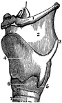
A Side View of the Cartilages of the Larynx
A side view of the cartilages of the larynx. Labels: *, The front side of the thyroid cartilage. 1,…
A Back View of the Cartilages and Ligaments of the Larynx
A back view of the cartilages and ligaments of the larynx. Labels: 1, The posterior face of the epiglottis.…
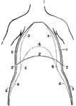
A Front View of the Chest and Abdomen in Respiration
A front view of the chest and abdomen in respiration. Labels: 1, The position of the walls of the chest…
A Side View of the Chest and Abdomen in Respiration
A side view of the chest and abdomen in respiration. Labels: 1, The cavity of the chest. 2, The cavity…
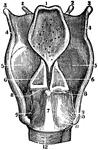
A View of the Larynx Showing the Vocal Ligaments
A view of the larynx showing the vocal ligaments. Labels: 1, The anterior edge of the larynx. 4, The…
A Back View of the Brain and Spinal Cord
A back view of the brain and spinal cord. Labels: 1, The cerebrum. 2, The cerebellum. 3, The spinal…
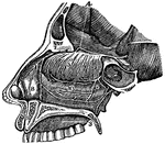
A Side View of the Passage of the Nostrils
A side view of the passage of the nostrils. 4, The distribution of the first olfactory pair of nerves.…
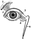
View of the Lachrymal Gland and Nasal Duct
View of the lachrymal gland and nasal duct. Labels: 1, The lachrymal gland. 2, Ducts leading from the…
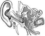
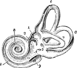
Parts of the Ear
A view of all the parts of the ear, Labels: 1, The tube that leads to the internal ear. 2, The membrana…
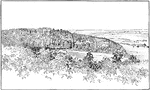
Fault Scarps
"Fault scarps of Orenaug Hill (floating block topography). The view is taken from the top of a cliff…
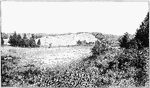
Fault Scarp Slope
"View of the southeastern slope of the eastern twin of Orenaug Hill. Fault scarps bound the hummocky…
Intrusion Machine
"The principal working parts of the apparatus are the sheet of boiler plate a (fig. 100), the cylinder…
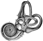
View of the Labyrinth Laid Open
A view of the labyrinth laid open. Labels: 1, The cochlea. 2, 3, Two channels that wind two and half…
View of the Auditory Nerve
A view of the auditory nerve. Labels: 1, The spinal cord. 2, The medulla oblongata. 3, The lower part…
A Magnified View of a Bone
A thin slice of bone, highly magnified, showing the lacunae, the tiny tubs (canaliculi) radiating from…
Microscopic View of a Muscle
A microscopic view of a muscle showing, at one end, the fibrillae; and at the other, the disk, or cells…
Magnified View of the Cuticle
Magnified views of the cuticle. Labels: A, represents a vertical section of the cuticle. B, the lateral…
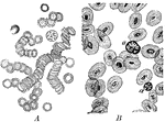
Corpuscles of Human and Animal Blood
A magnified view of corpuscles of human blood compared to animal blood. Labels: A, corpuscles of human…
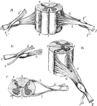
Nerve Fibers of the Sciatic Nerve
Two nerve fibers of the sciatic nerve. Labels: A. Node of Ranvier. B. Axis cylinder. C. Sheath of Schwann…
Behavior of the Nodes of Ranvier
Several fibers of a bundle of medullated nerve fibers acted upon by silver nitrate to show peculiar…
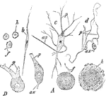
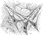
Different Forms of Ganglion Cells
Different forms of ganglion cells. A, a, round ball-shaped unipolar cell from the human Gasserian ganglion.…
Pacinian Corpuscle of a Cat
The Pacinian bodies or corpuscles are elongated oval bodies situated on some of the cerebrospinal and…
End-bulb of Krause
End-bulbs are oval or spheroidal, and are composed of medullated nerve fibers, which terminate in corpuscles…
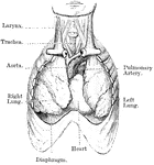
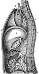
Heart and Lungs
View of heart and lungs in situ. The front portion of the chest wall, and the outer or parietal layers…
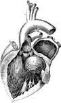
A Diagram of the Heart
The left auricle and ventricle opened and a part of their anterior and left walls removed. The pulmonary…
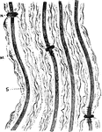
Surface View of an Artery
Surface view of an artery from the mesentery of a frog, ensheathed in a perivascular lymphatic vessel.…
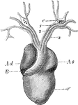
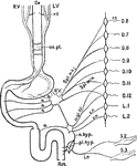
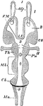
Heart of a Frog
The heart of a frog (Rana esculenta) from the front. Labels: V, ventricle, Ad, right auricle; As, left…
Front View of Respiratory Apparatus
Outline showing the general form of the larynx, trachea, and bronchi, as seen from front. Labels: h,…
Back View of Respiratory Apparatus
Outline showing the general form of the larynx, trachea, and bronchi, as seen from behind. Labels: h,…
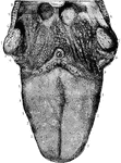
Tongue
The papillar surface of the tongue, with the fauces and tonsils. Labels: 1, circumvallate papillae,…
Nerves of the Alimentary Canal
Diagrammatic representation of the nerves of the alimentary canal. Oe to Rct, the various parts of the…
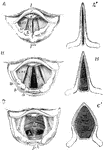
Cartilages from the Larynx
Cartilages from the larynx seen from the front. Labels: 1, vertical ridge of pomum Adami; 2, right ala;…
Movement of the Vocal Cords
Three laryngoscopic view of the superior aperture of the larynx and surrounding parts. Labels: A, the…
Upper Part of the Larynx
View of the upper part of the larynx as seen by means of the laryngoscope during the utterance of a…
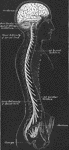
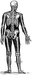
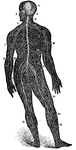
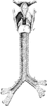
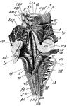
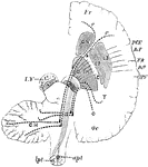
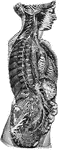
Central Nervous System
View of the cerebrospinal axis of the nervous system. The right half of the cranium and trunk of the…
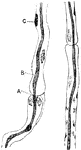
Views of a Spinal Cord
Different views of a portion of the spinal cord from the cervical region, with the roots of the nerves.…
Gray Matter of Spinal Cord
Section of gray matter of anterior cornu of a calf's spinal cord; a, nerve fibers of white matter in…
Medulla
Dorsal or posterior view of the medulla, fourth ventricle, and mesencephalon. Labels: p.n., line of…
Section across the Pons
Section across the pons, about the middle of fourth ventricle. py., pyramidal bundles; po., transverse…
Cerebral Hemisphere
Diagram of the motor tract as shown in a diagrammatic horizontal section through the cerebral hemispheres…
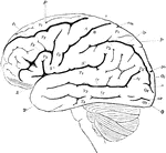
Lateral View of the Brain
Lateral view of the brain. F, Frontal lobe; P, Parietal lobe; O, Occipital lobe; T, Temporal lobe; S,…
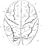
View of Brain from Above
View of brain from above. F, Frontal lobe; P, Parietal lobe; O, Occipital lobe; T, Temporal lobe; S,…
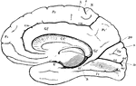
Right Hemisphere of the Brain
View of the right hemisphere in the median aspect. CC, corpus callosum longitudinally divided; Gf, gyrus…
Horizontal Section of a Vertebrate Brain
Diagrammatic horizontal section of a vertebrate brain. Mb, midbrain: what lies in front of this is the…
Vertical Section of a Vertebrate Brain
Longitudinal and vertical diagrammatic section of a vertebrate brain. Mb, midbrain: what lies in front…
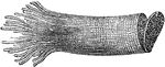
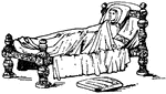
Medieval Bedstead
The Medieval bedstead of the 13th century was richly decorated with turned posts and carved sides. The…
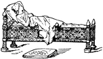
Medieval Bedstead
The Medieval bedstead of the 12th century was richly decorated with turned posts and carved sides. The…
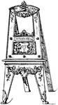
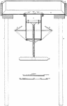
Modern Decorative Easel
The modern decorative easel is a sloping frame with three or four legs. The front and rear are often…
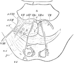
Sympathetic System
Diagrammatic view of the Sympathetic cord of the right side, showing its connections with the principal…
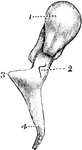
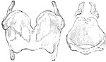
Malleus
The hammer-bone or malleus, seen from the front. 1, the head; 2, neck; 3, short process; 4, long process.
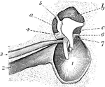
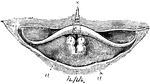
Tympanum
Interior view of the tympanum, with membrana tympani and bones in natural position. 1, Membrana tympani;…
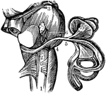
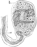
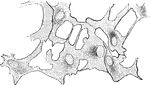
Interior of the Left Labyrinth
View of the interior of the left labyrinth. The bony wall of the labyrinth is removed superiorly and…
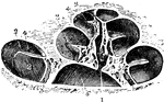
Osseous Cochlea
View of the osseous cochlea divided through the middle. Labels: 1, central canal of the modiolus; 2,…
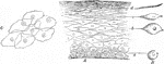
Lamella of Kitten's Cornea
Surface view of part lamella of kitten's cornea, prepared first with caustic potash and then with nitrate…
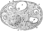
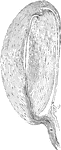
Ovary of the Cat
View of a section of the ovary of the cat. Labels: 1, outer covering and free border of the ovary; 1',…