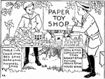
Illustration of a child buying toys from another boy. It can be used to write mathematics story problems involving addition, subtraction, money, and counting.

Child Buying Toys

Illustration of a child buying toys from another boy. It can be used to write mathematics story problems…

Illustration of a child with 8 quarts and 1 peck. He is using the baskets to practice measuring. It can be used to write mathematics story problems involving addition, subtraction, money, measurement, and counting.

Child Measuring Using Pecks And Quarts

Illustration of a child with 8 quarts and 1 peck. He is using the baskets to practice measuring. It…

Illustration showing the back side of two dimes.

2 Dimes

Illustration showing the back side of two dimes.

Illustration showing the back side of three dimes.

3 Dimes

Illustration showing the back side of three dimes.

Illustration showing the back side of four dimes.

4 Dimes

Illustration showing the back side of four dimes.

Illustration showing the back side of five dimes.

5 Dimes

Illustration showing the back side of five dimes.

"The repairing of a rent in the nest of the red tree ant (Ecophylla smaragdina). The nest is made of leaves, held together with a silken web. The figure shows the workers repairing a rent, one brigade of them being occupied in drawing the edges of the leaves together, while the others on the inside of the nest move the spinning larvae back and forth across the gap." -National Geographic, 1912

Red Tree Ants

"The repairing of a rent in the nest of the red tree ant (Ecophylla smaragdina). The nest is made of…

Side view of the muscles of the eye in their natural positions. Labels: a,b,c,d, the four straight muscles. a. is turned up to prevent the others from being hidden. e. the superior oblique muscle. f. the optic nerve. The other oblique muscle is not shown, but it lies on the other side of the ball, and comes round, and is inserted near the superior oblique; so that they pull opposite ways, like two men sawing timber. By the combined action of all these muscles, an infinite variety of motions of the eye can be produced.

Muscle of the Eye

Side view of the muscles of the eye in their natural positions. Labels: a,b,c,d, the four straight muscles.…

Muscles of the eyelids, the elevator passing back into the orbit; the sphincter, or orbicular muscle surrounding the eye.

Eyelid, Muscles of

Muscles of the eyelids, the elevator passing back into the orbit; the sphincter, or orbicular muscle…
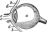
Side view of the eyeball. Labels: a, the eyeball, and b,b, are the upper and lower sides. Now in order to prevent dust and other bodies from working their way between the ball and the lid, through passages at c,c, into the socket of the eye where they would excite great inconvenience and pain, we find the common skin of the eye lids d,d, after covering their edges turn in a little way between the lid and the ball, and then turn back and are reflected over the surface of the cornea; where, to prevent the obstruction of vision, it becomes perfectly transparent.

Side View of the Eyeball

Side view of the eyeball. Labels: a, the eyeball, and b,b, are the upper and lower sides. Now in order…
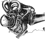
General sectional view of the structure of the ear. Labels: a, the meatus auditorius externus; b, the tympanum; c, the malleus; d, the incus, e, the os orbiculare; f, the stapes; g, the semicircular canals; h, the cochlea; i, the meatus auditorius internus; k, the eustachian tube.

Sectional View of the Ear

General sectional view of the structure of the ear. Labels: a, the meatus auditorius externus; b, the…

View of the diaphragm; 1, cavity of the thorax; 2, diaphragm separating the cavity of the thorax from that of the abdomen; 3, cavity of the pelvis.

Diaphragm

View of the diaphragm; 1, cavity of the thorax; 2, diaphragm separating the cavity of the thorax from…

Of the orpine family (Crassulaceae), the live-forever or garden orpine (Sedum purpureum).

Live-Forever or Garden Orpine

Of the orpine family (Crassulaceae), the live-forever or garden orpine (Sedum purpureum).
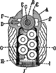
This image "represents a cross section through the ejector with the magazine loaded. The parts shown are receiver, A; bolt, B; firing pin, C; ejector, D; clip slots, E; bolt locking lug channels, F; magazine, G; follower, H; magazine spring, I; and floor plate, J." -Moss, 1914

Loaded Magazine

This image "represents a cross section through the ejector with the magazine loaded. The parts shown…
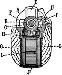
This image "shows a cross section through the magazine empty, and with cut-off "on," shown in projection. The parts are receiver, A; bolt, B; firing pin, C; cut-off, D; rear lug slot, E; bolt locking lug channels, F; magazine, G; follower, H; magazine spring, I; and floor plate, J." -Moss, 1914

Empty Magazine

This image "shows a cross section through the magazine empty, and with cut-off "on," shown in projection.…

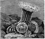
"Sea anemones on back of hermit crab." -Thomson, 1916

Anemone

"Sea anemones on back of hermit crab." -Thomson, 1916

"Life history of a coral, Monoxenia darwinii. A, B, Ovum. C, Division into two. D, four-cell stage. E, Blastula. F, Free-swimming blastula with cilia. G, Section of blastula. H, Beginning of invagination. I, Section of completed gastrula, showing ectoderm, and archenteron. K, Free-swimming ciliiated gastrula." -Thomson, 1916

Coral Stages

"Life history of a coral, Monoxenia darwinii. A, B, Ovum. C, Division into two. D, four-cell stage.…
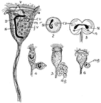
"Vorticella. 1. Structure. N., Macronucleus; n., micronucleus; c.v., contractile vacuole; m., mouth; f.v., food vacuole; v., vestibule. 2. Encysted individual. 3. Division. 4. Separation of a free-swimming unit--the result of a division. 5. Formation of eight minute units (mg.). 6. Conjugation of microzooid (mg.) with one normal size." -Thomson, 1916

Vorticella

"Vorticella. 1. Structure. N., Macronucleus; n., micronucleus; c.v., contractile vacuole; m., mouth;…
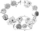
"Life history of Coccidium. 1. Sporozoite; 2. Sporozoite entering a cell and becoming a trophozoite; 3-4. Schizont, forming merozoites; 5. Merozoites entering another cell; 6a. Merozoite forming macrogamete; 6b. Merozoite forming microgametes; 7. Free microgamete; 8-9. Fertilisation of macrogamete by microgamete; 10. Zygote within oocyst; 11. Formation of spores within oocyst; 12. Spores forming sporozoites." -Thomson, 1916

Coccidium

"Life history of Coccidium. 1. Sporozoite; 2. Sporozoite entering a cell and becoming a trophozoite;…

"Diagrammatic representation of development of Oscarella lobularis. Bl., Free-swimming blastula with flagella; G., gastrula settled down. Next figure shows folding of inner layer (En.); Ec., outer layer. Lowest figure shows radial chambers (R.C.); Mesogloea (Mg.); inhalant pore (P.); exhalant osculum (O.)." -Thomson, 1916

O. Lobularis

"Diagrammatic representation of development of Oscarella lobularis. Bl., Free-swimming blastula with…

"Colony of Hydractinia on back of a Buccinum shell tenanted by a hermit-crab." -Thomson, 1916

Hydractinia

"Colony of Hydractinia on back of a Buccinum shell tenanted by a hermit-crab." -Thomson, 1916
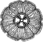
"Surface view of Aurelia. Showing four genital pockets in centre, much branched radial canals, eight peripheral niches for sense organs, and peripheral tentacles." -Thomson, 1916

Aurelia

"Surface view of Aurelia. Showing four genital pockets in centre, much branched radial canals, eight…

"Life history of liver fluke. 1. Developing embryo in egg-case; 2. free-swimming ciliated embryo; 3. sporocyst; 3a. shell of Limnaus truncatulus; 4. division of sporocyst; 5. sporocyst with rediae forming within it; 6. redia with more rediae forming within it; 7. tailed cercaria; 8. young fluke." -Thomson, 1916

Liver Fluke Stages

"Life history of liver fluke. 1. Developing embryo in egg-case; 2. free-swimming ciliated embryo; 3.…
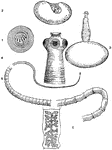
"Life history of Taenia solium. 1. Six-hooked embryo in egg-case; 2. proscolex or bladder-worm stage, with invaginated head; 3. bladder-worm with evaginated head; 4. enlarged head of adult, showing suckers and hooks; 5. general view of the tape-worm, from small head and thin neck to the rip joints; 6. a rip joint or proglottis with branched uterus; all other organs are now lost." -Thomson, 1916

Pork Tapeworm

"Life history of Taenia solium. 1. Six-hooked embryo in egg-case; 2. proscolex or bladder-worm stage,…

"Diagrammatic longitudinal section of a Nemertean (Amphiporum lactifloreus), dorsal view. p.p., Proboscis pore; b., brain giving off the lateral nerve-cords (n.); po., oesophageal pocket; p., proboscis lying within its sheath; st., stilet of proboscis; m., retractor muscles of proboscis; g., gut shown in outline at the sides of proboscis; e., the three main, longitudinal blood vessels, which unite both anteriorly and posteriorly." -Thomson, 1916

Nemertea

"Diagrammatic longitudinal section of a Nemertean (Amphiporum lactifloreus), dorsal view. p.p., Proboscis…
"Free-living Polychaete (Nereis cultrifera)." -Thomson, 1916

Polychaete

"Free-living Polychaete (Nereis cultrifera)." -Thomson, 1916

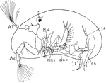
"Cypris, side view, after removal of one valve. e., Eye; A.1, first antennae; A.2, second antennae; MN., mandibles; mx.1, first maxilla; mx.2, second maxilla; f.1, f.2, thoracic legs; Ab., rudimentary abdomen." -Thomson, 1916

Cypris

"Cypris, side view, after removal of one valve. e., Eye; A.1, first antennae; A.2, second antennae;…
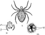
"Garden spider. I., Female garden spider; II., end view of head of the same showing the simple eyes, the poison fangs (ch.), and the pedipalps (p.); III., posterior end of body showing two pairs of spinnerets (sp.), with anus above." -Thomson, 1916

Garden Spider

"Garden spider. I., Female garden spider; II., end view of head of the same showing the simple eyes,…

The side view of Sepia officinalis, or the common cuttlefish.

Cuttlefish

The side view of Sepia officinalis, or the common cuttlefish.

View of the bronchial tubes, terminating in air vesicles. On the left is the external view (1, bronchial tube; 2, air vesicles). On the right is the bronchial tube and air vesicles laid open.

Bronchial Tubes Terminating in Air Vesicles

View of the bronchial tubes, terminating in air vesicles. On the left is the external view (1, bronchial…

"Diagram of Salpa africana. o.a., Oral aperture; d.t., dorsal tubercle; te., tentacle; g., ganglion; m., muscle bands; atr., atrium; b.v., blood-vessel; an., anus; a.a., exhalant aperture; v.n., visceral nucleus; h., heart st., stolon; d.l., dorsal lamina; E., endostyle; s.n.g., sub-neural gland; ph., pharynx; p.p.b., peri-pharyngeal band." -Thomson, 1916

Salpa Africana

"Diagram of Salpa africana. o.a., Oral aperture; d.t., dorsal tubercle; te., tentacle; g., ganglion;…

"Lateral view of Amphioxus. The notochord runs from tip to tip. t., Tentacular cirri; G., reproductive organs; a.p., atriopore; a., position of anus; 40 and 62, indicate number of myotomes." -Thomson, 1916

Lancelet

"Lateral view of Amphioxus. The notochord runs from tip to tip. t., Tentacular cirri; G., reproductive…
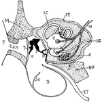
"Diagram showing the ear and related parts in a young cat. P., Pinna; Sq., squamosal: E.A.M., external auditory meatus; T., tympanum; M., malleus; I., incus; St., stapes abutting onforamen ovale; B., bulla of tympanic bone; Se., a septum in the bulla; E.T., eustachian tube leading from the tymmpanic cavity to the back of the mouth; B.O., basi-occipital; C., cochlea; S., sacculus; U., utriculus; D.E., ductus endolymphaticus; N., auditory nerve; S.C., semi-circular canal; PE., periotic bone." -Thomson, 1916

Cat Ear

"Diagram showing the ear and related parts in a young cat. P., Pinna; Sq., squamosal: E.A.M., external…

"Side view of skate's skull. l1., First labial cartilage; n.c., nasal capsule; a.o., antorbital; p.pt.q., palato-pterygo-quadrate; M.c., Meckel's cartilage; h.m., hyo-mandibular; e.h., epi-hyal; c.h., cerato-hyal; h.h., hypo-hyal; h.br.1-5, hypobranchials; c.br., ceratobranchial; e.br., epibranchial; p.br.1., first prebranchial; i.h., inter-hyal; m.pt., meta-pterygoid; 2, 5, 7, foramina of exit of the corresponding nerves." -Thomson, 1916

Skate Skull

"Side view of skate's skull. l1., First labial cartilage; n.c., nasal capsule; a.o., antorbital; p.pt.q.,…

"Side view of chief cranial nerves of Elasmobranchs." -Thomson, 1916

Shark Nerves

"Side view of chief cranial nerves of Elasmobranchs." -Thomson, 1916

"Lateral view of dogfish (Scyllium catulus). Note ventral mouth with naso-buccal groove, heterocercal tail, and unpaired fins. gs., Gill-slits; pc., pectoral fins; pv., pelvic fins." -Thomson, 1916

Dogfish

"Lateral view of dogfish (Scyllium catulus). Note ventral mouth with naso-buccal groove, heterocercal…
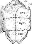
"Internal view of the plastron of the Greek tortoise. EP., Epiplastron (clavicle?); ENT., entoplastron (interclavicle?); HYO., hyoplastron; HYPO., hypoplastron; XIPHI., xiphiplastron." -Thomson, 1916

Greek Tortoise Plastron

"Internal view of the plastron of the Greek tortoise. EP., Epiplastron (clavicle?); ENT., entoplastron…
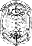
"Internal view of tortoise skeleton. H., humerus; SC., scapula running dorsally; PC., precoracoid; C., coracoid; EC., epicoracoid cartilage; P., pubis; IL., ilium running dorsally to sacral vertebrae; IS., ischium; DV., dorsal vertebrae fused in carapace; R., head of a rib; CEV., cervical vertebrae free; CAV., caudal vertebrae free." -Thomson, 1916

Tortoise Skeleton

"Internal view of tortoise skeleton. H., humerus; SC., scapula running dorsally; PC., precoracoid; C.,…
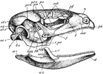
"Side view of skull of Lacerta. px., Premaxilla; mx., maxilla; l., lachrymal; j., jugal; t.pa., transpalatine; epg., epipterygoid; pg., pterygoid; bpg., basipterygoid; b.o., basioccipital; q., quadrate; oc.c., occipital condyle; sq., squamosal; pr.o., pro-otic; pt.o., postorbital; st.1, st.2, supratemporals; ps., presphenoid (the optic nerve is seen issuing in front of the end of the reference line); p.e., mesethmoid; s.ob., supraorbitals; pf., prefrontal; n., nasal; ar., articular; ag., angular; sag., surangular; cr., coranary; d., dentary." -Thomson, 1916

Lizard Skull

"Side view of skull of Lacerta. px., Premaxilla; mx., maxilla; l., lachrymal; j., jugal; t.pa., transpalatine;…

"Fore-limb and hind-limb compared. H., Humerus; R., radius; U., ulna; r., radiale; u., ulnare; C., distal carpals united to carpo-metacarpus; CC., the whole carpal region; MC.I., metacarpal of the thumb; I., phalanx of the thumb; MC.II., second metacarpus; II., second digit; MC.III., third metacarpus; III., third digit. F., femur; T.T., tibio-tarsus; Fi., fibula; Pt., proximal tarsals united to lower end of tibia; dt., distal tarsals nited to upper end of tarso-metatarsus (T.MT.); T., entire tarsal region; MT.I., first metatarsal, free; I.-IV., toes." -Thomson, 1916

Bird Limbs

"Fore-limb and hind-limb compared. H., Humerus; R., radius; U., ulna; r., radiale; u., ulnare; C., distal…

"A., Filoplume. B., very young feather within its sheath (sh.); c., the core of dermis; b., the barbs. C., the same, external view." -Thomson, 1916

Feathers

"A., Filoplume. B., very young feather within its sheath (sh.); c., the core of dermis; b., the barbs.…

"Bones of hind-limb of eagle. f., Femur; t.t., tibio-tarsus; fb., fibula; a., ankle-joint; m.t., tarso-metatarsus; m.t'., first metatarsal (free)." -Thomson, 1916

Eagle Hind-Limb

"Bones of hind-limb of eagle. f., Femur; t.t., tibio-tarsus; fb., fibula; a., ankle-joint; m.t., tarso-metatarsus;…

"Side view of rabbit's skull. Pmx., Premaxilla; Na., nasal; Fr., frontal; Pa., parietal; Sq., squamosal; S.O., supraoccipital; Per., periotic; T., tympanic (the reference line points to the bony external auditory meatus, beneath it lies the inflated bulla); PO., paroccipital process." -Thomson, 1916

Rabbit Skull

"Side view of rabbit's skull. Pmx., Premaxilla; Na., nasal; Fr., frontal; Pa., parietal; Sq., squamosal;…

"Dorsal view of rabbit's brain. olf.l., Olfactory lobes; c.h., cerebral hemispheres; o.l., optic lobes (corpora quadrigemina); cb., median part of cerrebellum; Fl., flocculus of cerebellum; 10, root of the tenth or vagus nerve; s.c., spinal cord." -Thomson, 1916

Dorsal View of Rabbit Brain

"Dorsal view of rabbit's brain. olf.l., Olfactory lobes; c.h., cerebral hemispheres; o.l., optic lobes…

"Side view of sheep's skull. PMX., Premaxilla; MX., maxilla; NA., nasal; J., Jugal; L., lachrymal; FR., frontal; PA., parietal; SQ., squamosal; CO., condyle; PP., paroccipital process." -Thomson, 1916

Sheep Skull

"Side view of sheep's skull. PMX., Premaxilla; MX., maxilla; NA., nasal; J., Jugal; L., lachrymal; FR.,…

"Side view of horse's skull. P., Parietal; FR., frontal; NA., nasal; PMX., premaxilla; MX., maxilla; J., jugal; L., lachrymal; SQ., squamosal; PP., paroccipital process; CO., condyle CA., canine." -Thomson, 1916

Horse Skull

"Side view of horse's skull. P., Parietal; FR., frontal; NA., nasal; PMX., premaxilla; MX., maxilla;…
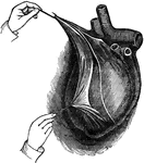
View of the heart enclosed in its bag, or pericardium, which is a serious membrane. It is here laid open and turned back.

Heart in the Pericardium

View of the heart enclosed in its bag, or pericardium, which is a serious membrane. It is here laid…
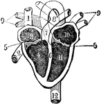
View of the heart with its several chambers exposed and the vessels in connection with them. Labels: 1, the superior vena cava. 2, the inferior vena cava. 3. the chamber called the auricle. 4. the right ventricle. 5. the line marking the passage between the two chambers, and the points of attachment of one margin of the valve. 6. the septum between the two ventricles. 7. the pulmonary artery, arising from the right ventricle, and dividing at 8 into right and left, of the corresponding lungs. 9. the four pulmonary veins, bringing the blood from the lungs into 10, the left auricle. 11. the left ventricle. 12. the aorta, arising from the left ventricle, and passing down behind the heart, to distribute blood to every part of the system. Thus the blood moves in a double circle, one from the heart to the body, and from the body back to the heart, called the systemic circle; the other, from the heart to the lung, and from the lung back to the heart, called the pulmonic circle.

Heart and its Chambers

View of the heart with its several chambers exposed and the vessels in connection with them. Labels:…

"C, more mature condition, in which the planula has become fixed: f, foot or attached end; o, oral or free end at which the tentacles and mouth will be developed." -Galloway, 1915

Mature Hydroid Planula

"C, more mature condition, in which the planula has become fixed: f, foot or attached end; o, oral or…

An illustration of a promissory note. "A promissory note is a written promise to pay a specified sum of money at a stated time."

Promissory Note

An illustration of a promissory note. "A promissory note is a written promise to pay a specified sum…

An illustration of a promissory note that has been indorsed. "A promissory note is a written promise to pay a specified sum of money at a stated time."

Indorsed Promissory Note

An illustration of a promissory note that has been indorsed. "A promissory note is a written promise…

An illustration of a demand note. A note in which the maker agrees to pay whenever the payee wishes the money and "demands" it.

Demand Note

An illustration of a demand note. A note in which the maker agrees to pay whenever the payee wishes…

An illustration of a commercial draft for $122.50. "A commercial draft is a written order by which one person directs a second person to pay a specified sum of money at a stated time, through a bank."

Commercial Draft

An illustration of a commercial draft for $122.50. "A commercial draft is a written order by which one…

"Two stages in the metamorphosis of the Mosquito. A, larva; B, pupa; C, ventral view of the oar-like appendages of the last segment of the pupa." -Galloway, 1915

Mosquito Metamorphosis

"Two stages in the metamorphosis of the Mosquito. A, larva; B, pupa; C, ventral view of the oar-like…
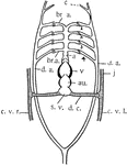
"Diagram of the heart, the branchial arches, and the principal veins in the Teleosts. Ventral view. The heart is represented without the sigmoid flexure; that is, with the auricle posterior. a, aorta; au., auricle; br.a., branchial arches of the aorta (1-4, numbering from the front); c, carotid; c.v., cardinal veins (right and left); d.a., dorsal arteries; j, jugular veins; d.c., ductus Cuvieri; s.v., sinus venosus; v, ventricle. Only four arterial arches are shown." -Galloway, 1915

Teleost Heart

"Diagram of the heart, the branchial arches, and the principal veins in the Teleosts. Ventral view.…

"Diagram of the principal vessels in the circulation of a Fish, lateral view. a, aorta; au., auricle; br.a., afferent branchial arteries; br.e., efferent branchial arteries; bu., bulbus (or conus) arteriosus; c, carotid; c.a., caudal artery; c.v.r., right cardinal vein; c.v.l., left cardinal vein; d.a., dorsal artery; d.c., ductus Cuvieri; g, gills; h.v., hepatic vein; h.p., hepatic portal vein; i, intestine; j, jugular vein; k, kidney; l, liver; r.p., renal portal vein; s.v., sinus venosus; v, ventricle." -Galloway, 1915

Fish Circulation Vessels

"Diagram of the principal vessels in the circulation of a Fish, lateral view. a, aorta; au., auricle;…

A lion toothed leaf.

Lion Toothed Leaf

A lion toothed leaf.

A lion, rabbit, hawk, all gather around the tree for a meeting held by the owl.

Animals Gathering

A lion, rabbit, hawk, all gather around the tree for a meeting held by the owl.

A thirsty lion slowly bringing himself to drink some water.

Thirsty Lion

A thirsty lion slowly bringing himself to drink some water.

Members of the royal family take a walk outside with the castle in the background.

Royalty

Members of the royal family take a walk outside with the castle in the background.