"Leaf of a live-forever (Sedum sp.), with a portion of the epidermis peeled back. Underneath the epidermis is the mesophyll." -Gager, 1916

Sedum Leaf

"Leaf of a live-forever (Sedum sp.), with a portion of the epidermis peeled back. Underneath the epidermis…
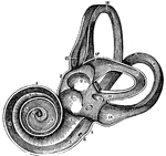
A view of the labyrinth laid open. Labels: 1, The cochlea. 2, 3, Two channels that wind two and half turns around a central point (5). 7, The central portion of the labyrinth called the vestibule. 11, 12, 13, 14, 15, 16, 17, 18, The semicircular canals.

View of the Labyrinth Laid Open

A view of the labyrinth laid open. Labels: 1, The cochlea. 2, 3, Two channels that wind two and half…
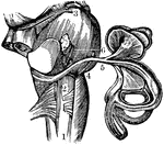
A view of the auditory nerve. Labels: 1, The spinal cord. 2, The medulla oblongata. 3, The lower part of the brain. 4, The auditory nerve. 5, A branch to the semicircular canals. 6, A branch to the cochlea.

View of the Auditory Nerve

A view of the auditory nerve. Labels: 1, The spinal cord. 2, The medulla oblongata. 3, The lower part…
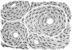
A thin slice of bone, highly magnified, showing the lacunae, the tiny tubs (canaliculi) radiating from them, and four Huversian canals, three seen crosswise and one lengthwise.

A Magnified View of a Bone

A thin slice of bone, highly magnified, showing the lacunae, the tiny tubs (canaliculi) radiating from…

The spine showing the seven vertebrae of the neck, cervical; the twelve of the back, dorsal; the five of the loins, lumbar; the sacrum (a), and the coccyx (b), comprising the nine "false vertebrae".

The Spine

The spine showing the seven vertebrae of the neck, cervical; the twelve of the back, dorsal; the five…

A microscopic view of a muscle showing, at one end, the fibrillae; and at the other, the disk, or cells of the fiber.

Microscopic View of a Muscle

A microscopic view of a muscle showing, at one end, the fibrillae; and at the other, the disk, or cells…
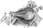
The muscles of the right eye. Labels: A, superior straight; B, superior oblique passing through a pulley, D; G, inferior oblique; H, external straight, and , back of it, the internal straight muscle.

Muscles of the Eye

The muscles of the right eye. Labels: A, superior straight; B, superior oblique passing through a pulley,…

Magnified views of the cuticle. Labels: A, represents a vertical section of the cuticle. B, the lateral view of the cells. C, flat side of scales like d, magnified, showing the nucleated cells transformed into broad scales.

Magnified View of the Cuticle

Magnified views of the cuticle. Labels: A, represents a vertical section of the cuticle. B, the lateral…
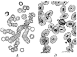
A magnified view of corpuscles of human blood compared to animal blood. Labels: A, corpuscles of human blood; B, corpuscles in the blood of an animal (a non-mammal).

Corpuscles of Human and Animal Blood

A magnified view of corpuscles of human blood compared to animal blood. Labels: A, corpuscles of human…

The vertical section of the stratified epithelium of the cornea of a rabbit. Labels: a, Anterior epithelium showing the different shapes of the cells at various depths from the free surface; b, a portion of the substance of cornea.

Epithelium of the Rabbit's Cornea

The vertical section of the stratified epithelium of the cornea of a rabbit. Labels: a, Anterior epithelium…

When a 2 percent fresh solution of tannic acid is applied to frog's blood it causes the appearance of a sharply defined little knob, projecting from the free surface (Robert's macula): the coloring matter becomes at the same concentrated in the nucleus, which grows more distinct. A somewhat similar effect is produced on the human blood corpuscle.

Effect of Tannin on Red Blood Cells

When a 2 percent fresh solution of tannic acid is applied to frog's blood it causes the appearance of…
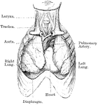
View of heart and lungs in situ. The front portion of the chest wall, and the outer or parietal layers of the pleurae and pericardium have been removed. The lungs are partly collapsed.

Heart and Lungs

View of heart and lungs in situ. The front portion of the chest wall, and the outer or parietal layers…

The left auricle and ventricle opened and a part of their anterior and left walls removed. The pulmonary artery has been divided at its commencement; the opening into the left ventricle is carried a short distance into the aorta between two of the segments of the semilunar valves; and the left part of the auricle with its appendix has been removed. The right auricle is out of view. Labels: 1, the two right pulmonary veins cut short; their opening are seen within the auricle; 1' , placed within the cavity of the auricle on the left side of the septum and on the right side and on the part which forms the remains of the valve of the foramen ovale, of which the crescentic fold is seen towards the left hand of 1'; 2, a narrow portion of the wall of the auricle and ventricle preserved round the auriculo-ventricular orifice; 3, 3', the cut surface of the walls of the ventricle, seen to become very much thinner towards 3", at the apex; 4, a small part of the anterior wall of the left ventricle which has been preserved with the principle anterior columna carnea or musculus papillaris attached to it; 5. ,musculi papillares; 5', the left side of the septum, between the two ventricles, within the cavity of the left ventricle; and above the three segments of its semilunar valve which are hanging loosely together; 7', the exterior of the great aortic sinus; 8, the root of the pulmonary artery and its semilunar valves; 8' the separated portion of the pulmonary artery remaining attached to the aorta by 9, the cord of the ductus arteriosus; 10, the arteries rising from the summit of the aortic arch.

A Diagram of the Heart

The left auricle and ventricle opened and a part of their anterior and left walls removed. The pulmonary…

Surface view of an artery from the mesentery of a frog, ensheathed in a perivascular lymphatic vessel. Labels: a, The artery, with its circular muscular coat (media) indicated by broad transverse markings, with an indication of the adventitia outside. 1. Lymphatic vessel: its wall is a simple endothelial membrane.

Surface View of an Artery

Surface view of an artery from the mesentery of a frog, ensheathed in a perivascular lymphatic vessel.…
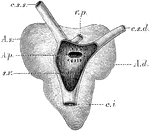
The heart of a frog (Rana esculenta) from the back. Labels: s.v., sinus venosus opened; c.s.s., left vena cava superior; c.s.d., right vena cava superior; c.i., vena cava inferior; v.p., vena pulmonales; A.d., right auricle; A.s., left auricle; A.p., opening of communication between the right auricle and the sinus venosus.

Heart of a Frog

The heart of a frog (Rana esculenta) from the back. Labels: s.v., sinus venosus opened; c.s.s., left…

Outline showing the general form of the larynx, trachea, and bronchi, as seen from front. Labels: h, the great cornu of the hyoid bone; e,epiglottis; t, superior and inferior cornu of the thyroid cartilage; c, middle of the cricoid cartilage; tr, the trachea, showing sixteen cartilaginous rings; b, the right bronchus; b1, the left bronchus.

Front View of Respiratory Apparatus

Outline showing the general form of the larynx, trachea, and bronchi, as seen from front. Labels: h,…

Outline showing the general form of the larynx, trachea, and bronchi, as seen from behind. Labels: h, the great cornu of the hyoid bone; e,epiglottis; t, superior cornu of the thyroid cartilage ; t1, inferior cornu of the thyroid cartilage; a, points to the back of both the arytenoid cartilages, which are surmounted by the cornicula; c, the middle ridge on the back of the cricoid cartilage; tr, the posterior membranous part of the trachea; b, b1, right and left bronchi.

Back View of Respiratory Apparatus

Outline showing the general form of the larynx, trachea, and bronchi, as seen from behind. Labels: h,…
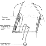
The stethometer consists of a frame formed of two parallel steel bars joined by a third at one end. At the free end of the bars is attached a leather strap, by means of which the apparatus may be suspended from the neck. Attached to the inner end of one bar is a tambour and ivory button, to the end of the other an ivory button. When in use, the apparatus is suspended with the transverse bar posteriorly, the button of the tambour is placed on the part of the chest the movement of which it is desire to record, and the other button is made to press upon the corresponding side of the chest, so that the chest is held between a pair of callipers.

Stethometer

The stethometer consists of a frame formed of two parallel steel bars joined by a third at one end.…

Section of a mucous gland from the tongue. Labels: A, opening of the duct on the free surface; C, basement membrane with nuclei; B, flattened epithelial cells lining duct. The duct divides into several branches, which are convoluted and end blindly, being lined throughout by columnar epithelium. D, lumen of one of the tubuli of the gland.

Mucous Gland from Tongue

Section of a mucous gland from the tongue. Labels: A, opening of the duct on the free surface; C, basement…

Section showing the pyloric glands. Labels: s, free surface; d, ducts of pyloric glands; n, neck of same; m, the gland alveoli; mm, muscularis mucosae.

Pyloric Gland

Section showing the pyloric glands. Labels: s, free surface; d, ducts of pyloric glands; n, neck of…

Types of microorganisms. Labels: a, micrococci arranged singly; in twos, diplococci- if all the micrococci at a were grouped together, they would be called staphylococci- and in fours, sarcinae; b, micrococci, in chains streptococci; c and d, bacilli of various kinds, one is represented with flagellum; e, various forms of spirilla; f, spores, either free or in bacilli.

Types of Microorganisms

Types of microorganisms. Labels: a, micrococci arranged singly; in twos, diplococci- if all the micrococci…

The hair is produced by a peculiar growth and modification of the epidermis. Externally it is covered by a layer of fine scales closely imbricated, or overlapping like the tiles of a house, but with the free edges turned upwards. It is called the cuticle of the hair. Shown in the magnified surface of a hair. The wave lines mark the upper or free edges of the cortical scales. B, separate scales.

Magnified Surface of a Hair

The hair is produced by a peculiar growth and modification of the epidermis. Externally it is covered…
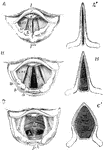
Three laryngoscopic view of the superior aperture of the larynx and surrounding parts. Labels: A, the glottis during the emission of a high note in singing; B, in easy and quite inhalation of air; C, in the state of widest possible dilation, as in inhaling a very deep breath. The diagrams A', B', and C', show in horizontal sections of the glottis in position of the vocal ligaments and arytenoid cartilages in the three several states represented in the other figures. In all the figures, so far as marked, the letters indicate the parts as follows, viz. : l, the base of the tongue; e, the upper free part of the epiglottis; e', the tubercle or cushion of the epiglottis; p h, part of the anterior all of the pharynx behind the larynx; in the margin of the aryteno-epiglottisdean fold w, the swelling of the membrane caused by the cartilages of Wrisberg; s. that of the cartilages of Santorini: a, the tip or summit of the arytenoid cartilages; c v, the true vocal cords r lips of the rima glottidis; c v s, the superior or false vocal cords; between them the ventricle of the larynx; in C, tr, is placed on the anterior wall of the receding trachea, and b indicated the commencement of the two bronchi beyond the bifurcation which may be brought into view in this state of extreme dilation.

Movement of the Vocal Cords

Three laryngoscopic view of the superior aperture of the larynx and surrounding parts. Labels: A, the…

View of the upper part of the larynx as seen by means of the laryngoscope during the utterance of a grave note. Labels: c, epiglottis; s, tubercles of the cartilages of Santorini; a, arytenoid cartilages; z, base of the tongue; ph, the posterior wall of the pharynx.

Upper Part of the Larynx

View of the upper part of the larynx as seen by means of the laryngoscope during the utterance of a…

View of the cerebrospinal axis of the nervous system. The right half of the cranium and trunk of the body have been removed by a vertical section; the membranes of the brain and spinal cord have also been removed, and the roots and first part of the fifth and ninth cranial, and of all the spinal nerves of the right side, have been dissected out and laid separately on the wall of the skull and on the several vertebrae opposite to the place of their natural exit from the craniospinal cavity.

Central Nervous System

View of the cerebrospinal axis of the nervous system. The right half of the cranium and trunk of the…
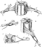
Different views of a portion of the spinal cord from the cervical region, with the roots of the nerves. A, the anterior surface of the specimen is shown; the anterior nerve root of its right side being divided. B, a view of the right side. C, the upper surface. D, the nerve roots and ganglion are shown from below. Labels: 1, The anterior median fissure; 2, posterior median fissure; 3, anterior lateral depression, over which the anterior nerve roots are seen to spread; 4, posterior lateral groove, into which the posterior roots are seen to sink; 5, anterior roots passing the ganglion; 5', the anterior root divided; 6, the posterior roots, the fibers of which pass into the ganglion 6'; 7, the united or compound nerve; 7', the posterior primary branch, seen is A and D to be derived in part from the anterior and in part from the posterior root.

Views of a Spinal Cord

Different views of a portion of the spinal cord from the cervical region, with the roots of the nerves.…

Dorsal or posterior view of the medulla, fourth ventricle, and mesencephalon. Labels: p.n., line of the posterior roots of the spinal nerves; p.m.f., posterior median fissure; f.g., funiculus gracilis; cl., its clava; f.c., funiculus cuneatus; f.R., funiculus of Rolando; r.b., restiform body; c.s., calamus scriptorius; l, section of ligula or taenia; part of choroid plexus is seen beneath t; l.r., lateral recess of the ventricle; str., striae acusticae; i.f., inferior fossa; s.f., posterior fossa; between it and the median sulcus is the fasciculus teres; cbl., cut surface of the cerebellar hemisphere; nd., central or gray matter; s.m.v., superior medullary velum; lng., ligula; s.c.p., superior cerebellar peduncle cut longitudinally; cr., combined section of the three cerebellar peduncles; c.q.s., c.q.i., corpora quadrigemina (superior and inferior); fr., fraenulum; f; f., fibers of the fillet seen on the surface of the tegmentum; c, crusti; l.g., lateral groove; c.g.i, corpus geniculum internus; th., posterior part of the thalamus; p., pineal body. The Roman numbers indicate the corresponding cranial nerves.

Medulla

Dorsal or posterior view of the medulla, fourth ventricle, and mesencephalon. Labels: p.n., line of…
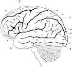
Lateral view of the brain. F, Frontal lobe; P, Parietal lobe; O, Occipital lobe; T, Temporal lobe; S, fissure of Sylvius; S', horizontal; S", ascending ramus of the same; c, sulcus centralis (fissure of Rolando); A, ascending frontal convolutions; fr, superior, f2, inferior frontal sulcus; f3, precentral sulcus; P1, superior parietal lobule; P2, inferior parietal lobule consisting of P2, supramarginal gyrus, and P2', angular gyrus; ip, interparietal sulcus; cm, termination of callosomarginal fissure; O1, first O2, second, 03, third occipitals inferior; T1, first T2, second, T3, third temporal convolutions; tr, first , t2, second temporal fissures.

Lateral View of the Brain

Lateral view of the brain. F, Frontal lobe; P, Parietal lobe; O, Occipital lobe; T, Temporal lobe; S,…
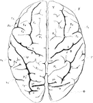
View of brain from above. F, Frontal lobe; P, Parietal lobe; O, Occipital lobe; T, Temporal lobe; S, fissure of Sylvius; S', horizontal; S", ascending ramus of the same; c, sulcus centralis (fissure of Rolando); A, ascending frontal convolutions; fr, superior, f2, inferior frontal sulcus; f3, precentral sulcus; P1, superior parietal lobule; P2, inferior parietal lobule consisting of P2, supramarginal gyrus, and P2', angular gyrus; ip, interparietal sulcus; cm, termination of callosomarginal fissure; O1, first O2, second, 03, third occipitals inferior; T1, first T2, second, T3, third temporal convolutions; tr, first , t2, second temporal fissures. Sr, end of horizontal ramus of fissure of Sylvius.

View of Brain from Above

View of brain from above. F, Frontal lobe; P, Parietal lobe; O, Occipital lobe; T, Temporal lobe; S,…

View of the right hemisphere in the median aspect. CC, corpus callosum longitudinally divided; Gf, gyrus fornicatus; H, gyrus hippocampi; h, sulcus hippocampi; U, uncinate gyrus; cm, calloso marginal fissure; F1, median aspect of first frontal convolution; c, terminal portion of sulcus centralis (fissure of Rolando); A, ascending frontal; B, ascending parietal convolution; P1', praecuneus; Oz, cuneus; po, parieto-occipital fissure; o, sulcus occipitalis transversus; oc, calcarine fissure; oc', superior; oc", inferior ramus of the same; D, gyrus descendens; T4, gyrus occipito-temporalis lateralis (lobulus fusiformis) ; T5, gyrus occipito-temporalis medialis (lobulus lingualis).

Right Hemisphere of the Brain

View of the right hemisphere in the median aspect. CC, corpus callosum longitudinally divided; Gf, gyrus…

The five layers of the cortical gray matter of the cerebrum. 1, Superficial layer with abundance of neuroglia and few small multipolar ganglion cells. 2, A thin layer of a large number of closely packed small ganglion cells of pyramidal shape. 3, The most important layer, and the thickest of all: it contains many large pyramidal ganglion cells, each with a process running off from the apex vertically towards the free surface, and lateral processes at the base which are always branched. 4, Numerous ganglion cells come are large and others small, forming the granular formation of Meynert. 5, Spindle-shaped and branched ganglion cells of moderate size arranged chiefly parallel to the free surface.

Cortical Gray Matter of the Cerebrum

The five layers of the cortical gray matter of the cerebrum. 1, Superficial layer with abundance of…
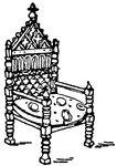
King David's Arm-chair was designed in the 13th century. The Arm-chair was made from a relief portal of a cathedral in Auxerre, France.

King David's Arm-Chair

King David's Arm-chair was designed in the 13th century. The Arm-chair was made from a relief portal…

The cane chair's seat or back is of woven cane-work or padded. The Chair is termed "cane" meaning upholstered.

Modern Cane Chair

The cane chair's seat or back is of woven cane-work or padded. The Chair is termed "cane" meaning upholstered.
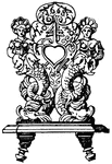
The cane chair's seat or back is of woven cane-work or padded. The Chair is termed "cane" meaning upholstered.

Modern cane chair

The cane chair's seat or back is of woven cane-work or padded. The Chair is termed "cane" meaning upholstered.

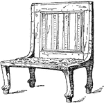
The back of the Egyptian chair is treated as a frame.

Egyptian Chair

The back of the Egyptian chair is treated as a frame.

The German 17th century chair had openings for the hand that were carved into the wooden back of the chair. This was done for convenience in moving the chair.

German 17th Century Chair

The German 17th century chair had openings for the hand that were carved into the wooden back of the…
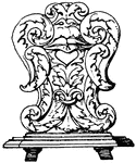
The German 17th century chair had openings for the hand that were carved into the wooden back of the chair. This was done for convenience in moving the chair.

German 17th Century Chair

The German 17th century chair had openings for the hand that were carved into the wooden back of the…
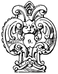
The German 17th century chair had openings for the hand that were carved into the wooden back of the chair. This was done for convenience in moving the chair.

German 17th Century Chair

The German 17th century chair had openings for the hand that were carved into the wooden back of the…

The modern chair's top of the back is horizontal and is crowned with a cornice or an ornament.

Modern Chair

The modern chair's top of the back is horizontal and is crowned with a cornice or an ornament.
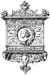
This modern Tablet Architectural frame was in the style of the Italian Renaissance. It had the general shape of an erect triangle that has a cresting feature, free-ending upwards.

Tablet Frame

This modern Tablet Architectural frame was in the style of the Italian Renaissance. It had the general…
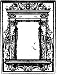
The pulpit Architectural frame was a German frame that was dated between 1595 to 1597. It had the general shape of an erect triangle that has a cresting feature, free-ending upwards.

Pulpit Frame

The pulpit Architectural frame was a German frame that was dated between 1595 to 1597. It had the general…

The Ancient Persian Throne was decorated to represent a king sitting on his throne borne-up by slaves.

Ancient Persian Throne

The Ancient Persian Throne was decorated to represent a king sitting on his throne borne-up by slaves.

Vertical section of dog's cerebellum. Labels: p m, pia mater; p, corpuscles of Purkinje, which are branched nerve cells lying in a single layer and sending single processed downwards and more numerous ones upwards, which branch continuously and extend through the deep "molecular layer" towards the free surface; g, dense layer of ganglionic corpuscles., closely resembling nuclear layers of retina; f, layer of nerve fibers, with a few scattered ganglionic corpuscles. This last layer (f) constitutes part of the white matter of the cerebellum, while the layer between it and the free surface are gray matter.

Cerebellum of Dog's Brain

Vertical section of dog's cerebellum. Labels: p m, pia mater; p, corpuscles of Purkinje, which are branched…

Diagrammatic view of the Sympathetic cord of the right side, showing its connections with the principal cerebrospinal nerves and the main prae-aortic plexuses. Cerebrospinal nerves. VI, a portion of the sixth cranial as it passes through the cavernous sinus, receiving two twigs from the carotid plexus of the sympathetic nerve; O, opthalmic ganglion connected by a twig with the carotid plexus; M, connection of the spheno-palatine ganglion by the Vidian nerve with the carotid plexus; C, cervical plexus; Br, brachial plexus; D 6, sixth intercostal nerve; D 12, twelfth; L 3, third lumbar nerve; S 5, fifth; Cr, anterior crural nerve winding round the subclavian artery. Sympathetic Cord. c, superior cervical ganglion; c', second or middle; c", inferior; from each of these ganglia cardiac nerves (all deep on this side) are seen descending to the cardiac plexus; d 1, placed immediately below the first dorsal sympathetic ganglion; d 6, is opposite he sixth; l 1, first lumbar ganglion. Preaortic and Visceral Plexuses. p, pharyngeal, and, lower down, laryngeal plexus; pl, post-pulmonary plexus spreading from the vagus on the back of the right bronchus; c a,on the aorta , the cardiac plexus, towards which , in addition to the cardiac nerves from the three cervical sympathetic ganglia, other branches are seen descending from the vagus and recurrent nerves; co, right or posterior and co1, left or an. coronary plexus; o, esophageal plexus in long meshes on the gullet; sp, great splanchnic nerve formed by branched from the fifth, sixth, seventh, eight, and ninth dorsal ganglia; +, small splanchnic from the ninth and tenth; ++, smallest or third splanchnic from the eleventh: the first and second of theses are shown joining the solar plexus, s o; the third descending to the renal plexus, r e; connecting branches between the solar plexus and the vagi are also represented; pn', above the place where the right vagus passes to the lower or posterior surface of the stomach; pn", the left distribution on the anterior side or upper surface of the cardiac portion of the organ: from the solar plexus large branches are seen surrounding the arteries of the coeliac axis, and descending from the solar and sup. mesenteric plexuses and from the lumbar ganglia; mi, the inf. mesenteric plexus surrounding the corresponding artery; hy, hypogastric plexus; pl, the right pelvic or inf. hypogastric plexuses; pl, the right pelvic plexus; from this the nerves descending are joined by those from the plexus on the sup. hemorrhoidal vessels, mi', by nerves from the third and fourth sacral spinal nerves, and there are thus formed the rectal, vesical, and other plexuses, which ramify upon the viscera, ,as towards ir, and v, the rectum and bladder.

Sympathetic System

Diagrammatic view of the Sympathetic cord of the right side, showing its connections with the principal…
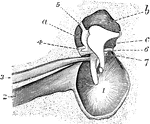
Interior view of the tympanum, with membrana tympani and bones in natural position. 1, Membrana tympani; 2, Eustachian tube; 3, tensor tympani muscle; 4, lig. mallei super; 6, corda-tympani nerve; a, b, and c, sinuses about ossicula.

Tympanum

Interior view of the tympanum, with membrana tympani and bones in natural position. 1, Membrana tympani;…
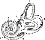
View of the interior of the left labyrinth. The bony wall of the labyrinth is removed superiorly and externally. Labels: 1, fovea hemielliptica; 2m fovea hemispherica; 3, common opening of the superior and posterior semicircular canals; 4, opening of the aqueduct of the vestibule; 5, the superior, 6, the posterior, and 7, the external semicircular canals; 8, spiral tube of the cochlea (scala tympani); 9, opening of the aqueduct of the cochlea; 10, placed on the lamina spiralis in the scala vestibuli.

Interior of the Left Labyrinth

View of the interior of the left labyrinth. The bony wall of the labyrinth is removed superiorly and…

View of the osseous cochlea divided through the middle. Labels: 1, central canal of the modiolus; 2, lamina spiralis ossea; 3, scala tympani; 4, scala vestibuli; 5, porous substance of the modiolus near one of the sections of the canalis spiralis modioli.

Osseous Cochlea

View of the osseous cochlea divided through the middle. Labels: 1, central canal of the modiolus; 2,…

Vertical section of rabbit's cornea. Labels: anterior epithelium, showing the different shapes of the cells at various depths from the free surface; b, portion of the substance of cornea.

Magnified Rabbit's Cornea

Vertical section of rabbit's cornea. Labels: anterior epithelium, showing the different shapes of the…

Surface view of part lamella of kitten's cornea, prepared first with caustic potash and then with nitrate of silver. By this method the branched cornea-corpuscles with their granular protoplasm and large oval nuclei are brought out.

Lamella of Kitten's Cornea

Surface view of part lamella of kitten's cornea, prepared first with caustic potash and then with nitrate…

Vertical section of rabbit's cornea, stained with gold chloride. Labels: e, Laminated anterior epithelium. Immediately beneath this is the anterior elastic lamina of Bowman. n, Nerves forming a delicate sub-epithelial plexus, and sending up fine twigs between the epithelial cells to end in a second plexus on the free surface; d, Descemet's membrane, consisting of a fine elastic layer, and a single layer of epithelial cells. the substance of the cornea, f, is seen to be fibrillated, and contains many layers of branched corpuscles, arranged parallel to the free surface, and here seen edgewise.

Section of Rabbit's Cornea

Vertical section of rabbit's cornea, stained with gold chloride. Labels: e, Laminated anterior epithelium.…
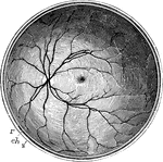
The posterior half of the retina of the left eye, viewed from before; s, the cut edge of the sclerotic coat; ch, the choroid; r, the retina; in the interior at the middle the macula lutea with the depression of the fovea centralis is represented by a slight oval shade; towards the left side the light spot indicates the colliculus or eminence at the entrance of the optic nerve, from the center of which the arteria centralis is seen spreading its branches into the retina, leaving the part occupied by the macula comparatively free.

Posterior Half of the Retina

The posterior half of the retina of the left eye, viewed from before; s, the cut edge of the sclerotic…
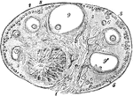
View of a section of the ovary of the cat. Labels: 1, outer covering and free border of the ovary; 1', attached border; 2, the ovarian stroma, presenting a fibrous and vascular structure; 3, granular substance lying external to the fibrous stroma; 4, blood-vessels; 5, ovigerms in their earliest stages occupying part of the granular layer near the surface; 6, ovigerms which have begun to enlarge and to pass more deeply into the ovary; 7, ovigerms round which the Graafian follicles and tunica granulosa are now formed and which have passed somewhat deeper into the ovary and are surrounded by the fibrous stroma; 8, more advanced Graafian follicle with the ovum imbedded in the layer of cells constituting the proligerous disc; 9, the most advanced follicle containing the ovum; 9', a follicle from which the ovum has accidentally escaped; IO, corpus luteum.

Ovary of the Cat

View of a section of the ovary of the cat. Labels: 1, outer covering and free border of the ovary; 1',…

"Hair-cap moss (Polytrichum commune). A, male plant; B, same, proliferating; C, female plant, bearing sporogonium; D, same; g, gametophyte; s, seta; c, capsule; o, operculum; a, calyptra, E, top view of male plant." -Gager, 1916

Hair-Cap Moss

"Hair-cap moss (Polytrichum commune). A, male plant; B, same, proliferating; C, female plant, bearing…

"Morphology of typical monocotyledonous plant. A, leaf, parallel-veined; B, portion of stem, showing irregular distribution of vascular bundles; C, ground plan of flower (the parts in 3's); D, top view of flower; E, seed, showing monocotyledonous embryo." -Gager, 1916

Monocotyledonous Morphology

"Morphology of typical monocotyledonous plant. A, leaf, parallel-veined; B, portion of stem, showing…

"Mountain laurel (Kalmia latifolia). a, stamens in their original position, with the anthers in the pouches; b, stamens inflexed (detail at e); c, side view; d, essential organs; f and g, stamens." -Gager, 1916

Mountain Laurel

"Mountain laurel (Kalmia latifolia). a, stamens in their original position, with the anthers in the…
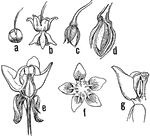
"Milkweed (Asclepias sp.). a, flower-bud; b, flower; c, very young pod; d, older pod in section, showing seeds; e, section of flower; f, top view of flower, showing the 5 hoods f the crown, each with a horn incurving to the stigma; between the horns are the cleft glands to which the pollinia are attached." -Gager, 1916

Milkweed Stages

"Milkweed (Asclepias sp.). a, flower-bud; b, flower; c, very young pod; d, older pod in section, showing…
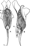
"Sections of flowers of the toad-flax (Linaria vulgaris). A, front view; a, anthers; s, stigma; n, nectar-gland. B, side view; o, ovary." -Gager, 1916

Common Toadflax

"Sections of flowers of the toad-flax (Linaria vulgaris). A, front view; a, anthers; s, stigma; n, nectar-gland.…
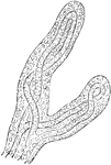
Very soon after the entrance of the ovum into the uterus, in the human subject, the outer surface of the chorion is found beset with fine processes, the so-called chorion villi, which give it a rough and shaggy appearance. Shown is a magnified view of chorion villi.

Magnified View of Chorion Villi

Very soon after the entrance of the ovum into the uterus, in the human subject, the outer surface of…
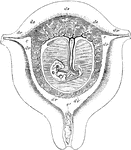
Diagrammatic view of a vertical transverse section of the uterus at the seventh week of pregnancy. Labels: c, c', cavity of uterus, which becomes the cavity of the decidua, opening at c, the cornua, into the Fallopian tubes, and at c' into the cavity of the cervix, which is closed by a plug of mucus; dv, decidua vera; dr, decidua reflexa, with the sparser villi imbedded in its substance; ds, decidua serotina, involving the more developed chorionic villi of the commencing placenta. The fetus is seen lying in the amniotic sac; passing up from the umbilicus is seen the umbilical pedicle of the yolk sac, which lies in the cavity between the amnion and chorion.

Uterus at Seventh Week of Pregnancy

Diagrammatic view of a vertical transverse section of the uterus at the seventh week of pregnancy. Labels:…
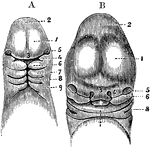
A, Magnified view of the head and neck of a human embryo of three weeks. Labels: 1, anterior cerebral vesicle or cerebrum; 2, middle cerebral vesicle; 3, middle or frontonasal process; 4, superior maxillary process, or first visceral arch, and below it the first cleft; 7, 8, 9, second, third, and fourth arches and clefts. B, Anterior view of the head of a human fetus of about the fifth week. 1, 2, 3, 5, the same parts as in A; 4, the external nasal or lateral frontal process: 6, the superior maxillary process; 7, the lower jaw; X, the tongue; 8, first branchial cleft becoming the meatus auditorius externus.

Head of an Embryo

A, Magnified view of the head and neck of a human embryo of three weeks. Labels: 1, anterior cerebral…