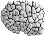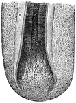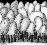
"<em>A, B,</em> glands seen in vertical section with their orifices at <em>C</em> opening upon the membrane between the villi; <em>D</em>, villus." &mdash; Blaisedell, 1904

Glands and villi of the small intestine

"A, B, glands seen in vertical section with their orifices at C opening upon the membrane…

"In the figure on the left are seen the artery and vein of a villus. In the right figure are represented the central lacteal of the villus, with the plexus of lymph vessels at the base. The mucous, submucuous, muscular, and serous coats are indicated at the base of the right-hand figure.  The epithelium of each villus is well shown." &mdash; Blaisedell, 1904

Transverse section of the small intestine

"In the figure on the left are seen the artery and vein of a villus. In the right figure are represented…
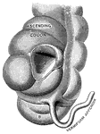
"<em>A</em>, a portion of the colon laid open to show the valve between the large and small intestine; <em>B</em>, the caecum." &mdash; Blaisedell, 1904

Vermiform appendix

"A, a portion of the colon laid open to show the valve between the large and small intestine;…

"<em>A</em>, a muscle which aids in pushing the food down the esophagus; <em>B</em>, esophagus; <em>C</em>, section of the right bronchus; <em>D</em>, two right pulmonary veins; <em>E</em>, great azygos vein crossing the esophagus to empty into the superior vena cava, <em>F</em>, thoracic duct; <em>H</em>, thoracic aorta; <em>K</em>, lower portion of the esophagus passing through the diaphragm; <em>L</em>, diaphragm as it appears in sectional view, enveloping the heart; <em>M</em>, inferior vena cava, passing through the diaphragm and emptying into the right auricle; <em>N</em>, right auricle; <em>O</em>, section of the right branch of the pulminary artery; <em>P</em>, aorta; <em>R</em>, superior vena cava; <em>S</em>, trachea." &mdash; Blaisedell, 1904

Lateral section of the chest

"A, a muscle which aids in pushing the food down the esophagus; B, esophagus; C,…

"Principal Muscles on the Right, Certain Organs of the Chest and Abdomen, and the Larger Blood Vessels on the Left." — Blaisedell, 1904

Muscles and Blood Vessels

"Principal Muscles on the Right, Certain Organs of the Chest and Abdomen, and the Larger Blood Vessels…

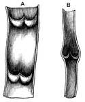
"<em>A</em>, part of a vein laid open, with two pairs of valves; <em>B</em>, longitudinal section of a vein, showing the valves closed." &mdash; Blaisedell, 1904

Vein

"A, part of a vein laid open, with two pairs of valves; B, longitudinal section of…

"The Right Axillary and Branchial Arteries, with Some of their Main Branches." &mdash; Blaisedell, 1904

Arteries

"The Right Axillary and Branchial Arteries, with Some of their Main Branches." — Blaisedell, 1904
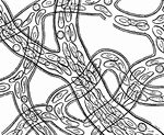
"Showing how the circulation of blood in the web of a frog's foot looks as seen under the microscope." &mdash; Blaisedell, 1904

Circulation of blood

"Showing how the circulation of blood in the web of a frog's foot looks as seen under the microscope."…
"The Internal, or Long Saphenous, Vein of the Left Leg." &mdash; Blaisedell, 1904

Leg vein

"The Internal, or Long Saphenous, Vein of the Left Leg." — Blaisedell, 1904

"The Femoral Artery of Right Leg." &mdash; Blaisedell, 1904

Leg artery

"The Femoral Artery of Right Leg." — Blaisedell, 1904

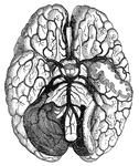
"Arteries and their Branches at the Base of the Brain." &mdash; Blaisedell, 1904

Blood vessels of the brain

"Arteries and their Branches at the Base of the Brain." — Blaisedell, 1904
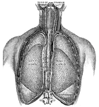
"Artery in the Front of the Right Leg." &mdash; Blaisedell, 1904

Leg artery

"Artery in the Front of the Right Leg." — Blaisedell, 1904

"<em>L.A.</em>, left ventricle; <em>B</em>, right ventricle; <em>F</em>, pulmonary artery; <em>G</em>, aorta; <em>H</em>, arch of the aorta; <em>K</em>, innominate artery; <em>L</em>, right common carotid artery; <em>M</em>, right subclavian artery; <em>N</em>, thyroid cartilage forming upper portions of the larynx; <em>O</em>, trachea." &mdash; Blaisedell, 1904

Lungs

"L.A., left ventricle; B, right ventricle; F, pulmonary artery; G,…

"Larynx, Trachea, and the Bronchi. (Front view.) <em>A</em>, epiglottis; <em>B</em>, thyroid cartilage; <em>C</em>, cricothyroid membrane, connecting with the cricoid cartilage below, all forming the larynx; <em>D</em>, rings of the trachea." &mdash; Blaisedell, 1904

Trachea

"Larynx, Trachea, and the Bronchi. (Front view.) A, epiglottis; B, thyroid cartilage;…

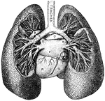
"<em>A</em>, division of left bronchus to upper lobe; <em>B</em>, left branch of the pulmonary artery; <em>C</em>, left bronchus; <em>D</em>, left superior pulmonary vein; <em>E</em>, left inferior pulmonary vein; <em>F</em>, left auricle; <em>K</em>, inferior vena cava; <em>L</em>; division of the right bronchus to lower lobe; <em>M</em>, right inferior pulmonary vein; <em>N</em>, right superior pulmonary vein, <em>O</em>, right branch of the pulmonary artery; <em>P</em>, division of right bronchus to upper lobe; <em>R</em>, left ventricle; <em>S</em>, right ventricle." &mdash; Blaisedell, 1904

Lungs

"A, division of left bronchus to upper lobe; B, left branch of the pulmonary artery;…

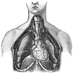
"Relative Postion of the Lungs, the Heart, and Some of the Great Vessels belonging to the latter. <em>A</em>, left common carotid artery; <em>B</em>, external carotid artery; <em>C</em>, internal carotid artery; <em>D</em>, left jugular vein; <em>E</em>, sterno-cleido muscle; <em>F</em>, right innominate artery with branches to head and arm; <em>G</em>, left subclavian artery; <em>H</em>, great azygos vein; <em>K</em>, thoracic duct." &mdash; Blaisedell, 1904

Lungs

"Relative Postion of the Lungs, the Heart, and Some of the Great Vessels belonging to the latter. A,…
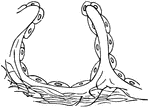
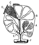
"<em>A</em>, an air sac; <em>B</em>, an air sac cut open; <em>C</em>, capillary network over an air sac; <em>D</em>, branch of pulmonary artery; <em>E</em>, branch of pulmonary vein; <em>F</em>, bronchial tube." &mdash; Blaisedell, 1904

Lobule of the lung

"A, an air sac; B, an air sac cut open; C, capillary network over an air…

"Lobules of various sizes are well shown." &mdash; Blaisedell, 1904

Portion of a child's lung

"Lobules of various sizes are well shown." — Blaisedell, 1904

"<em>A</em>, epithelial lining wall; <em>B</em>, partition between two adjacent sacs, in which run capillaries; <em>C</em>, fibers of elastic tissues." &mdash; Blaisedell, 1904

Diagrammatic view of an air sac

"A, epithelial lining wall; B, partition between two adjacent sacs, in which run capillaries;…
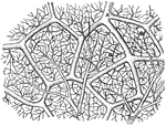
"Diagram showing the capillary network of the air sacs and origin of the pulmonary veins.. <em>A</em>, small branch of pulmonary artery; <em>B</em>, twigs of pulmonary artery; <em>C</em>, capillary network around the walls of the air sacs; <em>D</em>, branches of network converging to form the veinlets of the pulmonary veins." — Blaisedell, 1904

Capillaries of the Air Sac

"Diagram showing the capillary network of the air sacs and origin of the pulmonary veins.. A,…

"A Layer of the Outer Skin from the Palm of the Hand. Detached by maceration." &mdash; Blaisedell, 1904

Outer skin

"A Layer of the Outer Skin from the Palm of the Hand. Detached by maceration." — Blaisedell, 1904
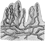
"In each papilla are seen vascular loops (dark lines) running up from the vascular network below; the tactile corpuscules (white lines) which supply the papillae are also shown." &mdash; Blaisedell, 1904

Papillae of the skin

"In each papilla are seen vascular loops (dark lines) running up from the vascular network below; the…

"Surface of Palm of the Hand, showing Openings of Sweat Glands and Grooves between Papillae of the Skin. In the smaller figure the same epidermal surface is shown as seen with the naked eye." &mdash; Blaisedell, 1904

Surface of the palm

"Surface of Palm of the Hand, showing Openings of Sweat Glands and Grooves between Papillae of the Skin.…

"<em>A</em>, outer layer of cuticle; <em>B</em>, deeper layer of cuticle; <em>C</em>, duct of sweat gland; <em>D</em>, true skin; <em>E</em>, sublayer of true skin, with columnar cells. The blood vessels are injected to show black." — Blaisedell, 1904

Cross-Section of the Skin

"A, outer layer of cuticle; B, deeper layer of cuticle; C, duct of sweat…

"Cross-section of One Half of a Human Hair. A hair is made up of horny cells of the outer layer of the skin altered in shape and structure." &mdash; Blaisedell, 1904

Cross-section of a human hair

"Cross-section of One Half of a Human Hair. A hair is made up of horny cells of the outer layer of the…

"<em>A</em>, membrane of hair follicle, showing cells with nuclei and pigmentary granules; <em>B</em>, external lining of root sheath; <em>C</em>, internal lining of root sheath; <em>D</em>, cortical or fibrous portion of hairshaft; <em>E</em>, medullary portion (pith) of shaft; <em>F</em>, hair bulb, showing its development from cells." — Blaisedell, 1904

Hair Follicle

"A, membrane of hair follicle, showing cells with nuclei and pigmentary granules; B,…

"Concave or Adherent Surface of the Nail. <em>A</em>, border of the root; <em>B</em>, whitish portion of semi-lunar shape; <em>C</em>, body of nail. The continuous line around border represents the free edge." &mdash; Blaisedell, 1904

Surface of a nail

"Concave or Adherent Surface of the Nail. A, border of the root; B, whitish portion…

"<em>A</em>, section of cutaneous fold turned back to show root of nail; <em>B</em>, cutaneous fold covering root of nail; <em>C</em>, semilunar whitish portion; <em>D</em>, body of nail." &mdash; Blaisedell, 1904

Nail in position

"A, section of cutaneous fold turned back to show root of nail; B, cutaneous fold…

"<em>A</em>, last bone of finger; <em>B</em>, true skin on the dorsal surface of finger; <em>C</em>, outer skin; <em>D</em>, true skin; <em>E</em>, bed of nail; <em>F</em>, true skin of pulp of finger." &mdash; Blaisedell, 1904

Longitudinal seciont of a fingernail

"A, last bone of finger; B, true skin on the dorsal surface of finger; C,…

"The convoluted gland is seen surrounded by fat cells and may be traced through the true skin to its outlet in the horny layers of the outer skin." &mdash; Blaisedell, 1904

Sweat gland

"The convoluted gland is seen surrounded by fat cells and may be traced through the true skin to its…
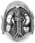
"The spinal column below the twelfth dorsal vertebra at <em>A</em> has been removed, as well as the various layers of the great muscles of the back. The two kidneys with the renal arteries and veins are plainly shown, in their normal positions. The relative positions of adjacent vessels and organs with their names printed upon them are also shown. <em>B</em>, portion of the diaphragm on the left side; <em>C</em>, receptaculum chyli; <em>D</em>, a part of the small intestine on the left side." — Blaisedell, 1904

Vertical Section of the Back

"The spinal column below the twelfth dorsal vertebra at A has been removed, as well as the…

"<em>A</em>, pyramids of the kidney; <em>B</em>, apices, or papillae, of the pyramids; <em>C</em>, pelvis of the kidney; <em>D</em>, upper end of the ureter</em>." &mdash; Blaisedell, 1904

Kidney

"A, pyramids of the kidney; B, apices, or papillae, of the pyramids; C, pelvis…

"Nerve tissue is really made up of a great number of distinctive units called <em>nerve cells</em>. Each cell usually contains a large nucleus and gives off one or more tiny branches, or processes. Thse cells vary more in shape and size than any other cell in the body. Each nerve cell has a number of short branches, and many have also one long branch which can be traced for some distance from the cell body." — Blaisedell, 1904

Nerve Cells

"Nerve tissue is really made up of a great number of distinctive units called nerve cells.…
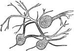
"Wherever nerve cells are abundant, the nerve tissue has a gray color; in other places, it looks white. Most of the gray matter of the brain is on the surface." — Blaisedell, 1904

Nerve Cells of the Brain

"Wherever nerve cells are abundant, the nerve tissue has a gray color; in other places, it looks white.…

"The axis cylinder is in the center. On either side is seen the medullary sheath, represented by dark lines. The primitive sheath, or neurilemma, is on the outside and represented by white lines in which is a nerve corpuscle with an oval nucleus." &mdash; Blaisedell, 1904

Portion of a medullated nerve fiber

"The axis cylinder is in the center. On either side is seen the medullary sheath, represented by dark…

"Showing a motor cell with its long, unbranched process (with two little lateral offshoots), with motor endings in striated, voluntary muscular tissue." &mdash; Blaisedell, 1904

Neuron

"Showing a motor cell with its long, unbranched process (with two little lateral offshoots), with motor…

"Diagram illustrating the General Arrangement of the Nervous System. (posterior view.)" — Blaisedell, 1904

Nervous System

"Diagram illustrating the General Arrangement of the Nervous System. (posterior view.)" — Blaisedell,…
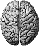
"The Upper Surface of the Cerebrum. Showing its division into two hemispheres, and also of the convolutions." &mdash; Blaisedell, 1904

Cerebrum

"The Upper Surface of the Cerebrum. Showing its division into two hemispheres, and also of the convolutions."…
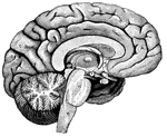
"<em>A</em>,frontal love of the cerebrum; <em>B</em>, parietal lobe; <em>C</em>, parieto-occipital lobe; <em>D</em>, occcipital lobe; <em>E</em>, cerebellum; <em>F</em>; arbor vitae; <em>H</em>, pons Varolii; <em>K</em>, medulla oblongata." — Blaisedell, 1904

Left Half of the Brain

"A,frontal love of the cerebrum; B, parietal lobe; C, parieto-occipital lobe;…
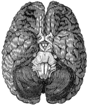
<em>A</em>, anterior lobe of the cerebellum; <em>B</em>, olfactory nerve; <em>C</em>, portion of the posterior lobe; <em>D</em>, optic chiasm; <em>E</em>, optic tract; <em>H, M,</em> hemispheres of the cerebellum; <em>K</em>, portion of the occipital lobe; <em>N</em>, medulla oblongata; <em>R</em>, pons Varolii."

Base of the brain

A, anterior lobe of the cerebellum; B, olfactory nerve; C, portion of the…

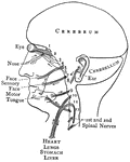
"The cranial nerves are thus arranged in pairs: 1, olfactory nerves, special nerves of smell; 2, optic nerves, passing to each eyeball, devoted to sight; 3, 4, and 6 control the muscles fo the eyes; 5, trifacial in three branches, which proceed mainly to the face, partly sensory and partly motor; 7, facial nerves, controlling the facial muscles; 8, auditory, or nerves of hearing; 9, glossopharyngeal nerves, partly sensory and partly motor: each nerve contains two roots, one a nerve of taste, the other a motor nerve, which controls the muscles engaged in swallowing; 10, pneumogastric nerves; 11, spinal accessory nerves, supplying some of the muscles of the neck and back; 12, hypoglossal nerves, controlling the movements of the tongue in speech and swallowing." — Blaisedell, 1904

Distribution of the Cranial Nerves

"The cranial nerves are thus arranged in pairs: 1, olfactory nerves, special nerves of smell; 2, optic…
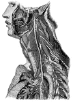
"Showing its distribution by its branches and ganglia to the larynx, pharynx, heart, lungs, and other parts." — Blaisedell, 1904

Trunk of the Pneumogastric Nerve

"Showing its distribution by its branches and ganglia to the larynx, pharynx, heart, lungs, and other…
"Illustrating the path of a simple nervous reflex action." — Blaisedell, 1904

Reflex Action

"Illustrating the path of a simple nervous reflex action." — Blaisedell, 1904

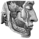
"Dental Branch of One of the Divisions of the Fifth Pair of Cranial Nerves, supplying the Lower Teeth. Branches from the motor root, distributed to various muscles, are also shown." — Blaisedell, 1904

Cranial Nerves

"Dental Branch of One of the Divisions of the Fifth Pair of Cranial Nerves, supplying the Lower Teeth.…

"The Cervical and Thoracic Portions of the Sympathetic Nerve and their Main Branches. In the center of the figure, running almost vertically, is shown the right pneumogastrix nerve. To the left may be seen a chain of ganglia of the sympathetic nerve, running along the vertebrae. The distribution of some smaller ganglia and nerve branches in the neck and chest regions are also shown." &mdash; Blaisedell, 1904

Sympathetic nerve

"The Cervical and Thoracic Portions of the Sympathetic Nerve and their Main Branches. In the center…
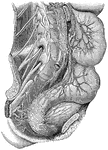
"Showing the distribution of some of the great plexuses of the sympathetic nerve in the lumbar and sacral regions." — Blaisedell, 1904

Plexuses of the Sympathetic Nerves

"Showing the distribution of some of the great plexuses of the sympathetic nerve in the lumbar and sacral…
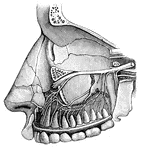
"Dental Branches of One of the Divisions of the Fifth Pair of Cranial Nerves, supply the Upper Teeth." — Blaisedell, 1904

Cranial Nerves

"Dental Branches of One of the Divisions of the Fifth Pair of Cranial Nerves, supply the Upper Teeth."…
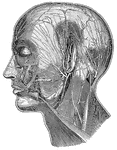
"Showing some of the superficial nerves on the left side of the neck and the head. A few superficial muscles and arteries in the same region are also shown." — Blaisedell, 1904

Superficial Nerves of the Head

"Showing some of the superficial nerves on the left side of the neck and the head. A few superficial…

"Superficial, or Cutaneous, Nerves on the Back of the Left Forearm and Hand." — Blaisedell, 1904

Superficial Nerves of the Forearm and Hand

"Superficial, or Cutaneous, Nerves on the Back of the Left Forearm and Hand." — Blaisedell, 1904

"The Main Nerve Trunks of the Right Forearm, showing the Accompanying Radial and Ulnar Arteries. (Anterior view.)" &mdash; Blaisedell, 1904

Nerve trunks

"The Main Nerve Trunks of the Right Forearm, showing the Accompanying Radial and Ulnar Arteries. (Anterior…

"A Great Nerve (Crural) and its branches on the Front of the Thigh. The femoral artery with its cut end is shown." — Blaisedell, 1904

Great Nerve

"A Great Nerve (Crural) and its branches on the Front of the Thigh. The femoral artery with its cut…

"A Great Nerve (Posterior Tibial) on the Back of the Leg, with its Accompanying Artery of the Same Name." — Blaisedell, 1904

Great Nerve

"A Great Nerve (Posterior Tibial) on the Back of the Leg, with its Accompanying Artery of the Same Name."…

"A Great Nerve (Plantar) and its Branches which supply the Bottom of the Feet. Note the cut tendons of the great muscles of the leg." — Blaisedell, 1904

Great Nerve

"A Great Nerve (Plantar) and its Branches which supply the Bottom of the Feet. Note the cut tendons…

"A Papilla of the Skin, with a Touch Corpucle. Highly magnified." &mdash; Blaisedell, 1904

Papilla of the skin

"A Papilla of the Skin, with a Touch Corpucle. Highly magnified." — Blaisedell, 1904
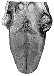
<em>A</em>, epiglottis; <em>B</em>, glands at the base of tongue; <em>C</em>, tonsil; The various kinds of papillae are plainly shown." &mdash; Blaisedell, 1904

Tongue

A, epiglottis; B, glands at the base of tongue; C, tonsil; The various kinds…

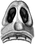
"Nasal Cavities, seen from Below. The sense of smell is located in the membrane which lines the cavities of the nose. This delicate membrane, over which the fibers of the olfactory nerves, or the nerves of smell, are distributed, is kept continually mois by the mucus which it secretes." &mdash; Blaisedell, 1904

Nasal cavities

"Nasal Cavities, seen from Below. The sense of smell is located in the membrane which lines the cavities…
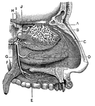
"<em>A</em>, branches of the nerves of smell; <em>B</em>, nerves of touch to the nostrils; <em>E, F, G,</em> nerves to the palate springing from a ganglion at <em>C</em>; <em>H</em>, a branch of the facial nerve, from which which other branches <em>D, I,</em> and <em>J</em>, spring to be distributed to the nostrils." — Blaisedell, 1904

Nerves of the Nostril

"A, branches of the nerves of smell; B, nerves of touch to the nostrils; E, F,

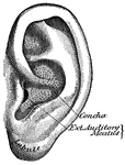
"The outer ear consists of a plate of gristle, shaped somewhat like a shell, known as the pinna, or auricle, and of a tube about an inch long, called the auditory canal." &mdash; Blaisedell, 1904

Pinna

"The outer ear consists of a plate of gristle, shaped somewhat like a shell, known as the pinna, or…
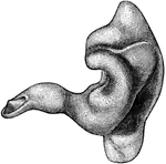
"A Cast of the External Auditory Canal. The auditory canal is a passage in the solid potion of the temporal bone. It is lined by skin on which there are fine hairs, and a set of glands secreting earwax, which serves to moisten the parts, catch particles fo dust, and keep away small insects." &mdash; Blaisedell, 1904

External auditory canal

"A Cast of the External Auditory Canal. The auditory canal is a passage in the solid potion of the temporal…
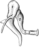
"1, malleus, or hammer; 2, incus, or anvil; 3, stapes, or stirrup." — Blaisedell, 1904
Bones of the Ear

"1, malleus, or hammer; 2, incus, or anvil; 3, stapes, or stirrup." — Blaisedell, 1904