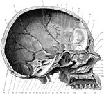
Shown is the inner aspect of the left half of the skull sagittally divided. Labels: 1, suture between parietal and temporal bone; 2, remains of subarcuate fossa; 3, grooves for branches of the middle meningeal artery; 4, dorsum sellae; 5, pituitary fossa; 6, anterior clinoid fossa; 7, optic foramen; 8, sphenoidal suture; 9, nasal surface of superior turbinated bone; 10, cribriform plate of ethmoid; 11, nasal surface of middle turbinated bone; 12, frontal sinus; 13, nasal spine; 14, nasal bone; 15, nasal process of superior maxilla; 16, middle meatus of nose; 17, directed towards opening of antrum; 18, nasal surface of inferior turbinated bone; 19, inferior meatus of nose; 20, anterior nasal spine; 21, anterior palatine canal; 22, palatal process of superior maxilla; 23, palatal process of palate bone; 24, posterior nasal spine; 25, hamular process of internal pterygoid plate; 26, external pterygoid plate; 27, superior meatus of nose; 28,sphenopalatine foramen; 29, pterygospinous ligament almost completely ossified to enclose a foramen; 30, styloid process of temporal bone; 31, alar spine of sphenoid; 32, mastoid process; 33, basion (midpoint of posterior border of foramen magnum); 34, internal auditory meatus; 35, anterior condylic foramen; 36, leading into jugular foramen; 37, opisthion (midpoint of posterior border of foramen magnum); 38, groove for sigmoid sinus; 39, opening of mastoid foramen; 40, for lateral sinus and attachment of tentorium cerebelli; 41, fossa for lodgment of cerebellar hemisphere; 42, internal occipital protuberance.

Skull Seen From Side

Shown is the inner aspect of the left half of the skull sagittally divided. Labels: 1, suture between…

Shown is a coronal section passing inferiorly through interval between between the first and second molar teeth. Labels: 1, groove of superior longitudinal sinus; 2,crest for attachment of falx cerebri; 3, crista galli of ethmoid; 4, cristiform plate of ethmoid; 5, perpendicular plate of ethmoid, assisting in the formation of nasal septum; 6, lateral mass of ethmoid consisting of the ethmoidal cells; 7, os planum of ethmoid; 8, middle meatus of nose; 9, middle turbinated bone; 10, opening from the middle meatus; 11, orbital plate of superior maxilla; 12, frontomalar suture; 13, infraorbital groove; 14, antrum or maxillary sinus; 15, canal for anterior dental nerve and vessels exposed; 16, inferior meatus of nose; 17, alveolar process of the superior maxilla; 18, nasal surface of inferior turbinated bone; 19, groove for anterior palatine nerve and vessels; 20, palatal process of superior maxilla; 21, maxillary crest forming part of nasal septum; 22, vomer forming part of nasal septum.

Coronal Section of Skull

Shown is a coronal section passing inferiorly through interval between between the first and second…

The lower extremity of the right humerus as seen from below.

Humerus, Lower End of

The lower extremity of the right humerus as seen from below.

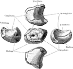
The right scaphoid bone.

Scaphoid Bone

The right scaphoid bone.

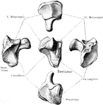
The right semilunar bone.

Semilunar Bone

The right semilunar bone.

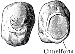
The right cuneiform bone.

Cuneiform Bone

The right cuneiform bone.

The right pisiform bone.

Pisiform Bone

The right pisiform bone.

The right trapezium bone.

Trapezium Bone

The right trapezium bone.

The right trapezoid bone.

Trapezoid Bone

The right trapezoid bone.

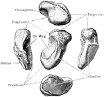
The right magnum bone.

Magnum Bone

The right magnum bone.

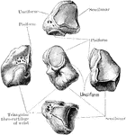
The right unciform bone.

Unciform Bone

The right unciform bone.

First right metacarpal bone.

First Metacarpal Bone

First right metacarpal bone.

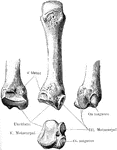
Second metacarpal bone.

Second Metacarpal Bone

Second metacarpal bone.

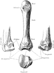
Third metacarpal bone.

Third Metacarpal Bone

Third metacarpal bone.

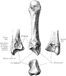
Fourth metacarpal bone.

Fourth Metacarpal Bone

Fourth metacarpal bone.

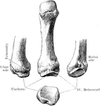
Fifth metacarpal bone.

Fifth Metacarpal Bone

Fifth metacarpal bone.

The phalanges of the fingers (palmar aspect).

Phalanges

The phalanges of the fingers (palmar aspect).

Lower end of right femur as seen from below.

Lower End of Femur

Lower end of right femur as seen from below.

The right astragalus. A, Upper surface. B, Under surface. Labels: 1, groove for flex, long, hallucis; 2, internal tubercle; 3, trochlear surface for tibia; 4, body; 5, for articulation with internal malleolus; 6, head; 7, for articulation with internal malleolus; 8, neck; 9, for articulation with external malleolus; 10, surface against which the inferior tibio fibular ligament rests; 11, external tubercle; 12, external tubercle; 13, posterior, middle, and anterior facets for os calcis; 14, for articulation with navicular; 15, surface resting on inferior calcaneo-navicular ligament; 16, interosseous groove; 17, internal tubercle; 18, groove for the flexor longus hallucis.

Astragalus

The right astragalus. A, Upper surface. B, Under surface. Labels: 1, groove for flex, long, hallucis;…

The right astragalus. A, As seen from the outer side. B, As seen from the inner side. Labels: 1, external tubercle; 2, groove for flexor longus hallucis; 3, internal tubercle; 4, surface against which the inferior tibio-fibular ligament rests; 5, trochlea for tibia; 6, for articulation with external malleolus; 7, neck; 8, head; 9, for articulation with navicular; 10, interosseous groove; 11, anterior, middle, and posterior facets for os calcis; 12, body; 13, surface resting on internal calcaneo navicular ligament; 14, for articulation with navicular; 15, head; 16, neck; 17, trochlea for tibia; 18, for articulation with internal malleolus; 19, body; 20, impression for internal lateral ligament; 21, internal tubercle; 22, groove for flexor longus hallucis; 23, external tubercle; 24, interosseous groove; 25, posterior and middle facets for os calis.

Astragalus

The right astragalus. A, As seen from the outer side. B, As seen from the inner side. Labels: 1, external…

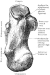
The right os calcis as seen from above.

Os Calcis Viewed from Above

The right os calcis as seen from above.

The right os calcis as seen from below.

Os Calcis Viewed from Below

The right os calcis as seen from below.

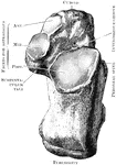
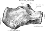
The os calcis as seen from the outer side.

Outer Side of Os Calcis

The os calcis as seen from the outer side.

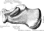
The os calcis as seen from the inner side.

Inner Side of Os Calcis

The os calcis as seen from the inner side.

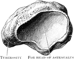
The right navicular bone as seen from behind.

Navicular Bone Viewed from Behind

The right navicular bone as seen from behind.

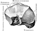
The right navicular bone viewed from the front.

Navicular Bone Viewed from the Front

The right navicular bone viewed from the front.

Anterior view of the three cuneiform bones of the right foot.

Cuneiform Bones

Anterior view of the three cuneiform bones of the right foot.

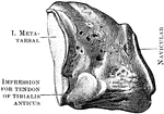
The right internal cuneiform (inner side).

Internal Cuneiform

The right internal cuneiform (inner side).

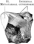
The right internal cuneiform (outer side).

Internal Cuneiform

The right internal cuneiform (outer side).

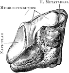
The right middle cuneiform (inner side).

Middle Cuneiform

The right middle cuneiform (inner side).

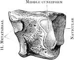
The right middle cuneiform (outer side).

Middle Cuneiform

The right middle cuneiform (outer side).

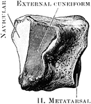
The right external cuneiform (inner side).

External Cuneiform

The right external cuneiform (inner side).

The right external cuneiform (outer side).

External Cuneiform

The right external cuneiform (outer side).

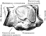
The right cuboid bone (outer side).

Cuboid

The right cuboid bone (outer side).

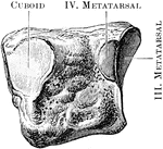
The right cuboid bone (inner side).

Cuboid

The right cuboid bone (inner side).

The first metatarsal bones of the right foot (plantar aspect).

First Metatarsal Bone

The first metatarsal bones of the right foot (plantar aspect).

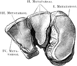
View of the bases and shafts of the second, third, and fourth metatarsal bones of the right foot.

Metatarsal Bones

View of the bases and shafts of the second, third, and fourth metatarsal bones of the right foot.

Fifth right metatarsal bone (dorsal aspect).

Fifth Metatarsal

Fifth right metatarsal bone (dorsal aspect).

The phalanges of the toes (dorsal aspect).

Phalanges

The phalanges of the toes (dorsal aspect).

Dissection from behind the ligaments connecting the occipital bone, the atlas, and the axis with each other.

Ligaments of the Occipital Bone

Dissection from behind the ligaments connecting the occipital bone, the atlas, and the axis with each…
Metacarpo phalangeal and interphalangeal joints.

Finger Joint

Metacarpo phalangeal and interphalangeal joints.

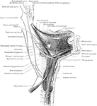
The muscles of the hyoid bone and styloid process, and the extrinsic muscles of the tongue, with their nerves.

Muscles of the Hyoid Bone

The muscles of the hyoid bone and styloid process, and the extrinsic muscles of the tongue, with their…
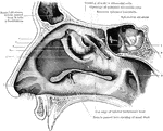
View of the outer wall of the nose. The turbinated bones having been removed. Labels: 1, vestibule, 2, hiatus semilunaris; 3, opening of antrum of highmore; 4, bulla ethmoidalis; 5, agger nasi; 6, opening of anterior ethmoidal cells; 7, cut edge of superior turbinated bone; 8, cut edge of middle turbinated bone; 9, pharyngeal orfice of Eustachian tube.

Outer Wall of Nose

View of the outer wall of the nose. The turbinated bones having been removed. Labels: 1, vestibule,…

Section through left temporal bone, showing inner wall of tympanic cavity.

Tympanic Cavity

Section through left temporal bone, showing inner wall of tympanic cavity.

Coronal section through larynx, to show its three compartments.

Coronal Section Through Larynx

Coronal section through larynx, to show its three compartments.

Mesial section through larynx, to show the outer wall of the right half.

Mesial Section Through Larynx

Mesial section through larynx, to show the outer wall of the right half.
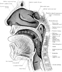
Sagittal section through mouth, tongue, larynx, pharynx, and nasal cavity. The section was slightly oblique, and the posterior edge of the nasal septum has been preserved. The specimen is viewed slightly from below, hence the apparently low position of the inferior turbinated bone.

Section of the Head and Neck

Sagittal section through mouth, tongue, larynx, pharynx, and nasal cavity. The section was slightly…

View of the outer wall of the middle ear. Section through the left temporal bone of a child to show the relations of the middle ear and the mastoid antrum to the middle and posterior fossae of the skull.

Outer Wall of Tympanic Cavity

View of the outer wall of the middle ear. Section through the left temporal bone of a child to show…

View of the inner wall of the middle ear. Section through the left temporal bone of a child to show the relations of the middle ear and the mastoid antrum to the middle and posterior fossae of the skull.

Inner Wall of Tympanic Cavity

View of the inner wall of the middle ear. Section through the left temporal bone of a child to show…

"Pectoral arch and fore limb of the pike (Esox lucius), an osseous fish, showing scapulocoracoid, composed of Scp, scapula or hypercoracoid; c, posterior end of the outer margin of the scapulocoracoid; b, b, 1, 2, 3, 4, 5, five fin-rays or radialia; a, actinosts or basalia." -Whitney, 1911

Pike Scapulocoracoid

"Pectoral arch and fore limb of the pike (Esox lucius), an osseous fish, showing scapulocoracoid, composed…
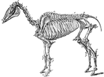
The skeleton of a horse. Axial Skeleton. The Skull. Cranial Bones: a, occipital, 1; b, wormian, 1; c, parietal, 2; d, frontal, 2; e, temporal, 2; sphenoid, 1; ethmoid, 1; auditory ossicles, 8. Facial bones: h, nasal, 2; g, lachrymal, 2; f, malar, 2; i, superior maxilla, 2; k, premaxila, 2; palatine, 2; pterygoid, 2; vomer, 1; tubinals, 4; l, inferior maxilla; 1; hyoid (segments), 5. Teeth: m, incisors, 12; n, canines, 4; o, molars, 24. The trunk: p-v, cervical vertebrae, 7; w, dorsal vertebrae, 18; x, lumbar vertebrae, 6; y, sacrum, 1; z, coccygeal vertebrae (variable), 18, d, d, ribs, 36; sternum (6-7 sternebrae), 7; *,coastal cartilages. Appendicular Skeleton. Pectoral Limb: e', scapula, 2; f', humerus, 2; g', radius, 2; h', ulna, 2. Carpus-i', trapesium, 2; k', cuneiform, 2; l', lunar, 2; m', scaphoid, 2; n', unciform, 2; o', magnum, 2; p', trapezoid, 2; q', pisiform, 2. Metacarpus-r', large bone, 2; s', small bone, 4. Digits- u', proximal phalanges, 2; v, median phalanges; w', distil phalanges, 2; t', large sesamoids, 4; small sesamoids, 2. Pelvic Limbs: Pelvis- b', ilium, 2; d' ischium, 2; c', pubis, 2. The limb- y', femur, 2; z', patella, 2; a', tibia, 2; b', fibula, 2. Tarsus- c', calcaneum, 2; d', astragalus, 2; e'. cuboid, 2; f', cuneiforme magnum, 2; g', cuneiforme medium, 2; h", cuneiforme parvum, 2. Metatarsus: large bone, 2; small bone, 4. Digit: proximal phalanges, 2; median phalanges, 2; distal phalanges, 2; large sesamoids, 4; small sesamoids, 2.

Skeleton of a Horse

The skeleton of a horse. Axial Skeleton. The Skull. Cranial Bones: a, occipital, 1; b, wormian, 1; c,…

"Schizorhinal skull of curlew (top view), showing the long cleft, a, between upper and lower forks of each nasal bone." -Whitney, 1911

Curlew Skull

"Schizorhinal skull of curlew (top view), showing the long cleft, a, between upper and lower forks of…

The skull of a horse seen from above. Labels: I, occipital lobe; II, parietal bone; III, squamosal bone; IV, frontal bone; V, nasal bone; VI, lachrymal bone; VII, malarbone; VIII, superior maxilla; IX, premaxilla; a, occipital crest; b, parietal crest; c, orbital process; d, supraorbital foramen.

Horse Skull Seen from Above

The skull of a horse seen from above. Labels: I, occipital lobe; II, parietal bone; III, squamosal bone;…

Inferior aspect of horse's skull, the mandible being removed. Above the line A is the posterior region or base between A and B the middle, and below B the anterior or nasal region. Labels: a, occipital condyle; B, foramen magnum; c, styloid process; d, temporal articular surface; e, basioccipital bone; f, basispenoid bone; i, temporal articular surface; k, condyloid foramen; k', condyloid notch; l, foramen lacerum basis cranii; m, pterygoid foramen; n, palatine suture; o, palatine foramen; p, molar teeth; q, incisive opening; r, foramen incisivum.

Inferior Aspect of Horse Skull

Inferior aspect of horse's skull, the mandible being removed. Above the line A is the posterior region…

Lateral aspect of horse's skull. Labels: 1, occipital bone; 2, parietal bone; 3, frontal bone; 9, nasal bone; 11, nasal peak; 10, premaxilla; 8, superior maxilla; 6, lachrymal bone; 7, malar bone; 5, zygomatic process of the squamosal bone; 4, petrosal bone; e, its external meatus; 12, inferior maxilla; h, coronoid process f inferior maxilla occupying the temporal fossa; k, temporomaxillary articulation; l, molar teeth.

Lateral Aspect of Horse Skull

Lateral aspect of horse's skull. Labels: 1, occipital bone; 2, parietal bone; 3, frontal bone; 9, nasal…

Longitudinal section of a horse's skull. Labels: 1, supraoccipital bone and crest; 2, parietal bone; 3, frontal bone; 4, frontal sinus; 5, nasal bone; 6, superior turbinal; 12, inferior turbinal; 14, superior maxilla; 13 and its palatine plate; 15, premaxilla; 11, palatine bone; 7, ethmoid bone and volutes; 8, basi and presphenoid bones; 9, basioccipital bone; 10, petrosal bone. The cranium is the cavity under 1 and 2.

Longitudinal Section of Horse Skull

Longitudinal section of a horse's skull. Labels: 1, supraoccipital bone and crest; 2, parietal bone;…

The hyoid series is composed of five distinct pieces- a body, or hyoid bone proper, two cornua or horns and two cornicula or lesser horns. Shown us the left aspect of the hyoid series of bones in a horse. Labels: a, proximal, end of left cornu; b, distal end of right cornu; c, proximal end of corniculum; d, hyoid bone. The left heel process points downwards and backwards, the spur process in the opposite direction.

Hyoid Series of Bones in a Horse

The hyoid series is composed of five distinct pieces- a body, or hyoid bone proper, two cornua or horns…

Front aspect of the right carpus of a horse. Labels: 1, cuneiform; 2, lunar; 3, scaphoid; 4, trapezium; 5, unciform; 6, magnum; 7, trapezoid; 8, small metacarpals, the large one lying between and before them. The uppermost bone is the distal end of the radius.

Carpus of a Horse

Front aspect of the right carpus of a horse. Labels: 1, cuneiform; 2, lunar; 3, scaphoid; 4, trapezium;…

External view of bones of right carpus, metacarpus, and digit of a horse. 1, distal end of radius; 2, groove for extensor tendons; 3, scaphoid; 4, lunar; 5, cuneiform;6, trapezium; 7, magnum; 8, unciform; 9, great metacarpal; 10, external small metacarpal (splint); 11, sesamoids; 12, proximal phalanx; 13, median phalanx; 5, distal phalanx; 16, basilar process.

Horse Leg

External view of bones of right carpus, metacarpus, and digit of a horse. 1, distal end of radius; 2,…

Posterior view of phalanges of a horse disarticulated. Labels: a, os suffraginis; b, os coronae; C, os pedis; D, os naviculare, or third sesamoid; 1, glenoid cavities joined by the groove, 2; 3, rough surface for V-shaped sesamoidean ligament; 4, condyles articulating with cavities, 5; 6, condyles of coronae articulating with glenoid cavities of pedal bone, 7; 8, pyramidal process; 9, basilar process; 10, retrosal processes; 11, articular surfaces between navicular and pedal bones; 12, surface, continues with glenoid cavities, 7; 13, posterior edge; 14, extremities for lateral ligaments of navicular bone.

Phalanges of a Horse

Posterior view of phalanges of a horse disarticulated. Labels: a, os suffraginis; b, os coronae; C,…