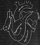
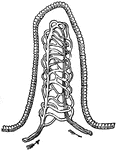
The arterial system.

The Arterial System

The arterial system.

The main arteries of the body. Labels: Crd, and Crs, right and left coronary arteries of the heart, cut short near their origin; Aa, and aA, aortic arch; At, thoracic aorta; Aab, abdominal aorta; K, renal artery; Sd, right, and Ssi, left subclavian; Cd, right, and Cs, left carotid; Ax, axillary artery; B, brachial artery; U, ulner artery; R, radial artery; Ai, common iliac artery; I, external iliac artery; C, femoral artery; Po, popliteal artery; Ta, anterior, and Tp, posterior tibial artery; Pe, peroneal artery.

The Main Arteries of the Body

The main arteries of the body. Labels: Crd, and Crs, right and left coronary arteries of the heart,…
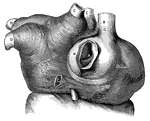
A portion of an artery.

Artery

A portion of an artery.

"The Femoral Artery of Right Leg." — Blaisedell, 1904

Leg artery

"The Femoral Artery of Right Leg." — Blaisedell, 1904

Relations of the artery, vein, and capillaries.

Relations of Artery, Vein, and Capillaries

Relations of the artery, vein, and capillaries.

<em>L.A.</em>, left auricle; <em>R.A.</em>, right auricle; <em>A</em>, opening of the inferior vena cava; <em>B</em>, superior vena cava; <em>C</em>, cardiac vein of right auricle laid open; <em>D</em>, left pulmonary veins; <em>E</em>, eight pulmonary veins; <em>F</em>, muscular fibers surrounding the openings between the left auricle and left ventricle, and the right auricle and right ventricle; <em>G, H, K,</em> and <em>L</em>, muscular fibers surrounding the great veins of the heart." &mdash; Blaisedell, 1904

Muscular fibers of the auricle

L.A., left auricle; R.A., right auricle; A, opening of the inferior vena…
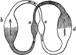
Diagram showing the circulation of blood in the heart. Let <em>a</em> represent the right side of the heart, <em>c</em>the left side, <em>b</em> the lungs, and <em>d</em> the general system of the body. The arrows point in the direction in which the blood flows. In all the shaded part the blood is dark (oxygen-poor), and in the part that is not shaded it is red (oxygen-rich).

Blood Circulation in the Heart

Diagram showing the circulation of blood in the heart. Let a represent the right side of the…
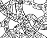
"Showing how the circulation of blood in the web of a frog's foot looks as seen under the microscope." &mdash; Blaisedell, 1904

Circulation of blood

"Showing how the circulation of blood in the web of a frog's foot looks as seen under the microscope."…

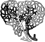
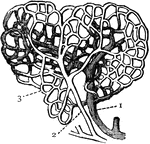
Capillaries. 1: Vein; 2: Artery; 3: Capillary.

Capillaries

Capillaries. 1: Vein; 2: Artery; 3: Capillary.

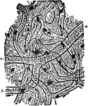
Isolated capillary network formed by the junction of several hallowed-out cells, and containing colored blood-corpuscles in a clear fluid. Labels: p, p, pointed cell-processes extending in different directions for union with neighboring capillaries.

Capillary Network

Isolated capillary network formed by the junction of several hallowed-out cells, and containing colored…

An ideal view of the circulation in the lungs and system.

Circulation

An ideal view of the circulation in the lungs and system.

Diagram of circulation. Labels: L, left side of heart; R, right side of heart; a,a,a arterial system; b,b, capillaries; c,c,c veins; Alim., alimentary canal; Liv., liver; p, portal vein; H,hepatic vein; Lymph., lymphatic duct and tributaries; Pulm., lungs; Pa, pulmonary artery; Pu, pulmonary vein.

Diagram of Circulation

Diagram of circulation. Labels: L, left side of heart; R, right side of heart; a,a,a arterial system;…

Circulation in frog's foot under a microscope. Labels: A, walls of capillaries; B, tissue of web lying between the capillaries; C, cells of epidermis covering web (these are only shown in the right-hand and lower part of the field; in the other parts of the field the focus of the microscope lies below the epidermis); D, nuclei of these epidermic cells; E, pigment cells contracted, not partially expanded; F, red blood corpuscle (oval in the frog) passing along capillary -- nucleus not visible; G, another corpuscle squeezing its way through a capillary, the canal of which is smaller than its own transverse diameter; H, another bending as it slides round a corner; K, corpuscle in capillary seen through the epidermis; I, white blood corpuscle.

Circulation in a Frog's Foot

Circulation in frog's foot under a microscope. Labels: A, walls of capillaries; B, tissue of web lying…

Circulation in the web of a Frog's foot.

Circulation in the Web of a Frog's Foot

Circulation in the web of a Frog's foot.

Diagram of the circulation in a crustacean. Labels: a, branchial; b, somatic circulation. On the right side is the heart with is single cavity, and on the left the venous lacunae.

Circulation of a Crustacean

Diagram of the circulation in a crustacean. Labels: a, branchial; b, somatic circulation. On the right…
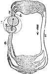
A diagram of the circulation of a fish. Labels: 1, The pericardium. 2, The single auricle. 3, The single ventricle. 4, The vessel that conveys the blood from the ventricle to the gills. 5, The vessel that conveys the blood from the body to the heart. The arrows show the direction of the blood.

Circulation of a Fish

A diagram of the circulation of a fish. Labels: 1, The pericardium. 2, The single auricle. 3, The single…

Diagram of the circulation of the fish. Labels: a, branchial circulation; b, somatic circulation; c, heart; d, auricle; e, ventricle; f, arterial trunk.

Circulation of a Fish

Diagram of the circulation of the fish. Labels: a, branchial circulation; b, somatic circulation; c,…

A diagram of the circulation of a frog. Labels: 1, The pericardium. 2, The single ventricle. 3, The right auricle. 4, The left auricle. The arrows indicate the direction of the blood.

Circulation of a Frog

A diagram of the circulation of a frog. Labels: 1, The pericardium. 2, The single ventricle. 3, The…

Diagram of the circulation of the reptile. Labels: a, pulmonic, and b, somatic circulation; c, heart showing the three cavities. The arrows indicate the course of the blood current.

Circulation of a Reptile

Diagram of the circulation of the reptile. Labels: a, pulmonic, and b, somatic circulation; c, heart…

Insects have neither arteries nor veins. The circulation, such as it is, is animated by the action of a vessel called dorsal, which is situated above the digestive tube. Labels: 1, Dorsal vessels in which the blood flows. 2,The lateral currents. The arrows show the outward and the inward current.

Diagram of the Circulation of an Insect.

Insects have neither arteries nor veins. The circulation, such as it is, is animated by the action of…
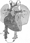
A diagram illustrating the circulation of the blood. Labels: A, vena cava descending (superior); Z, vena cava ascending (inferior); C, right auricle; D, right ventricle; E, pulmonary artery; F, P, lungs and pulmonary veins; G, left auricle; H, left ventricle; I, K, aorta.

Circulation of Blood

A diagram illustrating the circulation of the blood. Labels: A, vena cava descending (superior); Z,…

"<em>R.A.</em>, right auricle; <em>L.A.</em>, left auricle; <em>R.V.</em>, right ventricle; <em>L.V.</em>, left ventricle; <em>P.A.</i., pulmonary artery; <em>A</em>, pulmonary artery and vein of right lung; <em>B</em>, pulmonary artery and vein of left lung; <em>C</em>, carotid artery to head, showing branch of left subclavian artery; <em>D</em>, portal vein; <em>F</em>, hepatic artery; <em>G</em>, superior vena cava, bringing blood from head and upper limbs to right auricle." &mdash; Blaisedell, 1904

Diagram of the circulation of the blood

"R.A., right auricle; L.A., left auricle; R.V., right ventricle; L.V.,…

Diagram of circulation.

Diagram of Circulation

Diagram of circulation.

Diagram of circulation.

Diagram of Circulation

Diagram of circulation.

Diagram of fetal circulation.

Diagram of Fetal Circulation

Diagram of fetal circulation.

Plan of fetal circulation. In this plan, the figured arrows represent the kind of blood, as well as the direction which it takes in the vessels. Thus, arterial blood is figured ..... ; venous blood ----; mixed (arterial and venous) -...- .

Diagram of Fetal Circulation

Plan of fetal circulation. In this plan, the figured arrows represent the kind of blood, as well as…

The circulatory organs. Labels: 1, The left auricle. 2, The right auricle. 3, The left ventricle. 4, The right ventricle. 5, 6, The aorta. 7, The femoral (leg) artery. 8, The branchial (arm) arteries. 9, carotid arteries. 10, The jugular veins. 11, The branchial veins. 12, The femoral veins. 13, 14, The vena cavas. 15, The pulmonary artery. 16, The right and left pulmonary arteries. 17, The pulmonary veins. 18, The lacteals. 19, The thoracic duct. 20, The absorbents of the upper extremities. 22, The small intestine.

The Circulatory Organs

The circulatory organs. Labels: 1, The left auricle. 2, The right auricle. 3, The left ventricle. 4,…

Diagram of the circulatory system, showing that it forms a single closed circuit with two pumps in it, represented by the right and left halves of the heart, which are separated in the diagram. Labels: ra and rv, right auricle and ventricle; la and lv, left auricle and ventricle; ao, aorta; sc, systemic capillaries; vc, vena cava, pa, pulmonary artery; pc, pulmonary capillaries; pv, pulmonary veins.

Diagram of the Circulatory System

Diagram of the circulatory system, showing that it forms a single closed circuit with two pumps in it,…
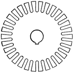
"The metal cut away near the center reduces the weight and provides passages for air circulation." &mdash; Hawkins, 1917

Solid core disk

"The metal cut away near the center reduces the weight and provides passages for air circulation." —…
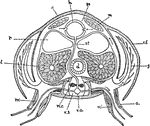
"Diagrammatic cross-section of Cray-fish in the thoracic region, to show relation of circulation and respiration. a, appendage; c, carapace; c.f., flap of carapace overhanging the gills; d, digestive tube; g, gill; h, heart; l, liver; m, body muscles; m', muscles of the appendages; n.c., nerve cord; p.s., paricardial sinus; r, reproductive glands; st, sternal artery; v.a., ventral artery; v.s., ventral blood sinus in which the nerve cord lies." &mdash; Galloway

Cray-fish

"Diagrammatic cross-section of Cray-fish in the thoracic region, to show relation of circulation and…
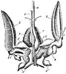
"Central organs of the circulaion, gills, and renal organs of Sepia officinalis. a, aorta; v, vena cava; v' visceral veins; c, systemic heart; d, dilatations of branchial veins on entering the heart; e, branchial hearts; b, branchiae; r, renal organs." &mdash; Encyclopedia Britannica, 1893

Cuttlefish Organs

"Central organs of the circulaion, gills, and renal organs of Sepia officinalis. a, aorta; v, vena cava;…
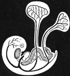
Diagram of young embryo and its vessels, showing course of circulation in the umbilical vesicle; and also that of the allantois (near the caudal extremity), which is just commencing.

Embryo Showing Course of Circulation

Diagram of young embryo and its vessels, showing course of circulation in the umbilical vesicle; and…
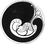
Diagram of embryo and its vessels, showing course of circulation. The large umbilical arteries are seen passing out in the placenta.

Embryo Showing Course of Circulation

Diagram of embryo and its vessels, showing course of circulation. The large umbilical arteries are seen…

"Diagram of the principal vessels in the circulation of a fish, ventral view. a, aorta; au., auricle; br.a., afferent branchial arteries; br.e., efferent branchial arteries; bu., bulbus (or conus) arteriosus; c, carotid; c.a., caudal artery; c.v.r., right cardinal vein; c.v.l., left cardinal vein; d.a., dorsal artery; d.c., ductus Cuvieri; g, gills; h.v., hepatic vein; h.p., hepatic portal vein; i, intestine; j, jugular vein; k, kidney; l, liver; r.p., renal portal vein; s.v., sinus venosus; v, ventricle." &mdash; Galloway

Fish Circulation

"Diagram of the principal vessels in the circulation of a fish, ventral view. a, aorta; au., auricle;…

The circulation of the blood in the web of a frog's foot. A, an artery; B, capillaries crowded with disks, owing to a rupture just above, where the disks are jammed into an adjacent mesh; C, a deeper vein; the black spots are pigments cells.

Circulation of Blood in a Frog's Foot

The circulation of the blood in the web of a frog's foot. A, an artery; B, capillaries crowded with…
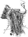
Arteries of the head. Labels: 1, common carotid; 2, internal carotid; 3, external carotid; 4, occipital; 5, superior thyroid; 6, trapezius; 7, lingual; 8, sterno-mastoid; 9, facial; 10, temporal; 11, submental; 12, transverse facial; 13, inferior labial; 15 inferior coronary; 17, superior coronary; 19, lateral nasal; 21, angular.

Arteries of the Head

Arteries of the head. Labels: 1, common carotid; 2, internal carotid; 3, external carotid; 4, occipital;…
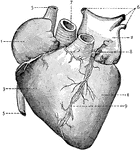
External view of the Heart. 1: Right Auricle; 2: Left Auricle; 3: Right Ventricle; 4: Left Ventricle; 5: Systemic Veins; 6: Pulmonary Veins; 7: Aorta; 8: Pulmonary Artery; 9: Coronary Vein and Artery.

Heart

External view of the Heart. 1: Right Auricle; 2: Left Auricle; 3: Right Ventricle; 4: Left Ventricle;…

In the class of Crustacea there is a single ventricle, which receives the blood from the gills and propels it to other parts of the body. Shown is the heart and arteries of a lobster. Labels: 1, The heart. 2, The abdominal artery. 5, Venous sinuses. 6, The branchia from which the blood returns to the hear.

The Heart and Arteries of a Lobster

In the class of Crustacea there is a single ventricle, which receives the blood from the gills and propels…

In high orders of Mollusca the circulation resembles that of fish. Shown is the heart and arteries of a snail. Labels: 2, The stomach. 3, Intestines. 5, Heart. 6, Aorta. 7, Pulmonary artery.

The Heart and Arteries of a Snail

In high orders of Mollusca the circulation resembles that of fish. Shown is the heart and arteries of…

The heart and blood vessels diagrammatically represented, showing the direction of blood flow.

Heart and Blood Vessels

The heart and blood vessels diagrammatically represented, showing the direction of blood flow.
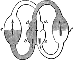
A diagram of the circulation through the heart.

Circulation Through the Heart

A diagram of the circulation through the heart.

Diagram showing the compartments of the heart. The smaller compartments are the auricles (also known as atria) and the larger compartments are the ventricles. The middle part of the figure represents the heart with its two sides; <em>a</em> being the right auricle, <em>b</em> the right ventricle, <em>d</em> the left auricle, and <em>e</em> the left ventricle. The blood is received in the right auricle, <em>a</em>, from the general system, <em>f</em>. It then passes into the right ventricle, <em>b</em>, and is forced by the contraction of it through arteries to the lungs, <em>c</em>. From the lungs it comes back to the heart, to the left side, and enters the left auricle, <em>d</em>. From this it passes into the left ventricle, <em>e</em>, from which it is sent all over the body, represented by <em>f</em>.

Heart, Compartments of

Diagram showing the compartments of the heart. The smaller compartments are the auricles (also known…
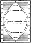
Diagram showing the circulation in an ideal ocean extending from pole to pole and covering one fourth the circumference of the earth.

Ideal Ocean Circulation

Diagram showing the circulation in an ideal ocean extending from pole to pole and covering one fourth…
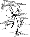
To show the sites at which communications occur between the portal and systemic circulations.

Communication of the Portal and Systemic Circulation

To show the sites at which communications occur between the portal and systemic circulations.

The portal vein and its branches. Labels: l, liver, under surface; gb, gall bladder; st, stomach; sp, spleen; p, pancreas; du, duodenum; ac, ascending colon; cd, descending colon; a,b,c,d,e, the portal vein and its branches. Portions of the duodenum and colon have been removed.

The Portal Vein and its Branches

The portal vein and its branches. Labels: l, liver, under surface; gb, gall bladder; st, stomach; sp,…
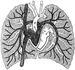
A diagram of pulmonary circulation. Labels: 1, Descending vena cava. 2, Ascending cava vein. 3, Chamber of right side of heart. 4, Chamber of left side of heart. 5, Aorta. 6, Arch of aorta. 7, Pulmonary artery. 8, Its left branch. 9, Its right branch. 10, Right pulmonary vein. 11, Left pulmonary vein.

A Diagram of Pulmonary Circulation

A diagram of pulmonary circulation. Labels: 1, Descending vena cava. 2, Ascending cava vein. 3, Chamber…
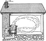
"Cold air flows up through the pipe a, and is heated by stobe b, inclosed in sheet iron c. The smoke stack e warms the air about it in brick flue f and thus draws the foul air through the opening d." —Davison, 1910

Ventilating room

"Cold air flows up through the pipe a, and is heated by stobe b, inclosed in sheet iron c. The smoke…

Semilunar valves of the heart.

Semilunar Valves

Semilunar valves of the heart.

"<em>A</em>, inferior vena cava, cut off just above the hepatic veins; <em>B</em>, superior vena cava, cut off just below the junction of the innominate veins; <em>C</em>, right and left innominate veins; <em>D</em>, left internal jugular vein. The veins, known as <em>azygos</em>, with some of their main branches, are shown. These veins connect the superior vena cava and the inferior vena cava." &mdash; Blaisedell, 1904

Vena cava

"A, inferior vena cava, cut off just above the hepatic veins; B, superior vena cava,…

"Ideal section, greatly enlarged, showing minute capillary circulations." &mdash;Brown, 1884

Villus

"Ideal section, greatly enlarged, showing minute capillary circulations." —Brown, 1884