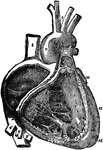
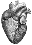
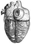
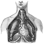
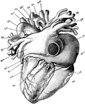
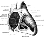
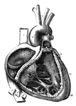
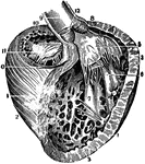
The right auricle (atrium) and ventricle of the heart opened, and a part of their right and anterior walls removed, so as to show their interior; 1/2, 1, superior vena cava; 2, inferior vena cava; 2', hepatic veins cut short; 3, right auricle; 3', placed in the fossa ovalis, below which is the Eustachian valve; 3", is placed close to the aperture of the coronary vein; +, +, placed n the auriculo-ventricular groove, where a narrow portion of the adjacent walls of the auricle and ventricle has been preserved; 4, 4, cavity of the right ventricle; the upper figure is immediately below the semilunar valves; 4', large columna carnea or musculus papillaris; 5, 5', 5", tricuspid valve; 6, placed in the interior o the pulmonary artery, a part of the anterior wall of that vessel having been removed, and a narrow portion of it preserved at its commencement where the semilunar valves are attached; 7, concavity of the aortic arch close to the cord of the ductus arteriosus; 8, ascending part of sinus of the arch covered at its commencement by the auricular appendix and pulmonary artery; 9, placed between the innominate and left carotid arteries; 10, appendix of the left auricle; 11, 11, the outside of the left ventricle, the lower figure near the apex.

Right Atrium and Ventricle of the Heart

The right auricle (atrium) and ventricle of the heart opened, and a part of their right and anterior…
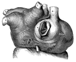
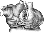
The arrangement of the muscles of the auricles.

Auricles Muscles

The arrangement of the muscles of the auricles.

<em>L.A.</em>, left auricle; <em>R.A.</em>, right auricle; <em>A</em>, opening of the inferior vena cava; <em>B</em>, superior vena cava; <em>C</em>, cardiac vein of right auricle laid open; <em>D</em>, left pulmonary veins; <em>E</em>, eight pulmonary veins; <em>F</em>, muscular fibers surrounding the openings between the left auricle and left ventricle, and the right auricle and right ventricle; <em>G, H, K,</em> and <em>L</em>, muscular fibers surrounding the great veins of the heart." &mdash; Blaisedell, 1904

Muscular fibers of the auricle

L.A., left auricle; R.A., right auricle; A, opening of the inferior vena…

This illustration shows a representation of the circulation of the blood, in its essential features. The arrows indicate the course of the blood. a, arteries; aur., auricle or receiving portion of the heart; d, digestive tract; c. d., capillaries of the digestive tract; c.r., capillaries of the respiratory organs; c.s., capillaries of the system; va., valves; ve, veins; vt, ventricle.

Blood Circulation

This illustration shows a representation of the circulation of the blood, in its essential features.…
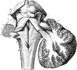
The cerebellum in section and fourth ventricle, with the neighboring parts. Labels: 1, median groove of fourth ventricle, ending below in the calamus scriptorius, with the longitudinal eminence formed by the fasciculi teretes, one on each side; 2, the same groove, at the place where the white streaks of th auditory nerve emerge from it to cross the floor of the ventricle; 3, in inferior crus or peduncle of the cerebellum, formed by the restiform body; 4, posterior pyramid; above this is the calamus scriptorius; 5, superior crus of cerebellum, or processus e cerebello ad cerebrum( or ad testes); 6, fillet to the side of the crura cerebri; 7, lateral grooves of the crura cerebri; 8, corpora quadrigemina.

The Cerebrum and Fourth Ventricle of the Brain

The cerebellum in section and fourth ventricle, with the neighboring parts. Labels: 1, median groove…
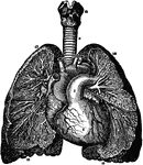
Branchi of the lungs, the heart, and blood vessels. Labels: 1, left auricle; 2, right auricle; 3, left ventricle; 5, pulmonary artery; 6, arch of the aorta; 7, superior vena cava; 8, arteria innominata; 9, left primitive carotid artery; 10, left subclavian artery; 11, trachea; 12, larynx; 13, upper lobe of right lung; 14, upper lobe of left lung; 15, trunk of right pulmonary artery; 16, lower lobes of the lungs.

Branchi and Blood Vessels

Branchi of the lungs, the heart, and blood vessels. Labels: 1, left auricle; 2, right auricle; 3, left…
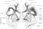
Two coronal sections through the cerebral hemisphere of an orangoutang, in the plan of the anterior commissure. A, Section through the left hemisphere n a plane a short distance behind B, which is a section through the right hemisphere.

Coronal Sections of the Cerebral Hemispheres

Two coronal sections through the cerebral hemisphere of an orangoutang, in the plan of the anterior…

"Cast or mold of the interior of the left ventricle of the heart of a deer. Shows that the left ventricular cavity in conical and spiral in its nature."&mdash;Pettigrew, 1874

Heart of a Deer

"Cast or mold of the interior of the left ventricle of the heart of a deer. Shows that the left ventricular…

"Frog with the left side cut away and some of the organs pulled downward. a, aorta leading from the ventricle; b, bladder; c, spinal cord surrounded by the vertebral column of bone; d, small intestine; e, opening of the lungs into the pharynx; h, ventricle of the heart; k, kidney; l, lung; liv, liver; n, brain; ov, ovary; ovd, oviduct; p, pancreas; r, rectum; sp, spleen; st, stomach; t, tongue; si, small intestine; u, left auricle; ur, ureter; v, vena cava; vc, body of vertebra." &mdash;Davison, 1906

Dissected Frog

"Frog with the left side cut away and some of the organs pulled downward. a, aorta leading from the…
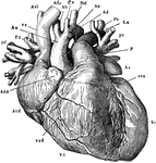
"The heart and the great blood-vessel attached to it, seen from the side towards the sternum. The left cavities and the vessels connected with them are colored red; the right black. Atd, right auricle; Adx and As, the right and left auricular appendages; Vd, right ventricle; Vs, left ventricle; Aa, aorta; Ab, innominate artery; Cs, left common carotid artery; Ssi, left subclavian artery; P, main trunk of the pulmonary artery, and Pd and Ps, its branches to the right and left lungs; cs, superior vena cava; Ade and Asi, the right and left innominate veins; pd, ps, the right and left pulmonary veins; crd and crs, the right and left coronary arteries." &mdash;Martin, 1917

Heart

"The heart and the great blood-vessel attached to it, seen from the side towards the sternum. The left…
"The heart vied from its dorsal aspect. ci, inverior vena cava; Vc, coronary vein; Atd, right auricle; Adx and As, the right and left auricular appendages; Vd, right ventricle; Vs, left ventricle; Aa, aorta; Ab, innominate artery; Cs, left common carotid artery; Ssi, left subclavian artery; P, main trunk of the pulmonary artery, and Pd and Ps, its branches to the right and left lungs; cs, superior vena cava; Ade and Asi, the right and left innominate veins; pd, ps, the right and left pulmonary veins; crd and crs, the right and left coronary arteries." &mdash;Martin, 1917

Heart

"The heart vied from its dorsal aspect. ci, inverior vena cava; Vc, coronary vein; Atd, right auricle;…

Front view of the heart and great vessels. The pulmonary artery has been cut short close to its origin. Labels: 1, right ventricle; 2, left ventricle; 3, root of the pulmonary artery; 4, 4', arch of the aorta; 4'', the descending thoracic aorta; 5, part of the right auricle; 6, part of the left auricle; 7, 7', innominate veins joining to form the vena cava superior; 8, inferior vena cava; 9, one of the large hepatic veins; X, placed in the right auriculo-ventricular groove, points to the right or posterior coronary artery; X, X, placed in the anterior interventricular groove, indicate the left or anterior coronary artery.

Heart

Front view of the heart and great vessels. The pulmonary artery has been cut short close to its origin.…
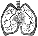
A diagram of the heart. Labels: 1. Left auricle. 2, Right auricle. 3, Left ventricle. 4, Right ventricle. 5, Right and left pulmonary veins. 6, Trachea.

The Heart

A diagram of the heart. Labels: 1. Left auricle. 2, Right auricle. 3, Left ventricle. 4, Right ventricle.…
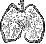
A diagram of the heart. Labels: 1. Right auricle. 2, Left auricle. 3, Right ventricle. 4, Left ventricle. 5, Right and left pulmonary veins. 6, Trachea.

The Heart

A diagram of the heart. Labels: 1. Right auricle. 2, Left auricle. 3, Right ventricle. 4, Left ventricle.…
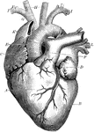
The heart. Labels: A, the right ventricle; B, the left ventricle; C, the right auricle; D, the left auricle.

Heart

The heart. Labels: A, the right ventricle; B, the left ventricle; C, the right auricle; D, the left…
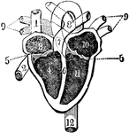
View of the heart with its several chambers exposed and the vessels in connection with them. Labels: 1, the superior vena cava. 2, the inferior vena cava. 3. the chamber called the auricle. 4. the right ventricle. 5. the line marking the passage between the two chambers, and the points of attachment of one margin of the valve. 6. the septum between the two ventricles. 7. the pulmonary artery, arising from the right ventricle, and dividing at 8 into right and left, of the corresponding lungs. 9. the four pulmonary veins, bringing the blood from the lungs into 10, the left auricle. 11. the left ventricle. 12. the aorta, arising from the left ventricle, and passing down behind the heart, to distribute blood to every part of the system. Thus the blood moves in a double circle, one from the heart to the body, and from the body back to the heart, called the systemic circle; the other, from the heart to the lung, and from the lung back to the heart, called the pulmonic circle.

Heart and its Chambers

View of the heart with its several chambers exposed and the vessels in connection with them. Labels:…
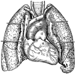
1, The trachea or windpipe; 2 and 3, right and left common carotid arteries; 4 and 5, right and left sub clavian arteries; 6, innominate artery; 7 and 8, right and left innominate veins; 9, Superior vena cava; 10, arch of the aorta; 11, ascending aorta; 12, pulmonary artery; 13, right atrium and auricula; 14 and 15, right and left lung; 16, conus arteriosus; 17, right ventricle; 18, left ventricle.

Heart and Lungs

1, The trachea or windpipe; 2 and 3, right and left common carotid arteries; 4 and 5, right and left…
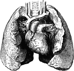
The heart and lungs. 1, right ventricle; 3, right auricle (atrium); 6, 7, pulmonary artery; 9, aorta; 10, superior vena cava; 11, innominate artery; 12, right subclavian vein; 14, innominate vein; 15, left common carotid; 17, trachea; 20, pulmonary veins; 22 to 25, lungs, partially turned back to show veins on left side.

Heart and Lungs

The heart and lungs. 1, right ventricle; 3, right auricle (atrium); 6, 7, pulmonary artery; 9, aorta;…
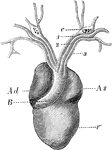
The heart of a frog (Rana esculenta) from the front. Labels: V, ventricle, Ad, right auricle; As, left auricle; B, bulbus arteriosus, dividing into right and left aorta.

Heart of a Frog

The heart of a frog (Rana esculenta) from the front. Labels: V, ventricle, Ad, right auricle; As, left…
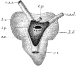
The heart of a frog (Rana esculenta) from the back. Labels: s.v., sinus venosus opened; c.s.s., left vena cava superior; c.s.d., right vena cava superior; c.i., vena cava inferior; v.p., vena pulmonales; A.d., right auricle; A.s., left auricle; A.p., opening of communication between the right auricle and the sinus venosus.

Heart of a Frog

The heart of a frog (Rana esculenta) from the back. Labels: s.v., sinus venosus opened; c.s.s., left…
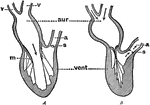
Diagram to illustrate the action (pumping) of the heart. Labels: aur., auricle; vent., ventricle; v, veins; a, aorta; m, mitral valve; s, semilunar valves. In A, auricle contracting, ventricle dilated, mitral valve open, semilunar valves closed. In B, auricle dilated, ventricle contracting, mitral valve closed, semilunar valves open.

Diagram Showing the Pumping of the Heart

Diagram to illustrate the action (pumping) of the heart. Labels: aur., auricle; vent., ventricle; v,…
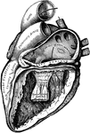
The left auricle and ventricle laid open, the posterior walls of both being removed.

Heart with Left Auricle and Ventricle Laid Open

The left auricle and ventricle laid open, the posterior walls of both being removed.
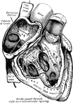
The right auricle and ventricle laid open, the anterior walls of both being removed.

Heart with Right Auricle and Ventricle Laid Open

The right auricle and ventricle laid open, the anterior walls of both being removed.
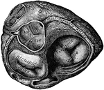
Base of ventricles exposed by removal of the auricles.

Heart with Ventricles Exposed

Base of ventricles exposed by removal of the auricles.

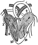
A diagram of the heart. Labels: 1, Right auricle. 2, Right ventricle. 9, Left auricle. 10, Left ventricle. 17, Tricuspid valves. 19, Mitral valves.

A Diagram of the Heart

A diagram of the heart. Labels: 1, Right auricle. 2, Right ventricle. 9, Left auricle. 10, Left ventricle.…
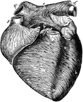
Anterior view of the heart, dissected, after long boiling to show the superficial muscular fibers. The aorta (b') and pulmonary artery (a') have been cut short close to the semilunar valves. Labels: a, right ventricle; b, left ventricle; c, c, groove between ventricles; d, d', right auricle (atrium); e, e', left auricle (atrium); f, superior vena cava; g', g", right and left pulmonary veins. The fibers are seen running a circular, oblique, transverse, and longitudinal direction.

Anterior View of the Heart

Anterior view of the heart, dissected, after long boiling to show the superficial muscular fibers. The…
"<em>A</em>, superior vena cava; <em>B</em>, right auricle; <em>C</em>, right ventricle; <em>D</em> left ventricle; <em>E</em>, left auricle; <em>F</em> pulminary vein; <em>H</em>, pulminary artery; <em>K</em> aorta; <em>L</em>, right subclavian artery; <em>N</em>, left common carotid artery." &mdash; Blaisedell, 1904

Anterior view of the heart

"A, superior vena cava; B, right auricle; C, right ventricle; D

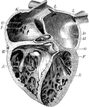
The cavities of the right auricle and right ventricle of the heart.

Auricle and Ventricle of the Heart

The cavities of the right auricle and right ventricle of the heart.

"Diagram of the rush of blood when the heart beats. The valves v open above are closed below while the valves s closed above are open below." &mdash;Davison, 1910

Beating heart

"Diagram of the rush of blood when the heart beats. The valves v open above are closed below while the…

"<em>A, B</em>, right pulmonary veins, <em>S</em>, openings of the left pulmonary veins; <em>E, D, C, aortic valves; <em>R</em>, aorta, <em>P</em>pulmonary artery, <em>O</em>, pulmonic valves, <em>H</em>, mitraal valve; <em>K</em>, columnae carneae, <em>M</em> right ventricular cavity." &mdash; Blaisedell, 1904

Cavities of the heart

"A, B, right pulmonary veins, S, openings of the left pulmonary veins; E, D, C,

The chambers of the heart. Labels: A, right ventricle; B, left ventricle; C, right auricle; D, left auricle; E, tricuspid valve; F, bicuspid valve; G, semilunar valves; H, valves of the aorta; I , inferior vena cava: K, superior vena cava; L, pulmonary veins.

Chambers of the Heart

The chambers of the heart. Labels: A, right ventricle; B, left ventricle; C, right auricle; D, left…

The left auricle and ventricle opened and a part of their anterior and left walls removed. The pulmonary artery has been divided at its commencement; the opening into the left ventricle is carried a short distance into the aorta between two of the segments of the semilunar valves; and the left part of the auricle with its appendix has been removed. The right auricle is out of view. Labels: 1, the two right pulmonary veins cut short; their opening are seen within the auricle; 1' , placed within the cavity of the auricle on the left side of the septum and on the right side and on the part which forms the remains of the valve of the foramen ovale, of which the crescentic fold is seen towards the left hand of 1'; 2, a narrow portion of the wall of the auricle and ventricle preserved round the auriculo-ventricular orifice; 3, 3', the cut surface of the walls of the ventricle, seen to become very much thinner towards 3", at the apex; 4, a small part of the anterior wall of the left ventricle which has been preserved with the principle anterior columna carnea or musculus papillaris attached to it; 5. ,musculi papillares; 5', the left side of the septum, between the two ventricles, within the cavity of the left ventricle; and above the three segments of its semilunar valve which are hanging loosely together; 7', the exterior of the great aortic sinus; 8, the root of the pulmonary artery and its semilunar valves; 8' the separated portion of the pulmonary artery remaining attached to the aorta by 9, the cord of the ductus arteriosus; 10, the arteries rising from the summit of the aortic arch.

A Diagram of the Heart

The left auricle and ventricle opened and a part of their anterior and left walls removed. The pulmonary…
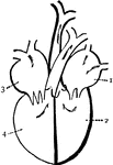
"Diagram of the passages of the heart. 1. left auricle. 2. left ventricle, 3. Right auricle. 4. Right ventricle." &mdash;Brown, 1884

Diagram of the heart

"Diagram of the passages of the heart. 1. left auricle. 2. left ventricle, 3. Right auricle. 4. Right…

A representation of the heart as it really appears showing the front view. At <em>a</em> is the right auricle that receives blood from all pats of the body by two large veins h and i. At <em>b</em> is the right ventricle, which receives the blood from the auricle and sends it to the lungs by the pulmonary artery <em>f</em>. At <em>c</em> is the left auricle, which receives the blood from the lungs by the pulmonary veins <em>g, g, g</em>. At <em>d</em> is the left ventricle which receives blood from the auricle and forces it all over the body through the aorta <em>e</em>.

Heart, Front View of

A representation of the heart as it really appears showing the front view. At a is the right…
Front view of the heart.

Front View of the Heart

Front view of the heart.

The interior of the heart is divided longitudinal into the right and left sections. Each right and left portion is divided into a upper and lower section.

Human Heart

The interior of the heart is divided longitudinal into the right and left sections. Each right and left…

1 Right pulmonary veins; 1' Cavity of the auricle; 2 Wall of the auricle; 3,3' Walls of the ventricle; 3'' Bottom of heart; 4 Anterior carnea of musculus papillares; 5,5 Musculi papillares; 5' Left side of the Septum; 6,6' Mitral valve; 7 Interior of the aorta; 7' Exterior of the aortic sinus; 8 Root of the pulmonary artery; 8' Separated portion of the pulmonary artery; 9 Cord of the ductus arteriosus; 10 Arteries rising from the summit of the aortic arch

Human Heart

1 Right pulmonary veins; 1' Cavity of the auricle; 2 Wall of the auricle; 3,3' Walls of the ventricle;…
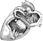
Left side of heart. Labels: 1, cavity of left auricle (atrium); 3, opening of right pulmonary veins; 5, left pulmonary veins; 5, left pulmonary veins; 6, auriculo-ventricular opening; 8, wall of left ventricle; 9, cavity of left ventricle; a, mitral valve (its flaps are attached by the chordae tendineae to the muscular pillars (b,b); d, arch of aorta); e, pulmonary artery.

Left Side of Heart

Left side of heart. Labels: 1, cavity of left auricle (atrium); 3, opening of right pulmonary veins;…
"<em>L.A.</em>, left auricle; <em>R.A.</em>, right auricle; <em>R.V.</em>, right ventricle; <em>A</em>, opening of the inferior vena cava; <em>B</em>, superior vena cava; <em>C</em>, right pulmonary veins; <em>D</em>, left pulmonary veins; <em>E</em>aorta; <em>F</em>, left branch of pulmonary artery; <em>G</em>, great cardiac vein; <em>H</em>middle cardiac vein. the cardiac lymphatics which follow the course of the cardiac veins are also shown." — Blaisedell, 1904

Posterior View of the Heart

"L.A., left auricle; R.A., right auricle; R.V., right ventricle; A,…

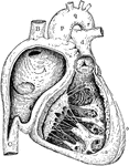
Right side of heart. Labels: A, cavity of right ventricle; B, superior vena cava; C, inferior vena cava; a, wall of right ventricle; b, c, columnae carneae; d, pulmonary vein; e, f, tricuspid valve; m, semilunar valve; o, wall of left ventricle; p, q, v, ascending aorta, arch and descending aorta.

Right Side of Heart

Right side of heart. Labels: A, cavity of right ventricle; B, superior vena cava; C, inferior vena cava;…
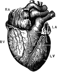
The heart. Labels: RA, right auricle, RV; right ventricle; LA, left auricle; LV, left ventricle.

The Heart

The heart. Labels: RA, right auricle, RV; right ventricle; LA, left auricle; LV, left ventricle.

The bases of the ventricle of the heart, showing the auriculoventricular, aortic, and pulmonary orifices and their valves.

Ventricle of the Heart

The bases of the ventricle of the heart, showing the auriculoventricular, aortic, and pulmonary orifices…
"<em>L.A.</em>, left ventricle; <em>B</em>, right ventricle; <em>F</em>, pulmonary artery; <em>G</em>, aorta; <em>H</em>, arch of the aorta; <em>K</em>, innominate artery; <em>L</em>, right common carotid artery; <em>M</em>, right subclavian artery; <em>N</em>, thyroid cartilage forming upper portions of the larynx; <em>O</em>, trachea." &mdash; Blaisedell, 1904

Lungs

"L.A., left ventricle; B, right ventricle; F, pulmonary artery; G,…

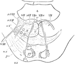
Section across the pons, about the middle of fourth ventricle. py., pyramidal bundles; po., transverse fibers passing po1, behind, po2, in front of py.; r., raphe; o.s., superior olive; a.V., bundles of ascending root of V. nerve enclosed in a prolongation of the substance of Rolando; VI., the sixth nerve, nVI., its nucleus; VII., facial nerve; VIII., auditory nerve, n.VIII., lateral nucleus of the auditory.

Section across the Pons

Section across the pons, about the middle of fourth ventricle. py., pyramidal bundles; po., transverse…
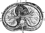
"The transverse section of the thorax. 1, anterior mediastinum; 2, internal mammary vessels; 3, triangularis sterni; 4, 5, phrenic nerves; 6, thoracic duct; 7, esophagus; 8, vena azygos major; 9, thoracic aorta; 19, sympathetic; R. V., right ventricle; R. A., right auricle; P.A., pulmonary artery; A., aorta; C., superior vena cava; V., dorsal vertebra." &mdash; Richardson, 1906

Thorax

"The transverse section of the thorax. 1, anterior mediastinum; 2, internal mammary vessels; 3, triangularis…
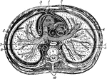
Transverse section of the thorax. Labels: 1, anterior mediastinum; 2, internal mammary vessels; 3, triangularis sterni; 4, 5, phrenic nerves; 6, thoracic duct; 7, esophagus; 8, vena azygos major; 9, thoracic aorta; 10, sympathetic; R. V., right ventricle; R. A., right auricle; P. A. pulmonary artery; A., aorta; C., superior vena cava; V., dorsal vertebra.

Transverse Section of the Thorax

Transverse section of the thorax. Labels: 1, anterior mediastinum; 2, internal mammary vessels; 3, triangularis…
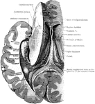
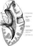
Dissection, to show the fornix and the posterior and descending cornua of the lateral ventricle of the left side.

Dissection to Show Ventricle and Fornix

Dissection, to show the fornix and the posterior and descending cornua of the lateral ventricle of the…
Dissection, to show the posterior and descending cornua of the lateral ventricle. Labels: B.G., Giacomini's band; F.D., gyrus dentatus; F, fimbria; H.C., hippocampal convolution.

Dissection to Show Ventricle

Dissection, to show the posterior and descending cornua of the lateral ventricle. Labels: B.G., Giacomini's…
A three-quarter view of the left ventricle after removal of its anterior parietes.

Left Ventricle of the Heart

A three-quarter view of the left ventricle after removal of its anterior parietes.

Transverse section through the middle of the ventricles (right and left) of a dog's heart in diastole (top) and in systole (bottom).

Transverse Section through the Ventricle of a Dog's Heart

Transverse section through the middle of the ventricles (right and left) of a dog's heart in diastole…
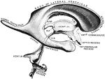
Cast of the ventricles of the brain. Labels" R.SP., recessus suprapinealis; R.P., recessus pinealis inferius; A.S., aqueduct of Sylvius; F.M., foramen of Monro.

Ventricles of the Brain

Cast of the ventricles of the brain. Labels" R.SP., recessus suprapinealis; R.P., recessus pinealis…
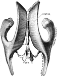
Drawing taken from a cast of the ventricular system of the brain, as seen from above. Vent. III, Third ventricle. Vent. IV, Fourth Ventricle. R.SP., recessus suprapinealis.

Ventricles of the Brain

Drawing taken from a cast of the ventricular system of the brain, as seen from above. Vent. III, Third…
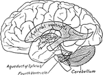
Scheme showing relations of the ventricles to the surface of the brain.

Relations of the Ventricles to the Surface of the Brain

Scheme showing relations of the ventricles to the surface of the brain.
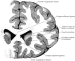
Coronal section through the frontal lobes and the anterior horns of the lateral ventricles.

Section Through Lateral Ventricles

Coronal section through the frontal lobes and the anterior horns of the lateral ventricles.

"Partial section of a Vertebrate brain (diagrammatic). OLF., Olfactory lobe; CH., cerebral hemispheres; C., wall of cerebrum cut to show ventricle, behind this the figure is that of a median section; PA., parietal organ arising from thalamencephalon; Pl., pineal organ; INF., infundibulum descending from thalamencephalon; H., hypophysis; OL., optic lobes; CB., cerebellum; CPL., choroid plexus on roof of fourth ventricle; MO., floor of the medulla oblongata; CC., central canal of spinal cord." -Thomson, 1916

Vertebrate Brain

"Partial section of a Vertebrate brain (diagrammatic). OLF., Olfactory lobe; CH., cerebral hemispheres;…
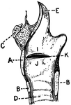
This illustration shows the vocal cords as seen from above during phonation (A. Thyroid Cartilage; B. Cricoid Cartilage; C. hyoid bone; D. Rings of trachea; E. Epiglottis; F. Arytenoid Cartilage; G. Thyro-arytenoid muscle; H. Adam's Apple; I. False Vocal Cords; J. True Vocal Cords; K. Ventricle; L. Rima Glottis).

Vocal Cords Seen from above During Phonation

This illustration shows the vocal cords as seen from above during phonation (A. Thyroid Cartilage; B.…
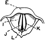
This illustration shows the vocal cords, seen from above during quiet breathing (A. Thyroid Cartilage; B. Cricoid Cartilage; C. hyoid bone; D. Rings of trachea; E. Epiglottis; F. Arytenoid Cartilage; G. Thyro-arytenoid muscle; H. Adam's Apple; I. False Vocal Cords; J. True Vocal Cords; K. Ventricle; L. Rima Glottis).

Vocal Cords Seen from above During Quiet Breathing

This illustration shows the vocal cords, seen from above during quiet breathing (A. Thyroid Cartilage;…