The Mammal Anatomy: Skeleton ClipArt gallery provides 277 views of bones, teeth, and skeletal system of various mammals.
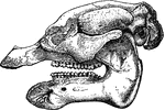
"Skull of African Manatee (Manatus senegatensis)." —The Encyclopedia Britannica, 1903

African Manatee

"Skull of African Manatee (Manatus senegatensis)." —The Encyclopedia Britannica, 1903
Anoplotherium, an extinct hoofed mammal.

Anoplotherium

Anoplotherium, an extinct hoofed mammal.

The skull of the Great Anteater (Myrmecophaga tridactyla).

Great Anteater Skull

The skull of the Great Anteater (Myrmecophaga tridactyla).

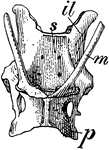
An illustration of the pelvis of a spiny anteater. "m, marsupial bones; il, ilium; p, pubis; s, sacrum." -Century, 1889

Pelvis of Spiny Anteater

An illustration of the pelvis of a spiny anteater. "m, marsupial bones; il, ilium; p, pubis; s, sacrum."…

The side view and base of the skull of an ape.

Ape Skull

The side view and base of the skull of an ape.

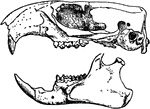
"Skull of Arciomys Monax." —The Encyclopedia Britannica, 1903

Arciomys Monax

"Skull of Arciomys Monax." —The Encyclopedia Britannica, 1903

An illustration of a pichiciago, a small burrowing armadillo. The front half of the animal is covered in the endoskeleton. The back half of the animal is stripped of the outer skeleton so that the exoskeleton or dermoskeleton can be viewed. The scientific name of the pichiciago is <i>Chlamydophorus truncatus</i>.

Armadillo - Endoskeleton and Exoskeleton or Dermoskeleton

An illustration of a pichiciago, a small burrowing armadillo. The front half of the animal is covered…

"Bones of fore foot of existing Artiodactyle. Pig (Sus scrofa)." &mdash;The Encyclopedia Britannica, 1903

Artiodactyles

"Bones of fore foot of existing Artiodactyle. Pig (Sus scrofa)." —The Encyclopedia Britannica,…

"Bones of fore foot of existing Artiodactyle. Red Deer (Cervus elaphus)." &mdash;The Encyclopedia Britannica, 1903

Artiodactyles

"Bones of fore foot of existing Artiodactyle. Red Deer (Cervus elaphus)." —The Encyclopedia Britannica,…
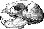
"Skull of the Aye-aye (Chiromys madagascariensis)." &mdash;The Encyclopedia Britannica, 1910

Aye-Aye Skull

"Skull of the Aye-aye (Chiromys madagascariensis)." —The Encyclopedia Britannica, 1910
Skull of a baboon, showing the brain cavity.

Baboon skull

Skull of a baboon, showing the brain cavity.

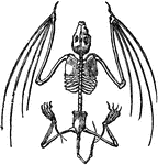
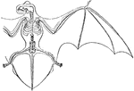
Skeleton of a bat.

Bat Skeleton

Skeleton of a bat.

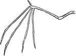
The skeleton of a bat's wing.

Bat Wing

The skeleton of a bat's wing.

Skeleton and outline of bat.

Horseshoe Bat

Skeleton and outline of bat.

The bat genus Nyctalus (Noctule bats) are Evening bats. They are distributed in the temperate and subtropical areas of Europe, Asia and North Africa.

Skeleton and Wing Membranes of the Noctule Bat

The bat genus Nyctalus (Noctule bats) are Evening bats. They are distributed in the temperate and subtropical…

"Palmar aspect of left fore foot of a black bear (Ursus americanus). scl, scapholunar; c, cuneiform; p, pisiform; tr, trapezium; t, trapezoid; m, magnum; u, unciform. The phalanges show a full series of sesamoid bones (unmarked)." -Whitney, 1911

Bear Foot

"Palmar aspect of left fore foot of a black bear (Ursus americanus). scl, scapholunar; c, cuneiform;…

This illustration shows the plantigrade leg of a bear. Plantigrade means that the animal walks flat on its foot.
P. Pelvis, FE. Femur, TI. Tibia, FI. Fibula, TA. Tarsus, MT. Metatarsus, PH. Phalanges, OC. Os Calcis

Leg of Bear

This illustration shows the plantigrade leg of a bear. Plantigrade means that the animal walks flat…

Close-up illustration of scalpriform incisor of a beaver. It is "chisel-shaped; having the character of a chisel-tooth; truncate at the end and beveled there to a sharp edge; specifically said of the incisor teeth of rodents, and the similar teeth of a few other mammals." &mdash;Whitney, 1889

Scalpriform, Left Lower Incisor of a Beaver

Close-up illustration of scalpriform incisor of a beaver. It is "chisel-shaped; having the character…

The skull of a beaver.

Beaver Skull

The skull of a beaver.

"Skull and teeth of Bennett's Kangaroo (Macropus bennettii). i1, i2, i3, first second and third upper incisors; pm, second or posterior premolar (the first having been already shed); m1, m2, m3, m4, the four true molars. The last, not fully developed, is nearly concealed by the ascending ramus of the jaw." &mdash;The Encyclopedia Britannica, 1910

Bennett's Kangaroo

"Skull and teeth of Bennett's Kangaroo (Macropus bennettii). i1, i2, i3, first second and third upper…

Bone

Bone

Bone

A firm, hard, whitish substance, composing the skeleton.

Bone

A firm, hard, whitish substance, composing the skeleton.

Homology of digits of odd toed mammals, showing gradual reduction in number and consolidation of bones. This figure displays a coney showing five digits.

Bones of Coney's Foot

Homology of digits of odd toed mammals, showing gradual reduction in number and consolidation of bones.…

Homology of digits of odd toed mammals, showing gradual reduction in number and consolidation of bones. This figure displays an elephant foot showing five digits.

Bones of Elephant's Foot

Homology of digits of odd toed mammals, showing gradual reduction in number and consolidation of bones.…

Homology of digits of odd toed mammals, showing gradual reduction in number and consolidation of bones. This figure displays a horse showing one digit.

Bones of Horse's Foot

Homology of digits of odd toed mammals, showing gradual reduction in number and consolidation of bones.…
Homology of digits of odd toed mammals, showing gradual reduction in number and consolidation of bones. This figure displays a rhinoceros showing three digits.

Bones of Rhonoceros' Foot

Homology of digits of odd toed mammals, showing gradual reduction in number and consolidation of bones.…

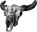
A buffalo skull

Buffalo

A buffalo skull

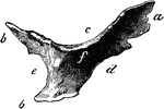
Right cardiac bone of an ox. Labels: a, anterior angle; b, posterior angles; c, superior border; d, anterior border; e, posterior border; f, right surface.

Cardiac Bone of an Ox

Right cardiac bone of an ox. Labels: a, anterior angle; b, posterior angles; c, superior border; d,…
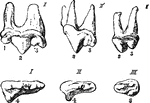
"Upper Sectorial Teeth of Carnivora. I, Felis; II, Canis; III, Ursus, 1, anterior, 2, middle, and 3, posterior in position, and without distinct root, characteristic of the Ursidae." &mdash;The Encyclopedia Britannica, 1903

Carnivora

"Upper Sectorial Teeth of Carnivora. I, Felis; II, Canis; III, Ursus, 1, anterior, 2, middle, and 3,…
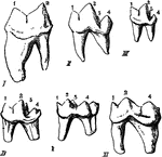
"Modifications of the Lower Sectorial Tooth in Carnivora. I, Felis; II, Cunis; III, Herpestes; IV, Lutra; V, Meles; VI, Ursus. 1, anterior lobe of blade; 2, posterior lobe of blade; 3, inner tubercle; 4, heel. It will be seen that the relative size of the two roots varies according to the development of the portion of the crown they have respectively to support." &mdash;The Encyclopedia Britannica, 1903

Carnivora

"Modifications of the Lower Sectorial Tooth in Carnivora. I, Felis; II, Cunis; III, Herpestes; IV, Lutra;…

The teeth of a carnivorous animal that lives on flesh alone. The front teeth are tearing ones, while the back teeth have sharp edges for cutting. The flesh is first torn by the front teeth, and then it is cut up by the back ones.

Carnivorous Skeleton

The teeth of a carnivorous animal that lives on flesh alone. The front teeth are tearing ones, while…

Front aspect of the right carpus of a horse. Labels: 1, cuneiform; 2, lunar; 3, scaphoid; 4, trapezium; 5, unciform; 6, magnum; 7, trapezoid; 8, small metacarpals, the large one lying between and before them. The uppermost bone is the distal end of the radius.

Carpus of a Horse

Front aspect of the right carpus of a horse. Labels: 1, cuneiform; 2, lunar; 3, scaphoid; 4, trapezium;…

Deep ligaments of the carpus- external view. Labels: a and b, deep portions of the external lateral ligament; c, superior, d, median, and e, inferior trapezial ligaments.

Deep Ligaments of the Carpus

Deep ligaments of the carpus- external view. Labels: a and b, deep portions of the external lateral…

Ligaments of the carpus-anterior aspect. Labels: a, internal lateral ligament; b, external lateral ligament; c, anterior carpal ligaments; d, anterior carpo-metacarpal ligaments.

Ligaments of the Carpus

Ligaments of the carpus-anterior aspect. Labels: a, internal lateral ligament; b, external lateral ligament;…

Ligaments of the carpus- postero-internal view. Label: a, b, deep portions of internal lateral ligament; c, oblique ligament; d, interosseous metacarpal ligament; e, external interosseous ligament of the upper row.

Ligaments of the Carpus

Ligaments of the carpus- postero-internal view. Label: a, b, deep portions of internal lateral ligament;…

Superficial ligaments of the carpus- posterior view. Labels: a, posterior annular ligament; b, posterior common ligament.

Superficial Ligaments of the Carpus

Superficial ligaments of the carpus- posterior view. Labels: a, posterior annular ligament; b, posterior…

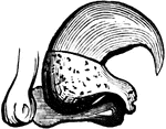
Cat's claw with theflexor muscle contracted.

Cat Claw

Cat's claw with theflexor muscle contracted.

Cat's claw with theflexor muscle extended.

Cat Claw

Cat's claw with theflexor muscle extended.

"Skull of Cat (Felis catus), showing the following bones, viz. : na, nasal; pm, premaxillary; m, maxillary; l, lacrymal; f, frontal; j, jugal; pa, palatine; p, parietal; sq, squamosal; ip, interparietal; so, supra-occipital; eo, exoccipital (the line leads to the occipital condyle); t, tympanic bulla; smf, stylomastoid foramen; mf, mental foramen; c, coronoid process of mandible; ar, ascending ramus of mandible; hr, horizontal ramus of mandible; an, angle of jaw." -Whitney, 1911

Domestic Cat Skull

"Skull of Cat (Felis catus), showing the following bones, viz. : na, nasal; pm, premaxillary; m, maxillary;…
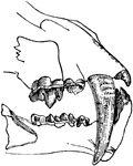
A cat having extremely long upper canine teeth.

Saber-Toothed Cat

A cat having extremely long upper canine teeth.

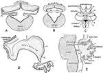
Showing the development of the cerebellum. A, Transverse section through the forepart of the cerebellum of a sheep embryo. B, Transverse section through the hinder part of the cerebellum of a sheep embryo. C, Cerebellum of a human fetus 17 cm long. D, Median section through cerebellum of sheep embryo 5 cm long.E, Median section through cerebellum of human fetus 17 cm long. Labels: 1, sulcus primarius; 2, sulcus above and afterwards behind the pyramid; 3, sulcus between pyramid and uvula; 4, great horizontal fissure; r.l., lateral recess ventricle IV; T, transverse groove in roof of ventricle IV.

Development of Cerebellum

Showing the development of the cerebellum. A, Transverse section through the forepart of the cerebellum…

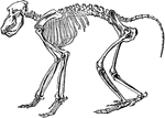
"The skeleton of the Chacma Baboon." &mdash; Encyclopedia Britanica, 1893

Chacma Baboon

"The skeleton of the Chacma Baboon." — Encyclopedia Britanica, 1893

The clavicle of a cat.

Clavicle of a Cat

The clavicle of a cat.

The clavicle of a dog.

Clavicle of a Dog

The clavicle of a dog.

Skeleton of the cow. 1: Frontal bone of the head. 2: Upper jaw, superior maxillary. 3: Lower jaw, inferior maxillary. 4: Cervical vertebrae. 5: Dorsal vertebrae. 6: Lumbar vertebrae. 7: Sacral vertebrae. 8: Caudal vertebrae. 9: Scapula. 10: Humerus 11: Radius and ulna. 12: Carpus. 13: Metacarpus. 14: Phalanges (toes). 15: Femur. 16: Tibia. 17: Tarsus. 18: Metatarsus. 19: Phalanges.

Cow Skeleton

Skeleton of the cow. 1: Frontal bone of the head. 2: Upper jaw, superior maxillary. 3: Lower jaw, inferior…
A skeleton of a cow shown to illustrate the internal skeleton which all vertebrates share. Labels: 1, Frontal bone of the head. 2, Upper jaw (superior maxillary). 3. Lower jaw (inferior maxillary). 4, Cervical vertebrae. 5, Dorsal vertebrae. 6, Lumbar vertebrae. 7 Sacral vertebrae. 8, Caudal vertebrae. 9; Scapula. 10, Humerus. 11, Radius and ulna. 12, Carpus. 13, Metacarpus. 14, Phalanges (toes). 15, Femur. 16, Tibia. 17, Tarsus. 18, Metatarsus. 19, Phalanges.

Skeleton of a Cow

A skeleton of a cow shown to illustrate the internal skeleton which all vertebrates share. Labels: 1,…
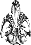
"Under Side of Skull of Cynomys Ludovicianus." &mdash;The Encyclopedia Britannica, 1903

Cynomys Ludovicianus

"Under Side of Skull of Cynomys Ludovicianus." —The Encyclopedia Britannica, 1903
Skull of a deer, showing the brain cavity.

Deer skull

Skull of a deer, showing the brain cavity.

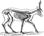
"The bones in the extremities of this the fleetest of quadrupeds are inclined very obliquely towards each other, and towards the scapular and iliac bones. This arrangement increases the leverage of the muscular system and confers great rapidity on the moving parts."&mdash;Pettigrew, 1874

Skeleton of the Deer

"The bones in the extremities of this the fleetest of quadrupeds are inclined very obliquely towards…

"Skull and Dentition of Dendrohyrax Dorsalis." &mdash;The Encyclopedia Britannica, 1903

Dendrohyrax Dorsalis

"Skull and Dentition of Dendrohyrax Dorsalis." —The Encyclopedia Britannica, 1903
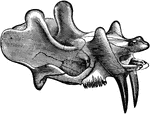
"Restoration of Dinoceras Mirabile." &mdash;The Encyclopedia Britannica, 1903

Dinoceras Mirabile

"Restoration of Dinoceras Mirabile." —The Encyclopedia Britannica, 1903

The skull and upper jaw of an early rhinoceros-like mammal from the Cenozoic time.

Dinoceras Mirabile

The skull and upper jaw of an early rhinoceros-like mammal from the Cenozoic time.
A restored skeleton of <em>Uintathcrium (Dinoceras) mirabile.</em>

Dinoceras Skeleton

A restored skeleton of Uintathcrium (Dinoceras) mirabile.

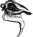
"Skull of Dinotherium Giganteum." &mdash;The Encyclopedia Britannica, 1903

Dinotherium Giganteum

"Skull of Dinotherium Giganteum." —The Encyclopedia Britannica, 1903

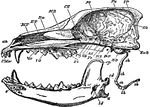
"Longitudinal and Vertical section of the skull of a dog, with mandible and hyoid arch. an, anterior narial aperture; MT, maxillo-turbinal bone; ET, the ethmo-turbinal; Fr, frontal; Pa, pariental; IP, interparietal; SO, supra-occipital; ExO, sphenoid; OS, orbito-sphenoid; PS, presphenoid; Pl, palatine; Vo, vomer; Mx, maxilla; PMx, premaxilla; sh, stylo-hyal; eh, epi-hyal; ch, cerato-hyal; bh, basihyal; th, thyro-hyal; s, symphysis of mandible; cp, coronoid process; cd, condyle; a, angle; id, inferior dental canal. The mandible is displaced downwards, to show its entire form; the * indicated the part of the cranium to which the condyle is articulated." &mdash;The Encyclopedia Britannica, 1903

Dog Skull

"Longitudinal and Vertical section of the skull of a dog, with mandible and hyoid arch. an, anterior…
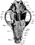
"Lower surface of dog's skull. o.c., Occipital condyle; B.O., basioccipital; T., tympanic bulla; m.c., postglenoid process behind fossa for condyle of mandible; B.S., basisphenoid; P.S., base of presphenoid; V., vomer; M.2, second molar; M.1, first molar; Pm. 1-4, premolars, the 4th the large carnassial; c., canine; I.1-3, incisors; Pmx., premaxilla; mx., maxilla; Pal., palatine; J., jugal; A.S., alisphenoid; Pt., pterygoid; Sq., squamosal (the reference line points to the glenoid fossa)." -Thomson, 1916

Dog Skull

"Lower surface of dog's skull. o.c., Occipital condyle; B.O., basioccipital; T., tympanic bulla; m.c.,…
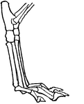
This illustration shows the leg of a dog. This leg is digitigrade. Animals with digitigrade legs walk on their toes (digits).
P. Pelvis, FE. Femur, TI. Tibia, FI. Fibula, TA. Tarsus, MT. Metatarsus, PH. Phalanges, OC. Os Calcis

Leg of Dog

This illustration shows the leg of a dog. This leg is digitigrade. Animals with digitigrade legs walk…

"Upper and lower teeth of one side of the mouth of a dolphin, as an example of homodont type of dentition. The bone covering the outer side of the roots of the teeth has been removed to show the simple character." &mdash;The Encyclopedia Britannica, 1903

Dolphin Teeth

"Upper and lower teeth of one side of the mouth of a dolphin, as an example of homodont type of dentition.…

"Skeleton of Dugong: In which the pelvis is a mere bony ring." &mdash; Chambers, 1881

Dugong Skeleton

"Skeleton of Dugong: In which the pelvis is a mere bony ring." — Chambers, 1881

The skeleton of a dugong, one of four living species in the order Sirenia.

Skeleton of the Dugong

The skeleton of a dugong, one of four living species in the order Sirenia.