This human anatomy ClipArt gallery offers 240 illustrations of the human digestive system. This includes views of the gastrointestinal tract and organs involved in the breakdown and absorption of food. Included in this category are the oral cavity, salivary glands, stomach, esophagus, intestines, colon, and gallbladder.

The stomach and intestines. Labels: 1, stomach; 2, duodenum; 3, small intestine; 4, termination of the ileum; 5, caecum; 6, vermiform appendix; 7, ascending colon; 8, transverse colon; 9, descending colon; 10, sigmoid flexure of the colon; 11, rectum; 12, spleen.

The Stomach and Intestines

The stomach and intestines. Labels: 1, stomach; 2, duodenum; 3, small intestine; 4, termination of the…

The Stomach and Liver. 1: Esophagus; 2: Cardiac entrance; 3: Large end of stomach; 4: Central portion; 5: Small end of stomach; 6: Pylorus; 7: Portion of Pancreas; 8: Duodenum; 9: Gall duct; 10: Hepatic artery; 11: Left lobe of liver; 12: Gall bladder; 13: Right lobe of liver.

Stomach and Liver

The Stomach and Liver. 1: Esophagus; 2: Cardiac entrance; 3: Large end of stomach; 4: Central portion;…

The stomach chamber and stomach bed shown after stomach has been removed.

Stomach Chamber

The stomach chamber and stomach bed shown after stomach has been removed.

Radiographic outline of the stomach of a patient who has taken a bismuth-laden diet.

Stomach in a Bismuth-Laden Diet

Radiographic outline of the stomach of a patient who has taken a bismuth-laden diet.

The three layers of the muscular coat of the stomach. A, Outer or longitudinal layer. B, Middle or circular layer. C, Internal or oblique layer. Labels: a, longitudinal fibers of esophagus; b, superficial circular fibers of esophagus passing into circular fibers of stomach in B; c, deep circular fibers of esophagus passing into oblique fibers of stomach in C; d, oblique fibers forming rings at the fundus; e, submucosa.

Stomach Muscles

The three layers of the muscular coat of the stomach. A, Outer or longitudinal layer. B, Middle or circular…

Stomach turned inside out, showing dissection of oblique and circular muscular coats.

Stomach Turned Inside Out

Stomach turned inside out, showing dissection of oblique and circular muscular coats.
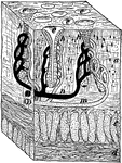
Wall of human stomach. Labels: E, epithelium; G, glands; Mm, muscularis mucosae.

Stomach Wall

Wall of human stomach. Labels: E, epithelium; G, glands; Mm, muscularis mucosae.

"A tiny block out of the stomach wall. a, the mucous membrane; c and d, the muscles; h, gastric glands; m and n, blood tubes to the glands; e, mouths of glands within the stomach." —Davison, 1910

Cross-Section of Stomach Wall

"A tiny block out of the stomach wall. a, the mucous membrane; c and d, the muscles; h, gastric glands;…
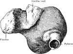
Section through wall of stomach, cardiac portion.

Section Through Stomach Wall

Section through wall of stomach, cardiac portion.

Stomach with puckered fundus, seen from behind and somewhat from left; hardened by formalin.

Stomach with Puckered Fundus

Stomach with puckered fundus, seen from behind and somewhat from left; hardened by formalin.
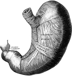
The middle and deep muscular layer of the stomach, viewed from above and in front.

Deep Muscles of the Stomach

The middle and deep muscular layer of the stomach, viewed from above and in front.

Moderately distended stomach, viewed A, from in front; B, from inner or right side; and C, from the outer or left side. The contents of the stomach were carefully removed, through an artificial opening , and replaced with gelatin, the stomach remaining in situ throughout the operation. After the jelly hardened, its exact orientation was carefully noted, and pins indicating the vertical, horizontal, and transverse planes, having been inserted, the organ was removed.

Distended Stomach

Moderately distended stomach, viewed A, from in front; B, from inner or right side; and C, from the…
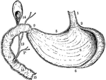
Vertical and longitudinal section of stomach, gall-bladder, and duodenum. Labels: 1, esophagus; 2, cardiac orifice of stomach; 5, lesser curvature; , greater curvature; 8, rugae in interior of stomach; 9, pyloric orifice; 10, 11, 13, interior of duodenum, showing valvulae conniventes; 12, duct conveying bile, and P, duct conveying pancreatic juice, into the duodenum; 14, gall-bladder; 15, commencement of jejunum.

Vertical and Longitudinal Section of Stomach, Gall-Bladder, and Duodenum

Vertical and longitudinal section of stomach, gall-bladder, and duodenum. Labels: 1, esophagus; 2, cardiac…
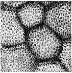
"The Inner Surface of the Stomach, from which the the Epithelium has been removed, showing the Openings of Gastric Glands." — Blaisedell, 1904

Inner surface of the stomach

"The Inner Surface of the Stomach, from which the the Epithelium has been removed, showing the Openings…
The interior of the stomach.

Interior of the Stomach

The interior of the stomach.

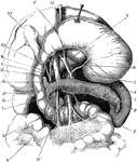
The stomach, pancreas, liver, and duodenum, with part of the rest of the small intestine and the mesentery; the stomach and liver have been turned up so as to expose the pancreas. Labels: V, stomach; D, D',D'', duodenum; L, spleen; P, pancreas; R, right kidney; T, jejunum; Vf, gall bladder; h, hepatic duct; c, cystic duct; ch, common bile duct; 1, aorta; 2, an artery (left coronary) of the stomach; 3, hepatic artery; 4, splenic artery; 5, superior mesenteric artery; 6, superior mesenteric vein; 7, splenic vein; Vp, portal vein.

The Stomach, Pancreas, Liver, and Duodenum

The stomach, pancreas, liver, and duodenum, with part of the rest of the small intestine and the mesentery;…
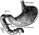
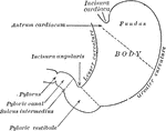
Outlines of the stomach, showing its anatomical landmarks.

Parts of the Stomach

Outlines of the stomach, showing its anatomical landmarks.

The opening part of the stomach where the esophagus joins it is called the cardiac opening; the one at the opposite end, where the small intestine joins it, is called the pylorus.

A Section of the Stomach

The opening part of the stomach where the esophagus joins it is called the cardiac opening; the one…
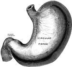
The superficial muscular layer of the stomach, viewed from above and in front.

Superficial Muscles of the Stomach

The superficial muscular layer of the stomach, viewed from above and in front.
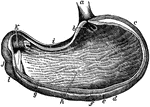
The stomach, the principal organ of digestion. It is a dilated part of the alimentary canal, situated between the termination of the esophagus and the beginning of the small intestine.

The Stomach

The stomach, the principal organ of digestion. It is a dilated part of the alimentary canal, situated…
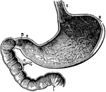
The inside of the stomach with the beginning of the intestines. At 3 is the left end and at 4 is the right end. At 1 is the opening of the gullet into the stomach. At 5 is a valve (the pylorus) which is sometimes shut, so as to prevent anything from passing from the stomach into the intestines. 8 indicates the inside lining of the stomach which produces gastric juices. 6 shows the duct from the liver.

The Stomach

The inside of the stomach with the beginning of the intestines. At 3 is the left end and at 4 is the…
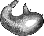
The stomach. Labels: d, lower end of the gullet; a, position of the cardiac aperture; b, the fundus; c, the pylorus; e, the commencement of the small intestine; along a, b, c, the great curvature; between the pylorus and d, the lesser curvature.

The Stomach

The stomach. Labels: d, lower end of the gullet; a, position of the cardiac aperture; b, the fundus;…
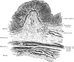
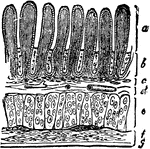
Transverse section of stomach (left end), showing general arrangement of coats.

Transverse Section of Stomach

Transverse section of stomach (left end), showing general arrangement of coats.
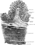
Transverse section of stomach, pyloric end; ruga is cut across, showing mucosa supported by core of submucous

Transverse Section of Stomach

Transverse section of stomach, pyloric end; ruga is cut across, showing mucosa supported by core of…
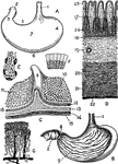
Views of the stomach. Labels: A. stomach (human). B. Same, anterior wall removed. C. Portion of stomach, pylorus and duodenum. D. Section through coats of stomach. E. Fundus of a cardiac gland. F. Epithelium from surface of stomach. G. Arterie sand veins of mucous membrane. w. Esophagus. 2. Duodenum. 3. Pylorus. $. Cardia. 5. Lesser curvature. 6. Greater curvature. 7. Anterior surface. 8. Pyloric valve. 9. Mucous membrane, with rugae. 10. Inner surface of gastric mucous membrane. 11. Mucous membrane of duodenum with villi. 12. Pyloric gastric glands. 13. Circular or deep muscular fibers. 17. Mucous membrane. 18. Muscularis mucosae. 19. Submucous coat. 20. Circular muscular layer. 21. Longitudinal muscular layer. 22. Serous coat. 23. Orifice of gland. 24. Fundus of gland.

Views of the Stomach

Views of the stomach. Labels: A. stomach (human). B. Same, anterior wall removed. C. Portion of stomach,…

The muscles involved in swallowing food and water.

Swallowing

The muscles involved in swallowing food and water.

"The circular muscle of the mouth (1) and the buccinator or trumpeter's muscle (2) help the tongue to push the food back to the upper margin of the gullet, where it is seized upon by the three constrictor muscles (3, 4 and 5) of the pharynx, and pushed down the gullet or esophagus, which is represented as being cut off at 6." — Richardson, 1906

Swallowing Muscles

"The circular muscle of the mouth (1) and the buccinator or trumpeter's muscle (2) help the tongue to…

The Thoracic Duct and Lacteals. 1: Mouth of thoracic duct; 2: Lower end of duct; 3: Mesenteries; 4: Lacteals; 5: Intestine.

Thoracic Duct

The Thoracic Duct and Lacteals. 1: Mouth of thoracic duct; 2: Lower end of duct; 3: Mesenteries; 4:…

"The lacteals conduct the chyle from the intestines into numerous glands nearby, called the mesenteries, from which a fewer number of lacteals lead the chyle into one large tube in front of the spinal column, called the thoracic duct." —Brown, 1884

Thoracic duct and lacteals

"The lacteals conduct the chyle from the intestines into numerous glands nearby, called the mesenteries,…
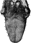
<em>A</em>, epiglottis; <em>B</em>, glands at the base of tongue; <em>C</em>, tonsil; The various kinds of papillae are plainly shown." &mdash; Blaisedell, 1904

Tongue

A, epiglottis; B, glands at the base of tongue; C, tonsil; The various kinds…

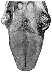
The upper surface of the tongue. Labels: 1,2, circumvallate papillae; 3, fungiform papillae; 4, filiform papillae; 6, mucous glands.

Tongue

The upper surface of the tongue. Labels: 1,2, circumvallate papillae; 3, fungiform papillae; 4, filiform…
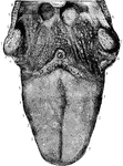
The papillar surface of the tongue, with the fauces and tonsils. Labels: 1, circumvallate papillae, in front of 2, the foramen caecum; 3, fungiform papillae; 4, filiform and conical papillae; 5, transverse and oblique rugae; 6, mucous glands at the base of the tongue and in the fauces; 7, tonsils; 8, part of the epiglottis; 9, median glosso-epiglottidean fold (fraenum epiglottidis).

Tongue

The papillar surface of the tongue, with the fauces and tonsils. Labels: 1, circumvallate papillae,…
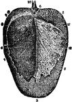
View of the back of the tongue, from which, by maceration, the periglottis has been removed and turned back on the right side.

Back View of the Tongue

View of the back of the tongue, from which, by maceration, the periglottis has been removed and turned…
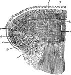
Coronal section of the tongue. Showing intrinsic muscles. Labels: a, Lingual artery; b, Inferior lingualis, cut through; c, Fibers of Hyoglossus; d, Oblique fibers of Styloglossus; e, Insertion of Transverse lingualis; f, Superior lingualis; g, Papillae of the tongue; h, Vertical fibers of Geniohyglossus intersecting Transverse lingualis; i, Septum.

Coronal Section of the Tongue

Coronal section of the tongue. Showing intrinsic muscles. Labels: a, Lingual artery; b, Inferior lingualis,…

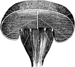
View of a section of the front part of the tongue, seen from behind.

Section of the Tongue

View of a section of the front part of the tongue, seen from behind.

A, Transverse vertical section through the tongue. B, Longitudinal vertical section through the tongue. C, A lymphoid follicle from back part of tongue.

Sections of the Tongue

A, Transverse vertical section through the tongue. B, Longitudinal vertical section through the tongue.…
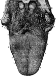
The upper surface of the tongue with part of the pillars of the fauces and the tonsils. Labels: 1, 2, circumvallate papillae; 3, fungiform papillae; 4, filiform papillae; 6, mucous glands; 7, tonsils; 8, tip of epiglottis.

Upper Surface of the Tongue

The upper surface of the tongue with part of the pillars of the fauces and the tonsils. Labels: 1, 2,…

Front view of the upper surface of the tongue; as also of the arch of the bone of the palate.

Upper Surface of the Tongue

Front view of the upper surface of the tongue; as also of the arch of the bone of the palate.
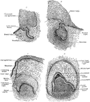
Frontal section showing four earl stages of tooth development.

Frontal Section Showing Tooth Development

Frontal section showing four earl stages of tooth development.

"Longitudinal section of a tooth, semi-diagrammatic. PH, pulp cavity; PH', opening of same; ZB, dentine; ZC, cement; ZS, enamel." -Parker, 1900

Tooth Section

"Longitudinal section of a tooth, semi-diagrammatic. PH, pulp cavity; PH', opening of same; ZB, dentine;…

A bicuspid tooth seen from its outer side; the inner cusp is, accordingly, not visible.

Bicuspid Tooth

A bicuspid tooth seen from its outer side; the inner cusp is, accordingly, not visible.

A canine or eyetooth.

Canine Tooth

A canine or eyetooth.

An incisor tooth.

Incisor Tooth

An incisor tooth.

A molar tooth.

Molar Tooth

A molar tooth.

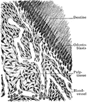
Section of periphery of pulp tissue of young tooth.

Section of Pulp Tissue of Tooth

Section of periphery of pulp tissue of young tooth.

Section through the abdomen, an inch and a half above the umbilicus. The liver is unusually large in this individual.

Cross Section of the Trunk Above Umbilicus

Section through the abdomen, an inch and a half above the umbilicus. The liver is unusually large in…

Diagram of the relations of the large intestine and kidneys, from behind.

Trunk Showing Organs of Digestion

Diagram of the relations of the large intestine and kidneys, from behind.

Section through the liver and stomach, at the level of the xiphoid process.

Cross Section of the Trunk Through the Liver and Stomach

Section through the liver and stomach, at the level of the xiphoid process.
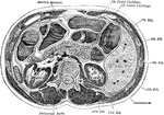
Transverse section through the middle of the first lumbar vertebra, showing the relations of the pancreas.
Transverse Section of the Trunk

Transverse section through the middle of the first lumbar vertebra, showing the relations of the pancreas.

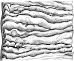
The valvulae conniventes are large folds or valvular flaps projecting into the lumen of the bowel. They are composed of reduplications or folds of the mucous membrane, the two layers of the fold being bound together by submucous tissue, they contain no muscular fibers, and, unlike the folds in the stomach, they are permanent, and not obliterated when the intestine is distended. Shown is valvulae conniventes is the upper part of the small intestine.

Valvulae Conniventes

The valvulae conniventes are large folds or valvular flaps projecting into the lumen of the bowel. They…
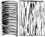
Valvulae Conniventes (folds) of the intestines.

Valvulae Conniventes of the Intestines

Valvulae Conniventes (folds) of the intestines.

Valvule conniventes. A, as seen in a bit of jejunum which has been filled with alcohol and hardened. B, a portion of fresh intestine spread out under water.

Valvule Conniventes

Valvule conniventes. A, as seen in a bit of jejunum which has been filled with alcohol and hardened.…

Piece of the small intestine cut open to show wrinkling of inner coat bearing villi.

Villi of Small Intestine

Piece of the small intestine cut open to show wrinkling of inner coat bearing villi.
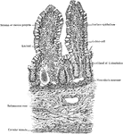
Transverse section of small intestine (jejunum), showing villi cut lengthwise.

Transverse Section of Villi of Small Intestine

Transverse section of small intestine (jejunum), showing villi cut lengthwise.
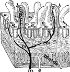
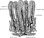
A tiny block cut from the wall of the intestine showing villi. Labels: a, mouths of glands; b, villus cut open; e, lacteal; m, blood tubes to absorb food.

Villi of the Intestine

A tiny block cut from the wall of the intestine showing villi. Labels: a, mouths of glands; b, villus…
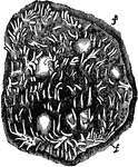
Villi of the small intestine. Villa are minute, vascular processes which project from the mucous membrane of the small intestine throughout its whole extend, giving to its surface a velvety appearance.

Villi of the Small Intestine

Villi of the small intestine. Villa are minute, vascular processes which project from the mucous membrane…
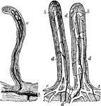
Villi of the small intestine; magnified about 80 diameters. In the right hand figure the lacteals, a, b, c, are filled with white injection; d, blood vessels. In the left-hand figure the lacteals alone are represented, filled with a dark injection. The epithelium covering the villi, and their muscular fibers, are omitted.

Villi of the Small Intestine

Villi of the small intestine; magnified about 80 diameters. In the right hand figure the lacteals, a,…
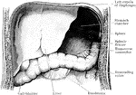
Vill of the small intestine.

Villi of the Small Intestine

Vill of the small intestine.

Villi of small intestine.

Villi of the Small Intestine

Villi of small intestine.

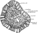
Transverse section of single intestinal villus, showing relation of epithelium, stroma, and vessels.
Transverse Section of Villus of Small Intestine

Transverse section of single intestinal villus, showing relation of epithelium, stroma, and vessels.