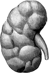
This human anatomy ClipArt gallery offers 82 illustrations of the human excretory system, including views of the systems involved in excreting waste that are not already included in the respiratory and digestive systems. Included here are the urinary tract, renal system (e.g., kidneys), and excretory glands of the skin (e.g., sebaceous and sweat glands).

Bladder
View looking into the pelvis from above and somewhat behind. The bladder has been artificially distended.
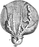
Bladder and Prostate Gland
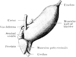
Dissection of the base of the bladder and prostate gland, showing the vesiculae seminales and vasa deferentia.…
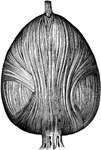
Fibers of the Bladder
The figure on the left shows fibers of the external longitudinal layer. The middle figure shows fibers…
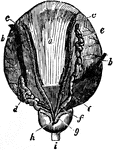
The Bladder
The bladder, a reservoir for urine, is a musculo-membranous sac, situated n the anterior portion of…

Under Aspect of Male Bladder
The under aspect of the empty male bladder from a subject in which the viscera has been hardened in…
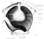
View of Male Pelvis Showing Bladder
View looking into the male pelvis from above and somewhat behind. From a specimen in which the bladder…
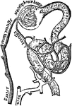
Plan of the Blood Vessels Connected with the Tubules
Plan of the blood vessels connected with the tubules. "The blood passes downwards in straight vessels…

Plan of the Blood Vessels Connected with the Tubules
Diagram of the course of two uriniferous tubules. Labels: M, Malpighian capsule, or dilated extremity,…

Epidermal Section
A section through the epidermis, somewhat diagrammatic, highly magnified. Below is seen a papilla of…

Epidermis and Sweat Gland
A section through the epidermis, highly magnified. Labels: Below is seen a papilla of the dermis, with…
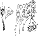
Epithelium of the Bladder
Epithelium of the bladder. Labels: a, one of the cells of the first row; b, a cell of the second row;…

Deep Layer of Bladder Epithelium
Deep layers of the epithelium of the bladder, showing large club-shaped cells above, and smaller, more…
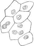
Superficial Layer of Bladder Epithelium
Superficial layer of the epithelium of the bladder. Composed of polyhedral cells of various sizes, each…

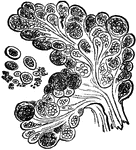
Compound Racemose Gland
Compound racemose gland. The resemblance to a bunch of fruit is very marked.

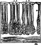
Compound Tubular Gland
Compound tubular gland. The upper part is the duct; the lower is the secreting portion.
Racemose Gland
A racemose gland, which is a gland where the ducts are branched and clustered like grapes.
Simple Gland
A simple secreting gland. Secreting glands serve to secrete (i.e., separate out some substance from…
Development of Glands
Diagram showing development of glands: A, a mere dimple in the surface; B, enlargement by division;…
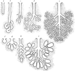
Types of Glands
Diagram showing types of glands. a-e, tubular; f-i, alveolar or saccular; a, simple; b, coiled; c-d,…

Kidney
Two glands having the function of secreting urine from the system, situated at the back of the abdominal…
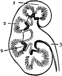
Kidney
Section of Kidney. 1: Body of Kidney; 2: Internal vessels; 3: Ureter, leading to the bladder.
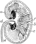
Kidney
Transverse section of the human kidney: "(a) cortex; (b) medulla; (c) small branch of the renal artery;…
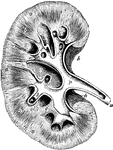
Kidney
Plan of a longitudinal section through the pelvis and substance of the right kidney. Labels: a, the…
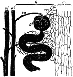
Kidney Circulation
Circulation in the kidney. Labels: ai, small branch of renal artery giving off the branch va, which…
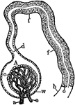
Kidney Glomerulus and Uriniferous Tubule
Diagram showing a kidney glomerulus and the commencement of an uriniferous tubule. Labels: a, afferent…
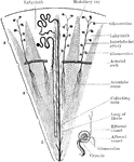
Structure of Kidney Lobe
Diagrammatic representation of the structure forming a kidney lobe. In the middle part of the figure…
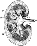
Section through the Kidney
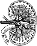
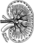
Section through the kidney showing the medullary and cortical portions, and the beginning of the ureter.…
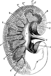
Kidney Section
Section through the right kidney from its outer to inner border. Labels: 1, cortex; 2, medulla; 2',…

Blood Supply of the Kidney
Vascular supply of the kidney. Labels: a, part of arterial arch; b, interlobular artery; c, glomerulus;…
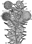
Diagram of the Structure of the Kidney
A diagram of the structure of the kidney. Labels: 1, Tubules or minute tubes. 2, Enlargement of a tubule…

Frontal Section Through Kidney
Frontal section through the right kidney and adjacent structures showing the renal fasciae and fatty…
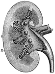
Longitudinal Section of a Kidney
A longitudinal section of a kidney. Labels: 1, 2, 3, Parts of the Kidney. 4, Pelvis. 5, Ureter. 6, Renal…

Minute Structure of the Kidney
The minute structure of the kidney, which commences in the cortical substance of the organ as the Malpighian…
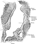
Sagittal Section Through Sinus of Kidney
Sagittal section through sinus of child's kidney, showing lower part of pelvis and commencement of ureter.
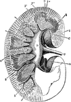
Section of the Right Kidney
Section through the right kidney from its outer to its inner border. Labels: 1, cortex; 2, medulla;…
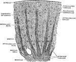
Section Through Kidney
Part of a section through the cortex of the kidney in the direction of the straight tubules.
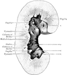
Section Through Kidney
Longitudinal section through the kidney. The vessels and fat have been removed to give a view of the…
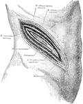
Surgical Incision to the Kidney
An incision in the right side above the kidney, showing a typical surgical approach to this organ, exposing…

The Kidney
The basic structure of the kidney, which consists of the cortical portion, the medullary substance,…

Kidneys
Kidneys and their vessels. 1: Left kidney; 2: Ascending vein; 3: Aorta; 4: Left ureter; 5: Bladder.

Kidneys from Behind
The kidneys viewed from behind. The dotted lines mark out the areas contact with the various muscle…
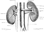
Kidneys from the Front
The kidneys and great vessels viewed from the front. The drawing was made before the removal of the…
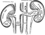
Anterior Surface of the Kidneys
The anterior surfaces of the kidneys, showing areas of contact of neighboring viscera.
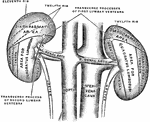
Posterior Surface of the Kidneys
The posterior surfaces of the kidneys, showing areas of relation to the parietes.
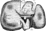
Liver
The under surface of the liver. Labels: G.B., gallbladder; H.D., common bile duct; H.A.; hepatic artery;…

Portion of a Lobule of Liver
Portion of a lobule of liver. Labels: a, bile capillaries between liver cells, the network which is…
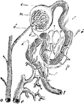
Malpighian Body
Relation of the Malpighian body to the uriniferous ducts and blood vessels. Labels: a, one of the interlobular…
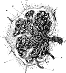
Malpighian Capsule
Malpighian capsule and tuft of capillaries, injected through the renal artery with colored gelatin.…
