The Reptile Anatomy ClipArt gallery provides 63 illustrations of reptile body parts, skeletons, organs, and other internal and external anatomical views.
Acrodont
"Skull of lizard with Acrodont Dentition. One of those lizards which have the edge of the jaw, without…
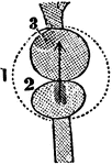
Fangs and Tongue of an Adder
"When the animal wishes to use these teeth, they issue from this fleshy sheath, somewhat in the same…
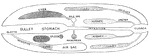
The Alimentary Canal of the Flying Lizard
In reptiles the alimentary canal differs much from that of mammals or birds. As a general rule, it is…
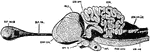
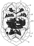
Alligator Brain
"Brain of alligator, from above. B. ol., olfactory bulb; G. p, epiphysis; HH, cerebellum; Med, spinal…
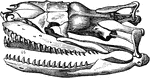
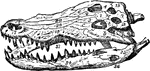
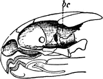
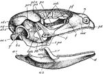
Alligator Skull
"Skull of a member of the family Alligatoridæ, or some American member of the Crocodilidæ."-Whitney,…
Boa
"In popular language, the name of all those large serpents which kill their prey by entwining themselves…
Boa Constrictor Skull
"Skull of Boa constrictor. a, quadrate bone; b, b, halves of lower jaw." -Cooper, 1887
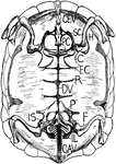
Brain of an Alligator
In Amphibians the nervous system is but slightly developed. The cerebrum is small; the cerebellum is…
Circulation of a Reptile
Diagram of the circulation of the reptile. Labels: a, pulmonic, and b, somatic circulation; c, heart…
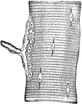
Skink Claw
In the U.S. the five-lined or blue-tailed skink is the common variety. This is a close-up of its claw.
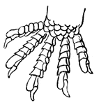
Crocodile Skeleton
"Skeleton of crocodile. C, causal region; D, thoracic region of spinal column; F, fibula; Fe, femur;…
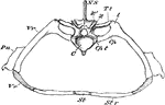
Crocodile Thoracic Region
"Segment of Endoskeleton from Thoracic Region of Crocodile. C, centrum of a vertebra, over which rises…
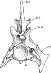
Crocodile Vertebra
"Cervical vertebra of crocodile. N.S., Neural spine; P.A., posterior articular process; A.A., anterior…
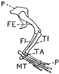
Leg of Crocodile
This illustration shows the leg of a crocodile. P. Pelvis, FE. Femur, TI. Tibia, FI. Fibula, TA. Tarsus,…
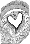
Eye of a Lizard
Longitudinal section through the pineal eye of a lizard. The eye is located in the middle of the dorsal…
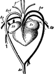
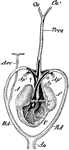
Heart and Blood Vessels of a Turtle
Heart and blood vessels of the turtle. Ad, right auricle; As, left auricle; Ao.d, right arch of the…
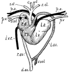
A Diagram of the Heart of a Reptile
A diagram of the heart of a reptile. Labels: 1, Pericardium. 2, Single ventricle. 3, Left auricle. 4,…
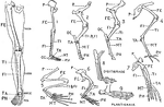
Human Leg (Front View), and Comparative Diagrams showing Modifications of the Leg
This illustration shows a human leg (front view), and comparative diagrams showing modifications of…
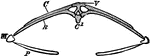
Dorsal Vertebra of the Hylonomus
Hylonomus was an early reptile. It lived 315 Ma (million years ago) during the Carboniferous period.…
Monitor Lizard Heart
"Heart of monitor (Varanus) dissected to show the cavity of the ventricle and the vessels leading out…
Nerve Ending in Muscle Fiber of Lizard
Nerve ending in muscular fiber of a lizard (Kühne). The end-plate, or motorial ending of the axone,…
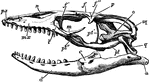
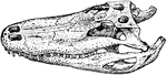
Lizard Skull
"Side view of skull of Lacerta. px., Premaxilla; mx., maxilla; l., lachrymal; j., jugal; t.pa., transpalatine;…
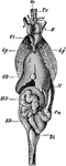
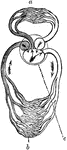
Sand Lizard Viscera
"Lacerta agilis. General view of the viscera in their naturaal relations. Bl, urinary bladder; Ci, post-caval…
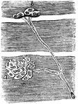
Muscular Fiber Cells of a Snake
From preparation of the nerve termination in the muscular fibers of a snake. A, End plate seen only…
Newt Anatomy
"Section through a young newt. c.t., Connective tissue; E., epidermis; D., dermis; S.C., spinal cord;…
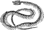
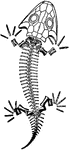
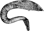
Rattlesnake Skeleton
"The vertebre of serpents are so formed as to admit a great pliancy of the body, which is capable of…
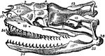
Rattlesnake Skull
This illustration shows the skull of a rattlesnake. ar, articular portion of lower jaw; de, dentary…
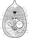
Reptile Development
"Origin of amnion and allantois. 1. Rise of amniotic folds (a.f.) around embryo (e); p.p., pleuro-peritoneal…
Tortoise Skeleton
Skeleton of a tortoise, seen from below, the plastron having been removed. (ca) carapace.
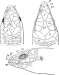
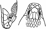
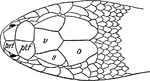
Skink Head
"Head-shields of Scinid Lizards. cs, chin-shields; d, disk on lower eylid; e, ear-opening; f, frontal;…
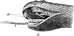
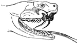
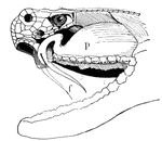
Snake Head
"Snake's head. dv., Poison fangs; b., sheath of fang; l., tongue; rl., muscles of tongue." -Thomson,…
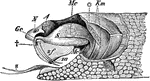
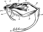
Rattlesnake Poison Apparatus
"Poison apparatus of rattlesnake. A, eye; Gc, poison-duct entering the poison-fang at +; Km, muscles…
Tortoise Heart
"Heart and associated vessels of tortoise. r.a., Right auricle; superior venae cavae (s.v.c.) and inferior…
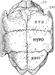
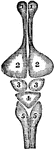
Greek Tortoise Plastron
"Internal view of the plastron of the Greek tortoise. EP., Epiplastron (clavicle?); ENT., entoplastron…
Greek Tortoise Plastron
"Scales on ventral surface of plastron of Greek tortoise. G., Gular; H., humeral; P., pectoral; Ab.,…
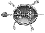
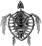
Skeleton of a Tortoise
A skeleton of a tortoise The ribs of which are expanded, forming the dorsal part of its shell. Labels:…
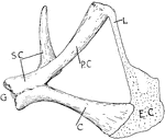
Tortoise Skeleton
"Internal view of tortoise skeleton. H., humerus; SC., scapula running dorsally; PC., precoracoid; C.,…
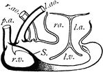
Turtle Heart
"Dissection of Chelonian heart. r.v., Right half of ventricle; S., septum; l.v., left half of ventricle;…
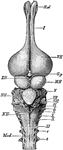
Turtle Hyoid
"Hyoid apparatus of a Chelonian. BH., Body of the hyoid (basihyal); H., representing another part of…
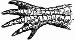
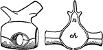
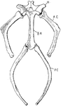
Turtle Pectoral Girdle
"Pectoral girdle of a Chelonian. G., Glenoid cavity; SC., scapula; P.C., procoracoid fused to the scapula;…
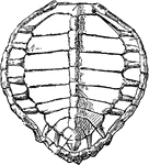
Green Turtle Skeleton
"Chelone midas. Transverse section of skeleton. C, costal plate; C', centrum; M, marginal plate; P,…
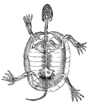
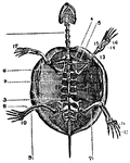
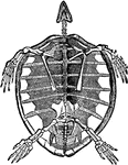
Marsh Turtle Skeleton
"Cistudo lutaria. Skeleton seen from below; the plastron has been removed and is represented on one…
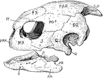
Turtle Skull
"Skull of turtle. "S.O., supra-occipital; PAR., parietal; FR., frontal; P.F., pre-frontal; PO.F., post-frontal;…
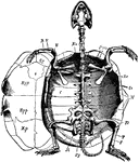
Skeleton of Turtle
"Turtle is, in zoology, the popular name for any species of the Cheloniidæ. They may be distinguished…
Skeleton of a Turtle
"The Tortoise will continue to live on for six months after it is deprived of its brain."