An illustration of a female nautilus without the shell. "c, points to the concave margin of the mantle-skirt leading into the sub-pallial chamber; g, the mid-foot or siphon; k, the superficial origin of its retractor muscles closely applied to the shell and serving to hold the animal in tis place; l, the sipjuncular pedicle of the visceral hump broken off short; v,v, the superior and inferior ophthalmic tentacles." (Britannica, 1910)

Female Nautilus without Shell

An illustration of a female nautilus without the shell. "c, points to the concave margin of the mantle-skirt…
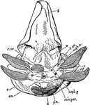
An illustration of the postero-ventral view female nautilus without the shell. "a, Muscular band passing from the mid-foot to the integument; b, The valve on the surface of the funnel, partially concealed by the inrolled lateral margin of the latter; c, The mantle-skirt retroverted; an, The median anus; x, Post-anal papilla of unknown significance; g.n., Nidamental gland ; r.ov, Aperture oft he right oviduct; l.ov, Aperture of the rudimentary left oviduct; neph.a, Aperture of the left anterior renal sac; neph.p, Aperture of the left posterior renal sac; viscper, Left aperture of the viscero-pericardial sac; olf, The left osphradium placed near the base of the anterior gill-plume." (Britnnica, 1910)

Female Nautilus without Shell

An illustration of the postero-ventral view female nautilus without the shell. "a, Muscular band passing…
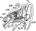
"Transverse Section through Side Walls of Skull, showing the Inner Parts of the Ear. Co, concha or external ear, or pinna; EM, external auditory meatus; TyM, tympanic membrane; Inc, incus; Mall, malleus; ASC, PSC, ESC, anterior, posterior, and external semicircular canals; Coc, cochlea; Eu, Eustachian tube; IM, internal auditory meatus, through which the auditory nerve passes to the organ of hearing." -Whitney, 1911

Inner Ear

"Transverse Section through Side Walls of Skull, showing the Inner Parts of the Ear. Co, concha or external…
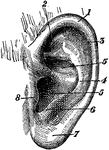
"External Ear, or Pinna. 1, helix; 2, fossa of antihelix, or fossa triangularis; 3, fossa of helix, or fossa scaphoidea; 4, antihelix; 5, 5, concha; 6, antitragus; 7, lobule; 8, tragus." -Whitney, 1911

External Ear

"External Ear, or Pinna. 1, helix; 2, fossa of antihelix, or fossa triangularis; 3, fossa of helix,…
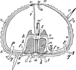
"Diagram of an Echinus (stripped of its spines). a, mouth; a', gullet; b, teeth; c, lips; d, alveoli; e, falces; f, f, auriculariae; g, retractor, and h, protractor, muscles of Aristotle's lantern; i, madreporic canal; k, circular ambulacral vessel; l, Polian pedicels; r, r, spines; s, tubercle; s', tubercle to which a spine is articulated; t, t, pedicellariae; u, anus; v, madreporic tubercle; x, ocular spot." -Whitney, 1911

Sea Urchin Section

"Diagram of an Echinus (stripped of its spines). a, mouth; a', gullet; b, teeth; c, lips; d, alveoli;…
The skull of the Great Anteater (Myrmecophaga tridactyla).

Great Anteater Skull

The skull of the Great Anteater (Myrmecophaga tridactyla).

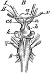
"Brain of Skate (Raia batis), an elasmobranchiate fish. B, from below, in part enlarged: ch, optic chiasm; h, pituitary body; n and v, vessels connected with h; k, saccus vasculosus; ß, pyrimids of medulla oblongata; a, I, II, V, same as in A." -Whitney, 1911

Skate Brain

"Brain of Skate (Raia batis), an elasmobranchiate fish. B, from below, in part enlarged: ch, optic chiasm;…
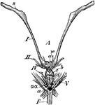
"Brain of Skate (Raia batis), an elasmobranchiate fish. A, from above; s, olfactory bulbs; a, cerebral hemispheres, united in the middle line; b, thalamencephalon; c, mesencephalon; d, cerebellum; aa, plaited bands formed by the restiform bodies; I, II, IV, V, first (olfactory), second (optic), fourth, and fifth pairs of cerebral nerves; f, medulla oblongata; w, a blood-vessel. " -Whitney, 1911

Skate Brain

"Brain of Skate (Raia batis), an elasmobranchiate fish. A, from above; s, olfactory bulbs; a, cerebral…
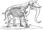
"Skeleton and Outline of African Elephant (Elephas or Loxodon africanus). fr, frontal; ma, mandible; ma', malar; fi, "finger" at end of trunk; C, cervical vertebrae; D, dorsal vertebrae; D, dorsal vertebrae; pe, pelvis; sc, scapula; st, sternum; hu, humerus; ul, ulna; ra, radius; mc, metacarpus; fe, femur; pat, patella; tib, tibia; fib, fibula; met, metatarsus." -Whitney, 1911

African Elephant Skeleton

"Skeleton and Outline of African Elephant (Elephas or Loxodon africanus). fr, frontal; ma, mandible;…
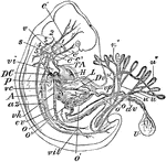
"Early Human Embryo, giving diagrammatically the principal vessels antecedent to the establishment of the regular fetal circulation. H, heart; P, lungs; L, liver; T A, the aortic trunk or cardiac aorta; c, c', c", common, external, and internal carotids; s, subclavian artery; v, vertebral artery; 1, 2, 3, 4, 5, the aortic arches (the persistent left aortic arch hidden); A, subvertebral aorta; o, o', omphalomeseraic artery and vein, to and from U, the umbilical vesicle with its vitelline duct, dv; u, u, the two hypogastric or umbilical arteries, with the ramifications, u", u", in the placenta; u', umbilical vein; vh, hepatic vein; cv, inferior vena cava; vil, iliac veins; az, an azygous vein; vc, a posterior cardinal vein; vi, innominate vein; vp, portal vein; Dv, the ductus venosus; DC, a ductus Cuvieri. The anterior cardinal vein is seen beginning in the head and running down to the ductus Cuvieri, on the under side of the numbers 1, 2, 3, 4, 5." -Whitney, 1911

Human Embryo

"Early Human Embryo, giving diagrammatically the principal vessels antecedent to the establishment of…

"Diagram of Vertebrate Encephalon ... in longitudinal vertical section. Mb, mid-brain; in front of it all is forebrain, behind it all is hind-brain; laminaterminalis, represented by the heavy black line in upper figure; Olf, olfactory lobes; Hmp, cerebral hemispheres; ThE, thalamencephalon; Py, pituitary body; FM, foramen of Monro; CS, corpus striatum; Th, optic thalamus; CQ, corpora quadrigemina; CC, crura cerebri; Cb, cerebellum; PV, pons Varolii; MO, medulla oblongata; I, olfactory nerves; II, optic nerves; III, point of ext from brain of the oculimotores; IV, of the pathetici; VI, of the abducentes; V-XII, origins of other cerebral nerves; 1, olfactory ventricle; 2, lateral ventricle; 3, third ventricle; 4, fourth ventricle." -Whitney, 1911

Encephalon

"Diagram of Vertebrate Encephalon ... in longitudinal vertical section. Mb, mid-brain; in front of it…

"Diagram of Vertebrate Encephalon ... in horizontal section. Mb, mid-brain; in front of it all is forebrain, behind it all is hind-brain; Lt, laminaterminalis; Olf, olfactory lobes; Pn, pineal body, or conarium; M, foramen of Monro; CS, corpus striatum; Th, optic thalamus; Cb, cerebellum; MO, medulla oblongata; I, olfactory nerves; II, optic nerves; 1, olfactory ventricle; 2, lateral ventricle; 3, third ventricle; 4, fourth ventricle; + is in the iter e tertio ad quartum ventriculum." -Whitney, 1911

Encephalon

"Diagram of Vertebrate Encephalon ... in horizontal section. Mb, mid-brain; in front of it all is forebrain,…
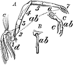
"A, Developed Endopodite, or ordinary ambulatory leg of the crawfish as a thoracic appendage: ab, the whole extent of the endopodite with seven joints; 1, coxopodite; 2, basipodite; 3, ischiopodite; 4, meropodite; 5, capopodite; 6, propodte; 7, dactylopodite; e, filaments borne on coxopodite; d, an epipodite. B and C, appendages respectively of first and second abdominal somite of th male: ab, endopodite; c, exopodite." -Whitney, 1911

Crawfish Leg

"A, Developed Endopodite, or ordinary ambulatory leg of the crawfish as a thoracic appendage: ab, the…

"Segment of Endoskeleton from Thoracic Region of Crocodile. C, centrum of a vertebra, over which rises the neural arch, inclosing the neural canal and ending in NS, the neural spine; Z, prezygapophysis; Z', postzygapophysis; Tt, transverse process which articulates with t, tubercle of a rib; Cpt, that which articulates with Cp, capitulum of a rib; Vr, ossified vertebral rib, or pleurapophysis; Vr', cartilaginous part of same; Str, sternal rib, or hemapophysis; St, segment of sternum; Pu, uncinate process of a rib or epipleura. From Cpt to St, on either side, is the hemal arch." -Whitney, 1911

Crocodile Thoracic Region

"Segment of Endoskeleton from Thoracic Region of Crocodile. C, centrum of a vertebra, over which rises…
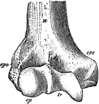
"Anterior View, Distal End, of Right Humerus of a Man. H, humerus; epc, epicondyle, or external supracondyloid protuberance; ept, epitrochlea, or internal supracondyloid protuberance; cp, capitellum, or convex articular suface for head of radius; tr, trochlea, or transversely concave articular surface for the ulna; epc and cp are together the ectocondyle, and ept and tr are together the entocondyle." -Whitney, 1911

Humerus

"Anterior View, Distal End, of Right Humerus of a Man. H, humerus; epc, epicondyle, or external supracondyloid…

The bill of the gull (Laridae) is epignathous: "hook-billed; having the end of the upper mandible decurved over and beyond that of the lower one." -Whitney, 1911

Gull Bill

The bill of the gull (Laridae) is epignathous: "hook-billed; having the end of the upper mandible decurved…
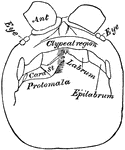
"Head of Scolopendra, from below, showing the epilabrum, the protomala with its cardo (Card), and stipes (St); Ant, antenna." -Whitney, 1911

Centipede Head

"Head of Scolopendra, from below, showing the epilabrum, the protomala with its cardo (Card), and stipes…

"Right Femur of a Youth. E, E, epiphyses; gtr, ltr, greater and lesser trochanter; h, head; et, it, external and internal tuberosity; ec, ic, external and internal condyle; n, neck." -Whitney, 1911

Youth Femur

"Right Femur of a Youth. E, E, epiphyses; gtr, ltr, greater and lesser trochanter; h, head; et, it,…

"Epipleurae.-- Thorax, scapular arch, and part of pelvic arch of a bobolink (Dolichonyx oryzivorus). ep, four epipleurae or uncinate processes of as many ribs; pl, pleurapophysial parts of seven ribs; hp, hemapopysial parts of six ribs; v, dorsolumbar vertebrae; st, sternum (the letters are on the carina or keel); m, manubrium sterni; cp, costal process of sternum, bearing f, furcula; ec, epiclidium of furcula; h, hypoclidium of fucula; gl, glenoid fossa, formed by coracoid and scapula; il, ilium; is, ischium; p, pubis; a, acetabulum." -Whitney, 1911

Bobolink Epipleurae

"Epipleurae.-- Thorax, scapular arch, and part of pelvic arch of a bobolink (Dolichonyx oryzivorus).…

Also known as fish lice, this is a species of copepod, a parasitic crustacean. "Female of Chondracathus gibbosus, enlarged; and example of the crustaceous Epizoa. B, ventral view; a, head; b, c, appendages; d, d, mediodorsal processes; e, e, medioventral processes; f, i, h, lateral processes; g, ovisacs; k, terminal segment; l, minute male lodged in vulva of female; m, n, mediodorsal ovarian tubes; p, lateral ovarian tubes; o, o, oviduct; 2, 3, antennules; 4, 5, 6, antennae, gnathites." -Whitney, 1911

Copepod

Also known as fish lice, this is a species of copepod, a parasitic crustacean. "Female of Chondracathus…
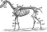
"Skeleton of Horse (Equus caballus). fr, frontal bone; C, cervical vertebrae; D, dorsal vertebrae; L, lumbar vertebrae; cd, caudal vertebrae; sc, scapula; pe, pelvis; ma, mandible; hu, humerus; ra, radius; cp, carpus; mc, metacarpus; fe, femur; tib, tibia; ca, calcaneum; tar, tarsus; mt, metatarsus; p, phalanges." -Whitney, 1911

Horse Skeleton

"Skeleton of Horse (Equus caballus). fr, frontal bone; C, cervical vertebrae; D, dorsal vertebrae; L,…

"Cartilaginous Cranium of the Pike (Esox lucius), with its intrinsic ossifications. A, top view; 3, small ossifications in the rostrum; N, N, nasal fossae; I Or, interorbital septum; Eth, ethmoid; Prf, Ptf, prefrontal and postfrontal; EpO, epiotic; OpO, opisthotic; Pt, pteriotic; EO, exoccipital." -Whitney, 1911

Pike Cranium

"Cartilaginous Cranium of the Pike (Esox lucius), with its intrinsic ossifications. A, top view; 3,…

"Cartilaginous Cranium of the Pike (Esox lucius), with its intrinsic ossifications. B, side view: V, VII, exits of trigeminal and of pneumogastric nerves; 3, small ossifications in the rostrum; I Or, interorbital septum; Eth, ethmoid; Prf, Ptf, prefrontal and postfrontal; PrO, proötic; EpO, epiotic; OpO, opisthotic; Pt, pteriotic; EO, exoccipital; BO, basioccipital; BS, basisphenoid; AS, alisphenoid." -Whitney, 1911

Pike Cranium

"Cartilaginous Cranium of the Pike (Esox lucius), with its intrinsic ossifications. B, side view: V,…
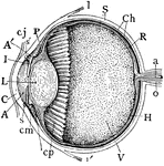
"Human Eye, in Median Vertical Anteroposterior Section. (Ciliary processes shown, through not all lying in this section.) A, anterior, and A', posterior chambers of aqueous humor; a, central artery of retina; C, cornea; Ch, choroid; cj, conjunctiva; cm, ciliary muscle; cp, ciliary processs; H, hyaloid; I, iris; L, crystalline lens in its capsule (the reference-line passes through the pupil); l, l', insertion of tendon of superior and inferior rectus muscles; o, optic nerve; P, canal of Petit; R, retina; S, sclerotic; s, s', circular sinus or canal of Schlemm; V, vitreous body filling back part of the eye." -Whitney, 1911

Median Vertical Anteroposterior Section of Eye

"Human Eye, in Median Vertical Anteroposterior Section. (Ciliary processes shown, through not all lying…
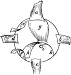
"Right Eyeball of Bird, seen from behind, showing the following muscles; a, rectus superior; b, rectus externus; c, rectus inferior; d, rectus internus; e, obliquus superior; f, obliquus inferior; g, quadratus; h, pyramidalis, with its tendon, k, passing through a pulley in the quadratus (as shown by dotted line) to keep it off the optic nerve, i, then passing around the edge of the ball to its insertion in the nictitating membrane." -Whitney, 1911

Right Eyeball of Bird

"Right Eyeball of Bird, seen from behind, showing the following muscles; a, rectus superior; b, rectus…
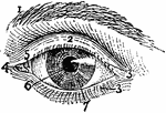
"Exterior of Left Human Eye. 1, supercilium, or eyebrow; 2, palpebra superior, or upper eyelid; 3, 3, cilia, or eyelashes; 4, caruncula lacrymalis; 5, plica semilunaris; 6, pupil; 7, iris." -Whitney, 1911

Exterior of Left Human Eye

"Exterior of Left Human Eye. 1, supercilium, or eyebrow; 2, palpebra superior, or upper eyelid; 3, 3,…
The side view of the compound eye of the housefly.

Compound Eye of Housefly

The side view of the compound eye of the housefly.

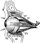
"Muscles of Left Human Eyeball. so, superior oblique, passing through a trochlea or pulley; io, inferior oblique; sr, superior rectus; ifr, interior rectus; ir, internal rectus; er, external rectus; f, frontal sinus; m, maxillary sinus; o, optic nerve." -Whitney, 1911

Muscles of Left Eyeball

"Muscles of Left Human Eyeball. so, superior oblique, passing through a trochlea or pulley; io, inferior…

"Skull of Cat (Felis catus), showing the following bones, viz. : na, nasal; pm, premaxillary; m, maxillary; l, lacrymal; f, frontal; j, jugal; pa, palatine; p, parietal; sq, squamosal; ip, interparietal; so, supra-occipital; eo, exoccipital (the line leads to the occipital condyle); t, tympanic bulla; smf, stylomastoid foramen; mf, mental foramen; c, coronoid process of mandible; ar, ascending ramus of mandible; hr, horizontal ramus of mandible; an, angle of jaw." -Whitney, 1911

Domestic Cat Skull

"Skull of Cat (Felis catus), showing the following bones, viz. : na, nasal; pm, premaxillary; m, maxillary;…

"Anterior View of Human Right Femur. ec, external condyle; etu, external tuberosity; ic, internal condyle; itu, internal tuberosity; ltr, lesser trochanter; gtr, great trochanter; h, head; n, neck." -Whitney, 1911

Anterior View of Human Right Femur

"Anterior View of Human Right Femur. ec, external condyle; etu, external tuberosity; ic, internal condyle;…

"Posterior View of Left Femur of a Horse. h, head; gtr, great trochanter; ttr, third trochanter; ltr, lesser trochanter; p, pit for round ligament; itf, intertrochanteric fossa; d, a depression or fossa; etu, itu, external and internal tuberosities; cc, the two condyles." -Whitney, 1911

Posterior View of Left Femur of Horse

"Posterior View of Left Femur of a Horse. h, head; gtr, great trochanter; ttr, third trochanter; ltr,…

Cypris, a modern ostracod. Female before sexual maturity, right valve of shell removed to show internal anatomy.

Cypris

Cypris, a modern ostracod. Female before sexual maturity, right valve of shell removed to show internal…

"In ornithology, pinnatiped; having pinnate feet, the toes being separately furnished with flaps, as in the grebes, coots, phalaropes, fin-foots, etc." -Whitney, 1911

Fin-Footed Coot Foot

"In ornithology, pinnatiped; having pinnate feet, the toes being separately furnished with flaps, as…

"Skeleton of Fish (Perch). a, intermaxillaries; b, nasal region; c, dentary bone of mandible; d, orbit of eye; e, supraoccipital crest; f, preoperculum; g, g', vertebral column; h, pectoral fin; i, ventral fin; k, first dorsal fin; l, second dorsal fin; m, anal fin; n, n, caudal fin, making a homocercal tail." -Whitney, 1911

Skeleton of Perch

"Skeleton of Fish (Perch). a, intermaxillaries; b, nasal region; c, dentary bone of mandible; d, orbit…
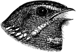
"In ornithology, having the beak broad and deeply cleft, as a swallow, swift, or goatsucker" or nightjar. -Whitney, 1911

Fissirostral Bill of Nightjar

"In ornithology, having the beak broad and deeply cleft, as a swallow, swift, or goatsucker" or nightjar.…

"Bones of Human Foot, or Pes, the third principal segment of the hind limb, consisting of tarsus, metatarsus, and phalanges. a, astralagus; ca, calcaneum; n, navicular, or scaphoid; co, cuboid; c1, c2, c3, entocuneiform, mesocuneiform, and ectocuneiform, or inner, middle, and outer cuneiform bones. The foregoing seven bones constitute the tarsus, and m1 to m5, first to the fifth metatarsal, constitue the metatarsus. The remaining fourteen bones are the phalanges, three to each digit excepting the great toe; h, distal phalanx of the hallux or great toe." -Whitney, 1911

Bones of Human Foot

"Bones of Human Foot, or Pes, the third principal segment of the hind limb, consisting of tarsus, metatarsus,…

"Heterocercal Caudal Fin of a Sturgeon (Acipenser brevirostris), showing the series of fulcrums, Fl, along the dorsal border." -Whitney, 1911

Shortnose Sturgeon Tail

"Heterocercal Caudal Fin of a Sturgeon (Acipenser brevirostris), showing the series of fulcrums, Fl,…

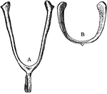
"A, Furcula or Merry-thought of a Domestic Fowl. ... h, hypoclidium." -Whitney, 1911

Furcula

"A, Furcula or Merry-thought of a Domestic Fowl. ... h, hypoclidium." -Whitney, 1911

An illustration of a cows horns.

Cow Horns

An illustration of a cows horns.

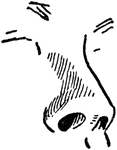
An illustration of a human nose.

Human Nose

An illustration of a human nose.

"Typical Skull of Common Fowl (Galliformes). A, side view: sa, surangular bone of mandible; ar, articular of mandible; d, dentary; f, frontal; j, jugal; l, lacrymal; me, mesethmoid; mx, maxillary; p, parietal; pf, postfrontal process; pt, pterygoid; px, premaxillary; q, quadrate; qj, quadratojugal; sq, squamosal; v, vomer." -Whitney, 1911

Skull of Common Fowl

"Typical Skull of Common Fowl (Galliformes). A, side view: sa, surangular bone of mandible; ar, articular…

"Typical Skull of Common Fowl (Galliformes). B, vertical longitudinal section: sa, surangular bone of mandible; ar, articular of mandible; d, dentary; f, frontal; me, mesethmoid; p, parietal; pf, postfrontal process; px, premaxillary; sq, squamosal; v, vomer; as, alisphenoid; bo, basioccipital; so, supraoccipital; os, orbitosphenoid; p', prootic; pf, pituitary fossa; sp, splenial bone." -Whitney, 1911

Skull of Common Fowl

"Typical Skull of Common Fowl (Galliformes). B, vertical longitudinal section: sa, surangular bone of…
An illustration of a human toes.

Toes

An illustration of a human toes.

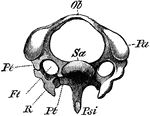
"Third cervical vertebra of Woodpecker (Picus viridis). (Viewed anteriorly.) Ft, vertebrarterial foramen; Ob, upper arch ; Pa, articular process; Psi, haemal spine ; Pt, Pt, the two bars of the transverse process, shewn on one side ancylosed with the cervical rib (R); Sa articular surface of centrum. (From Wieersheim.) A. H. Evans, 1900

The Diagram of a Third Cervical Vertebra of a Woodpecker

"Third cervical vertebra of Woodpecker (Picus viridis). (Viewed anteriorly.) Ft, vertebrarterial foramen;…

"Skeleton of the trunk of a Falcon. Ca, coracoid, which articulates with the sternum (St) at ; Cr, keel of sternum; Fu (Cl), furcula (clavicles); G, glenoid cavity for humerus; S, scapula; Un uncinate process; V, vertebral, and Sp, sternal, portion of rib. (From Wiedersheim.)" A. H. Evans, 1900

The Skeleton of the Trunk of a Falcon

"Skeleton of the trunk of a Falcon. Ca, coracoid, which articulates with the sternum (St) at ; Cr, keel…
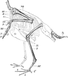
"Skeleton of the Limbs and Tail of a Carinate Bird. (The skeleton of the body is indicated by dotted lines.) F, digits; Fi, fibula; HW, carpus; MF, tarsometatarsus; MH, carpometacarpus; OA, humerus; OS, femur; Py, pygostyle; R, coracoid; Ul, ulna; Sch, scapula; St, sternum, with its keel (Cr); T, tibiotarsus; Rd, radius; Z, Z, digits of foot. (From Wiedersheim.)" A. H. Evans, 1900

Skeleton of the Limbs and Tail of a Carinate Bird

"Skeleton of the Limbs and Tail of a Carinate Bird. (The skeleton of the body is indicated by dotted…

"Pelvis of Apteryx austrlis. Lateral view. a, Acetabulum; il, ilium; is, ischium; p, pectineal process of pubis; p, pubis." (From Wiedersheim.)" A. H. Evans, 1900

Diagram of the Pelvis of a Kiwi

"Pelvis of Apteryx austrlis. Lateral view. a, Acetabulum; il, ilium; is, ischium; p, pectineal process…

"Skull of a Wild Duck (Anus boscas), from the side. ag, Angular; als, alisphenoid; ar, articular; bt, basitemporal; d, dentaryl en, external nostrils, e.o, exoccipital; eth, ethmoid; fr, frontal; j, jugal; lc, lacrymal; mx, maxilla; mx.p, maxillopalatine process; n, nasal, parietal; pg, pterygoid; pl, palatine; ps, presphenoid; px, premaxilla; q, quadrate; q.j, quadratojugal; s.ag, supra-angular; s.o, supraoccipital; sq, squamosal; ty, tympanic cavity; v, vomer; II, foramen for optic nerve; V, for trigeminal." (From Wiedersheim.)" A. H. Evans, 1900

Diagram of the Skull of a Wild Duck

"Skull of a Wild Duck (Anus boscas), from the side. ag, Angular; als, alisphenoid; ar, articular; bt,…
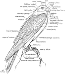
"The annexed figure explains the nomenclature of most of the outward of a Bird, but some further explanations may be given." A. H. Evans, 1900

A Labeled Diagram of a Falcon to Show the Nomenclature of the External Parts

"The annexed figure explains the nomenclature of most of the outward of a Bird, but some further explanations…
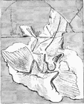
"Archaeornithes is at present represented by but one member, the first undoubted fossil Bird, made known in 1861 by Andreas Wagner form the Jurassic slate formation of Solenhofen in Bavaria, and now preserved in a British Museum. This he described under the name of Griphosaurus; but as Hermann von Meyer had already bestowed the title of Archaeopteryx Lithographica upon a bird, presumably identical, a feather of which had been obtained from the above system." A. H. Evans, 1900 This sample was obtained from the Limestone in Berlin

Archaeopteryx Lithographica

"Archaeornithes is at present represented by but one member, the first undoubted fossil Bird, made known…
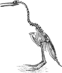
"Hesperornis regalis, (a fossilized restoration) which stood about three feet high, had blunt teeth in the grooves of both maxilla and mandible, the number being thirty or more below, but considerably less above, where they did not reach to the exterior extremity. The bill was long and pointed, the rami of the lower jaw being entirely separate; the head was rather small, the neck long, and the quadrate bone articulated with the skull by one knob only. The sternum was long, broad, and flat, without keel; the furcula was decidedly reduced, the metatarsus, being little more than a humerus; the tail was fairly long and broad, but had no pygostyle." A. H. Evans, 1900

The Restoration of the Hesperornis Regalis

"Hesperornis regalis, (a fossilized restoration) which stood about three feet high, had blunt teeth…

"Ichthyornis victor and I. dispar, ...were small forms of about the size of a Partridge, with the habits and appearance, it is presumed, of Terns or Gulls. The head was extremely large in proportion to the remainder of the skeleton; the beak was long and pointed, with entirely separate rami to the mandible; the sharp teeth, fixed regularly in distinct sockets, were inclined backwards, and occupied the whole of the lower and at least the posterior half of the upper jaw; the keel of the sternum was large and broad; the dorsal and cervico-dorsal vertebrae were biconcave. the quadrate articulated to the skull by one knob,... the metatarsus was short and the whole foot small; a furcula was probably present; the wings were well developed, indicating great powers of flight; while the tail was comparatively short, and ended in a pygostyle." A. H. Evans, 1900

Skeleton Head of a Ichthyornis

"Ichthyornis victor and I. dispar, ...were small forms of about the size of a Partridge, with the habits…

Flatworms are flattened, leaf-like forms living in damp places on land, in freshwater streams of ponds, or along the seacoast while a variety of other species are parasitic. The free forms are usually small, barely reaching a length greater than five or seven cetimeters (2 to 3 inches). Some of the parasitic species attain the great length of six to thirteen meters (20 to 40 feet). This is a fresh water flatworm. This is an anatomy of freshwater flatworm (Planaria), showing the excretory system, with flame-cell. The alimentary canal is stippled.

Flatworm

Flatworms are flattened, leaf-like forms living in damp places on land, in freshwater streams of ponds,…

The earthworms are also known as megadriles, in the families Tubificidae, Lumbriculidae, and Enchytraeidae. Earthworms have a pair of kidneys to every segment, each consisting of a coiled tube wrapped in a mass of small blood vessels, and at its inner end communicating with the body cavity by means of a funnel-shaped opening. Here is a diagram of an earthworm kidney., showing (b) blood vessel, (f) funnel opening into body cavity, (o) outer opening, (s)septum, (w) body wall

Earthworm Anatomy

The earthworms are also known as megadriles, in the families Tubificidae, Lumbriculidae, and Enchytraeidae.…
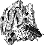
Beetles are the group of insects with the largest number of known species. The general anatomy of beetles is quite uniform, although specific organs and appendages may vary greatly in appearance and function between the many families in the order. Like all insects, beetles' bodies are divided into three sections: the head, the thorax, and the abdomen. We see here the long-horned borer beetle showing the larva, pupa, and adult insect.

Beetle

Beetles are the group of insects with the largest number of known species. The general anatomy of beetles…

"Trachea or windpipe of the red breasted merganser, Mergus serrator, about half natural size, viewed from above (behind); after Newton, A, tongue; B B, its attachments; C C; windpipe, dilated in the middle and swelling below into a bony box, D; E E, bronchial tubes, going to the lung" Elliot Coues, 1994

the Windpipe of a Male Red Breasted Merganser

"Trachea or windpipe of the red breasted merganser, Mergus serrator, about half natural size, viewed…
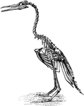
"Hesperornis regalis, (a fossilized restoration) which stood about three feet high, had blunt teeth in the grooves of both maxilla and mandible, the number being thirty or more below, but considerably less above, where they did not reach to the exterior extremity. The bill was long and pointed, the rami of the lower jaw being entirely separate; the head was rather small, the neck long, and the quadrate bone articulated with the skull by one knob only. The sternum was long, broad, and flat, without keel; the furcula was decidedly reduced, the metatarsus, being little more than a humerus; the tail was fairly long and broad, but had no pygostyle." A. H. Evans, 1900

Restoration of Hesperornis regalis

"Hesperornis regalis, (a fossilized restoration) which stood about three feet high, had blunt teeth…
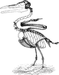
"Ichthyornis, though the wings are well developed, with fused metacarpals, and the sternum is keeled, the vertebrae present the extraordinary primitive character of being biconcave." Elliot Coues, 1884. This bird is believed to come from the Cretaceous of North America and is a seabird."

Restoration of Ichthyornis

"Ichthyornis, though the wings are well developed, with fused metacarpals, and the sternum is keeled,…
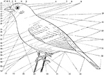
"fig. 25 - Topography of a Bird. 1, forehead (frons). 2, lore. 3, circumocular region. 4, crown (vertex). 5, eye. 6, hind head (occiput). 7, nape (nucha). 8, hind neck (cervix). 9, side of neck. 10, interscapular region. 11, dorsum, or back proper, including 10. 12, notaeum, or upper part of body proper, including 10, 11, and 13. 13, rump (uropygium). 14, upper tail-coverts. 15, tail. 16, under tail-coverts (crissum). 17, tarsus. 18, abdomen. 19, hind toe (hallux). 20, gastraeum, including 18 and 24. 21, outer and fourth toe. 22, middle and third toe. 23, side of the body. 24, breast (pectus). 25, primaries. 26, secondaries. 27, tertiaries; nos. 25, 26, 27 are all coverts. 28, primary coverts. 29, alula, or bastard wing. 30, greater coverts. 31, median coverts. 32, lesser coverts. 33, the "throat," including 34, 37, 38. 34, jugulum or lower throat. 35, auriculars. 36, malar region. 37, gula, or middle throat. 38, mentum, or chin. 39, angle of commissure, or corner of mouth. 40, ramus of under mandible. 41, side of under mandible. 42, gonys. 43, apex, or tip of bill. 44, tomia, or cutting edges of the bill. 45, culmen, or ridge of upper mandible, corresponding to gonys. 46, side of upper mandible. 47, nostril. 48, passes across the bill a little in front of it base." Elliot Coues, 1884

Topography of a Bird

"fig. 25 - Topography of a Bird. 1, forehead (frons). 2, lore. 3, circumocular region. 4, crown (vertex).…
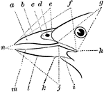
"Fig. 26 - Parts of a Bill. a, side of upper mandible; b, culmen; c, nasal fossa; d, nostril; e(see below); f, gape, or whole commissural line; g, rictus; h, commissural point or angle of the mouth; i, ramus of under jaw; j, tomia of under mandible (the reference lines e should have been drawn to indicate the correct tomia of upper mandible):k, angle of gonys; l, gonys; m, side of under mandible; n, tips of mandibles." Elliot Coues, 1884

The Parts of a Bird Bill

"Fig. 26 - Parts of a Bill. a, side of upper mandible; b, culmen; c, nasal fossa; d, nostril; e(see…