Cerebral convolutions of the brain.

Cerebral Convolutions

Cerebral convolutions of the brain.

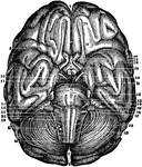
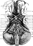
The base of brain. Labels: 1. Olfactory Bulb; 2. Second, or Optic Nerves; 3. Anterior Perforated Space; 4. Optic Tract; 5. Crus Cerebri; 6. 3rd Nerve; 7. 4th Nerve.; 8. 5th Nerve; 9. 6th Nerve; 10. Pyramid; 11. Olivary Body; 12. Vertebral Artery; 13. Anterior Spinal Artery; 14. Anterior Cerebral Artery; 15. Lamina Cinerea; 16. Middle Cerebral Artery; 17. Tuber Cinereum; 18. Corpora Albicantia; 19. Posterior Perforated Space; 20. Posterior Cerebral Artery; 21. Superior Cerebral Artery; 22. Pons Varolii; 23. Inferior Cerebellar Artery; 24. 7th and 8th Nerves; 25. 9th, 10th, and 11th Nerves; 26. 12th Nerve; 27. Cerebellum.

Base of the Brain

The base of brain. Labels: 1. Olfactory Bulb; 2. Second, or Optic Nerves; 3. Anterior Perforated Space;…
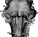
The medulla oblongata (brain stem).

The Medulla Oblongata

The medulla oblongata (brain stem).

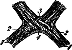
The optic chiasma or commissure, which is seen at the base of the brain in front of the tuber cinereum and behind the lamina cinerea. It contains 4 sets of fibers.

The Optic Chiasma

The optic chiasma or commissure, which is seen at the base of the brain in front of the tuber cinereum…
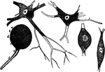
Forms of nerve cells. They each have a nucleus and nucleolus and are connected to each other by means of numerous branches and are found in the brain, spinal cords, and other nerves.

Nerve Cells

Forms of nerve cells. They each have a nucleus and nucleolus and are connected to each other by means…

Anterior view of the brain and spinal marrow. Labels: 1, 1, hemispheres of the cerebrum; 2, great middle fissure; 3, cerebellum; 4, olfactory nerves; 5, optic nerves; 6, corpora albicantia; 7, motor oculi nerves; 8, pons Varolii; 9, fourth pair of nerves; 10, lower portion of the medulla oblongata; 11, 11, medulla spinalis in its whole length; 12, 1, spinal nerves; 13, cauda equina.

Brain and Spinal Cord

Anterior view of the brain and spinal marrow. Labels: 1, 1, hemispheres of the cerebrum; 2, great middle…
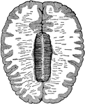
Diagram of the human nervous system showing the brain, spinal cord, and nerves.

Nervous System

Diagram of the human nervous system showing the brain, spinal cord, and nerves.

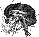
The general arrangement of the brain and attached nerves and blood vessels. At <em>a b</em> and <em>c</em> is the cerebrum; at <em>f</em> is represented the corpus callosum that joins the two hemispheres together; at d is the cerebellum; at <em>g</em> is the beginning of the optic nerves and at <em>l</em> is the olfactory nerve.

The Brain

The general arrangement of the brain and attached nerves and blood vessels. At a b and c

Cross-section of the brain. Here the upper half of the brain is cut off, and you see the upper cut surface of the lower half. The outer shaded part is the gray matter, and the inner lighter area is the white matter. In the center is the corpus callosum, a type of white matter that connects the two hemispheres together.

Cross-Section of the Brain

Cross-section of the brain. Here the upper half of the brain is cut off, and you see the upper cut surface…

Diagrammatic longitudinal section of the Body. Labels: a, the neural tube, with its upper enlargement in the skull cavity at a'; N, the spinal cord; N', the brain; ee, vertebrae forming the solid partition between the dorsal and ventral cavities; b, the pleural, and c, the abdominal division of the ventral cavity, separated from one another by the diaphragm, d; i, the nasal, and o, the mouth chamber, opening behind into the pharynx, from which one tube leads to the lungs, l, and the heart; k, a kidney; s, the sympathetic nervous chain. From the stomach, f, the intestinal tube leads through the abdominal cavity to the posterior opening of the alimentary canal.

Longitudinal Section of the Body

Diagrammatic longitudinal section of the Body. Labels: a, the neural tube, with its upper enlargement…
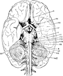
The base of the brain. The cerebral hemispheres are seen overlapping all the rest. Labels: I, olfactory lobes; II, optic tract passing to the optic chiasma from which the optic nerves proceed; III, the third nerve or motor oculi; IV, the fourth nerve or patheticus; V, the fifth nerve or trigminalis; VI, the sixth nerve or abducens; VII, the seventh or facial nerve; VIII, the auditory nerve; IX, the ninth or glossopharyngeal; X, the tenth or pneumogastric or vagus; XI, the spinal accessory; XII, the hypoglossal; ncI, the first cervical spinal nerve.

Base of the Brain

The base of the brain. The cerebral hemispheres are seen overlapping all the rest. Labels: I, olfactory…
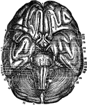
Base of the Brain. Labels: 1,2, longitudinal fissure; 3, anterior lobes cerebrum; 4, middle lobe; 5, fissure Sylvius; 6, posterior lobe; 7, infundibulum; 8, its body; 9, corporo albicantia; 10, cineritious matter; 11, crura cerebri; 12, pons Varolii; 13, medulla oblongata; 14, posterior prolongation of pons; 15, middle of cerebellum; 16, anterior part of cerebellum; 17, its posterior part and fissure; 18, medulla spinalis; 19, middle fissure medulla; 20, pyramidal body; 21, retiform body; 22, olivary body; 23, olfactory nerve; 24, its bulb; 25, its external root; 26, middle root; 27, internal root; 28, 29, optic nerve; 30, third nerve; 31, fourth nerve; 32, fifth nerve; 33, sixth nerve; 34, facial nerve; 35, auditory nerve; 36, 37, 38, eighth nerve.

Base of the Brain

Base of the Brain. Labels: 1,2, longitudinal fissure; 3, anterior lobes cerebrum; 4, middle lobe; 5,…

The brain and spinal cord. Labels: 1, 1, hemispheres of cerebrum; 2, great middle fissure; 3, cerebellum; 4, olfactory nerves; 5, optic nerves; 6, corpora albicanlia; 7, motor oculi; 8, pons Varolii; 9, fourth nerve; 10, medulla oblongata; 11, 11, medulla spinalis; 12, 12, spinal nerves; 18, cauda equina.

Brain and Spinal Cord

The brain and spinal cord. Labels: 1, 1, hemispheres of cerebrum; 2, great middle fissure; 3, cerebellum;…
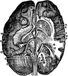
View of the course of the front columns of the spinal marrow terminating in the hemispheric ganglions of the brain.

Course of the Spinal Marrow

View of the course of the front columns of the spinal marrow terminating in the hemispheric ganglions…
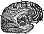
Middle vertical section of the callous body (corpus callosum). The inner left side of the brain is also seen.

Corpus Callosum

Middle vertical section of the callous body (corpus callosum). The inner left side of the brain is also…
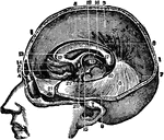
Base of the brain and cerebellum, together with the cranial nerves.

Base of Brain and Cerebellum

Base of the brain and cerebellum, together with the cranial nerves.

Section of the head showing the greater scythe, the horizontal apophysis of the dura mater between the brain and the cerebellum and other parts under the middle line of the head.

Head, Section of

Section of the head showing the greater scythe, the horizontal apophysis of the dura mater between the…

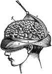
View of the appearance of the tortuous elevations (convolutions) of the brain, seen from above.

Convolutions of the Brain

View of the appearance of the tortuous elevations (convolutions) of the brain, seen from above.
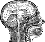
Vertical middle section of head and neck showing the opening through the Eustachian tube, and its relations with the pharynx.

Head and Neck, Section of

Vertical middle section of head and neck showing the opening through the Eustachian tube, and its relations…
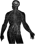
View of the nervous system in man, showing the nervous centers (the brain and the spinal near row) where the other nerves begin which provide the whole body.

Nervous System

View of the nervous system in man, showing the nervous centers (the brain and the spinal near row) where…
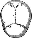
Sutures of the skull. Labels: a,a, the coronal suture, from the Latin corona, crown, so called from its situation on that part of the head, upon which the ancients placed the laurel, or olive crown, given to the victors in their games. It connects the frontal to the parietal bones; b, the sagittal suture, from a Latin word, signifying arrow, from its straight course. It runs from the middle of the frontal to the angle of the occipital bone, connecting the two parietals; c, the lambdoidal suture, extending from the sagittal suture down to the base of the brain on each side; e,e, the scaly overlapping of the temporal upon the parietal bones; hence called squamous suture.

Skull Sutures

Sutures of the skull. Labels: a,a, the coronal suture, from the Latin corona, crown, so called from…
1. Hemispheres of the brain proper, or cerebrum. 2. Hemispheres of the smaller brain, or cerebellum. 3. Spinal cord continuous with the brain, and the spinal nerves proceeding from it on each side.

Brain Hemispheres and Spinal Cord

1. Hemispheres of the brain proper, or cerebrum. 2. Hemispheres of the smaller brain, or cerebellum.…

The brain and spine.

Brain and Spine

The brain and spine.

"Diagrammatic longitudinal section of a Nemertean (Amphiporum lactifloreus), dorsal view. p.p., Proboscis pore; b., brain giving off the lateral nerve-cords (n.); po., oesophageal pocket; p., proboscis lying within its sheath; st., stilet of proboscis; m., retractor muscles of proboscis; g., gut shown in outline at the sides of proboscis; e., the three main, longitudinal blood vessels, which unite both anteriorly and posteriorly." -Thomson, 1916

Nemertea

"Diagrammatic longitudinal section of a Nemertean (Amphiporum lactifloreus), dorsal view. p.p., Proboscis…

"Partial section of a Vertebrate brain (diagrammatic). OLF., Olfactory lobe; CH., cerebral hemispheres; C., wall of cerebrum cut to show ventricle, behind this the figure is that of a median section; PA., parietal organ arising from thalamencephalon; Pl., pineal organ; INF., infundibulum descending from thalamencephalon; H., hypophysis; OL., optic lobes; CB., cerebellum; CPL., choroid plexus on roof of fourth ventricle; MO., floor of the medulla oblongata; CC., central canal of spinal cord." -Thomson, 1916

Vertebrate Brain

"Partial section of a Vertebrate brain (diagrammatic). OLF., Olfactory lobe; CH., cerebral hemispheres;…

"Median longitudinal section of anterior region of Myxine. B., Barbule; N., nasal aperture; NT., nasal tube with rings of cartilage; NC., nasal capsule; BR., brain; SC., spinal cord; N., notochord; G., gut; T., cartilage of "tongue"; MU., muscle; NTT., posterior part of nasal sac; TP., a tooth plate; MT., median tooth on roof of mouth (M.)." -Thomson, 1916

Hagfish Anterior

"Median longitudinal section of anterior region of Myxine. B., Barbule; N., nasal aperture; NT., nasal…

"Longitudinal vertical section of frog embryo, shortly before closure of blastopore. FB., fore-brain; EC., ectoderm; N., notochord; SC., canal of spinal cord; NE., nerenteric canal; B., blastopore; M., mesoderm; P., beginning of pituitary invagination." -Thomson, 1916

Frog Embryo

"Longitudinal vertical section of frog embryo, shortly before closure of blastopore. FB., fore-brain;…
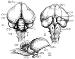
"Brain of pigeon (I. dorsal, II. ventral, III. lateral aspects). OLF.L., Olfactory lobes; C.H., cerebral hemispheres; PB., pineal body; OL., optic lobes; CB., cerebellum; FL., flocculus or lateral extension of cerebellum; M.O., medulla oblongata; PIT., pituitary body at end of infundibulum (INF.); O.N., optic nerves crossing in the chiasma." -Thomson, 1916

Pigeon Brain

"Brain of pigeon (I. dorsal, II. ventral, III. lateral aspects). OLF.L., Olfactory lobes; C.H., cerebral…

"Dorsal view of rabbit's brain. olf.l., Olfactory lobes; c.h., cerebral hemispheres; o.l., optic lobes (corpora quadrigemina); cb., median part of cerrebellum; Fl., flocculus of cerebellum; 10, root of the tenth or vagus nerve; s.c., spinal cord." -Thomson, 1916

Dorsal View of Rabbit Brain

"Dorsal view of rabbit's brain. olf.l., Olfactory lobes; c.h., cerebral hemispheres; o.l., optic lobes…

"Under surface of rabbit's brain. olf.l., Olfactory lobes; o.t., olfactory tract; f.l., frontal lobe of cerebral hemisphere; ch., optic chiasma; l.c., infundibulum; c.m., corpus mammillare; 3, root of oculomotor; 4, root of pathetic; 5, root of trigeminal; 6, root of abducenss; 7-8, roots of facial and auditory; Fl., flocculus of cerebellum; 9, root of glosso-pharyngeal; 10, roots of vagus; H., 12th , or hypoglossal; p.v., pons Varolii." -Thomson, 1916

Under Surface of Rabbit Brain

"Under surface of rabbit's brain. olf.l., Olfactory lobes; o.t., olfactory tract; f.l., frontal lobe…

"The nervous system of the Clam,&mdash;from the dorsal aspect. a, anterior; o, mouth; c.g., cerebral ganglia (brain); p.g., pedal ganglia; v.g., visceral ganglia." -Galloway, 1915

Clam Nervous System

"The nervous system of the Clam,—from the dorsal aspect. a, anterior; o, mouth; c.g., cerebral…

"The central nervous system in a leech. g, dorsal ganglia (brain); g', ventral chain of ganglia; o, mouth." -Galloway, 1915

Leech Nervous System

"The central nervous system in a leech. g, dorsal ganglia (brain); g', ventral chain of ganglia; o,…

"Diagrammatic sagittal section of Microstomum, showing a chain of four zooids produced by fission. b, brain of the original zooid (the exponents indicating corresponding structures of the more recently formed zooids); c, ciliated pit; d, dissepiments indicating different stages in the separation of the zooids; e, eye-spot; ent, entoderm; g, gut; gl., glandular cells about the mouth; m., mouth of the original worm." -Galloway, 1915

Microstomum

"Diagrammatic sagittal section of Microstomum, showing a chain of four zooids produced by fission. b,…
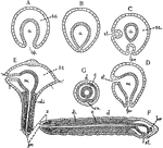
"Diagrams of stages in the metamorphosis of Polygordius, a primitive annelid. Ectoderm throughout is represented as nucleated without cell boundaries; the entoderm has the cell-boundaries shown, and the mesoderm is diagonally shaded. A, gastrula; B, same with blastopore closed; C and D represent formation of stomodaeum and proctodaeum from ectoderm; E, Trochosphere stage showing formation of segments in the posterior portion; F, adult (sagittal); G, adult (transverse). a, archenteron; bp., blostopore; br, brain; c, coelom; d, dorsal; di, dissepiments; m, mesenteron; pr., proctodaeum; s.c., segmentation cavity; st, stomodaeum; v.n., ventral nerve chain; z, zone of formation of nerve segments." -Galloway, 1915

Polygordius

"Diagrams of stages in the metamorphosis of Polygordius, a primitive annelid. Ectoderm throughout is…
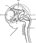
"Diagram of head and brain of human foetus six weeks old (heavy boundaries). The dotted line indicates the outline of the brain of a foetus three months old. Note thee great growth of the hemisphere (h). cer, cerebellum; med, medulla oblongata; mes, mesencephalon; p, pituitary body; pr, prosencephalon; s.c., spinal cord; th, thalamencephalon; 1, olfactory nerve; 2, optic nerve." -Galloway, 1915

Human Fetus

"Diagram of head and brain of human foetus six weeks old (heavy boundaries). The dotted line indicates…
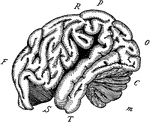
"The outline of the brain of an orang outang. Front portion F to O, cerebrum; C, cerebellum; M, medulla and spinal cord; F, the frontal lobe; P, the parietal lobe; O, the occipital lobe; T, the temporal lobe; R, the fissure of Rolando; S, the fissure of Silvius." -Galloway, 1915

Orangoutang Brain

"The outline of the brain of an orang outang. Front portion F to O, cerebrum; C, cerebellum; M, medulla…
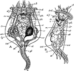
"Brachionus rubens. A, from the dorsal aspect; B, from the right side. a, anus; br, brain; d. f. dorsal feeler; c. gl, cement gland; cl, cloaca; c. l, ciliary lobes; c. v, contractile vesicle; e, eye-spot; int, intestine; lr, lorica; l. f, lateral feeler; m, muscular bands; nph, nephridial tubes; ov, ovary; ph, pharynx; st, stomach; t, tail; tr, d, trochal disc; vt, vitellarium." -Parker, 1900

Brachionus Rubens

"Brachionus rubens. A, from the dorsal aspect; B, from the right side. a, anus; br, brain; d. f. dorsal…

"Nervous system of Astacus fluviatilis. bg, sub-oesphageal ganglion; cs, commissural ganglion; g, brain; s, visceral nerve; sc, oesophageal connective; y, post-oesophageal commissure; IV-VIII, thoracic ganglia; 1-6, abdominal ganglia." -Parker, 1900

Crayfish Nervous System

"Nervous system of Astacus fluviatilis. bg, sub-oesphageal ganglion; cs, commissural ganglion; g, brain;…

"Periplaneta. General view of the nervous system. abd. 6, sixth abdominal ganglion; ant, antennary nerve; br, brain; conn, oesophageal connective; inf, sub-oesophageal ganglion; opt, optic nerve; thor. 1, thor. 2, thor. 3, first, second, and third thoracic ganglia." -Parker, 1900

Cockroach Nervous System

"Periplaneta. General view of the nervous system. abd. 6, sixth abdominal ganglion; ant, antennary nerve;…

The dissection of a female sea lamprey or Petromyzon marinus. ad, anterior dorsal cartilage; an, annular cartilage; au, auricle; bd, bile-duct; bp', basal plate; br, brain; cd, cardinal vein; d.ao, dorsal aorta; dm, dorsal muscles; ea, external gill aperture; ft, fibrous tissue; gr, groove canal; hp, basal plate to fontanelle; hv, hepatic vein; i, integument; i.ju; inferior jugular vein; int, intestine; ju, jugular; lg, lingual cartilage; lr, liver; m, buccal vavity; mv, median ventral cartilage; my, spinal cord; na, olfactory sac; na', pituitary pouch; na'', nasal aperture; na.c, nasal capsule; nc, notochord; n.ca, spinal canal; oc, cranial roof; oes, gullet; o.f, buccal funnel; ov, ovary; pc, pericardium; pd, posterior dorsal cartilage; pmt, protractor muscle of tongue; rmt; retractor of tongue; rt, respiratory tube; ss, lymph sinus; sv, sinus venosus; t, tongue; v, ventricle; v.ao, ventral aorta; vl, velum; vm, ventral muscles. -Parker, 1900

Lamprey Anatomy

The dissection of a female sea lamprey or Petromyzon marinus. ad, anterior dorsal cartilage; an, annular…
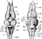
"Rana esculenta. The brain. A, from above; B, from below. ch. opt, optic chiasma; HH, cerebellum; Hyp, pituitary body; Inf, infundibulum; L. ol, olfactory lobe; Med, spinal cord; MH, mid-brain; NH, medulla oblongata; Th. opt, optic thalamus; Tr. opt, optic tract; VH, cerebral hemisphere; ZH, diencephalon; I-X, cerebral nerves; XII. (1), hypoglossal (first spinal) nerve." -Parker, 1900

Edible Frog Brain

"Rana esculenta. The brain. A, from above; B, from below. ch. opt, optic chiasma; HH, cerebellum; Hyp,…

"Brain of alligator, from above. B. ol., olfactory bulb; G. p, epiphysis; HH, cerebellum; Med, spinal cord; MX, optic lobes; NH, medulla oblongata; VH, cerebral hemispheres; I-XI, cranial nerves; 1, 2, first and second spinal nerves." -Parker, 1900

Alligator Brain

"Brain of alligator, from above. B. ol., olfactory bulb; G. p, epiphysis; HH, cerebellum; Med, spinal…
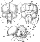
"Columba livia. The brain; A, from above; B, from below; C, from the left side. cb, cerebellum; c. h, cerebral hemispheres; f, flocculus; inf, infundibulum; m. o, medulla oblongata; o. l, optic lobes; o. t, optic tracts; pn, pineal body; II-XIII, cerebral nerves; sp. 1, first spinal nerve." -Parker, 1900

Rock Pigeon Brain

"Columba livia. The brain; A, from above; B, from below; C, from the left side. cb, cerebellum; c. h,…
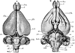
"Lepus cuniculus. Brain. A, dorsal view; B, ventral; b. o, olfactory lobe; cb', median lobe of cerebellum (vermis); cb'', lateral lobe of cerebellum; cr, cauro-cerebri; ep, epiphysis; f. b, parencephala; f. p, longitudinal fissure; h. b, hind-brain; hp, hypophysis; m. b, mid-brain (corpora quadrigemina); md, medulla oblongata; p.v, pons Varolii; i-xii, cranial nerves." -Parker, 1900

Rabbit Brain

"Lepus cuniculus. Brain. A, dorsal view; B, ventral; b. o, olfactory lobe; cb', median lobe of cerebellum…

"Lepus cuniculus. Longitudinal vertical sectioin of the brain. cb, cerebellum, showing arbor vitae; c. c, crus cerebri; ch1, parencephalon; ch2, temporal lobe; c. ma, corpus mammillare; f. m, foramen of Monro; inf, infundibulum; ly, lyra; m. o, medulla oblongata; o. ch, optic chiasma; olf, olfactory lobe; pty, pituitary body; vl. ip, velum interpositum; v. vn, valve of Vieussens; II, optic nerve." -Parker, 1900

Rabbit Brain

"Lepus cuniculus. Longitudinal vertical sectioin of the brain. cb, cerebellum, showing arbor vitae;…
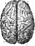
Labels: 1, longitudinal fissure separating the hemispheres; 2, frontal lobes of the cerebrum; 3, posterior lobes.

Brain Seen from Above

Labels: 1, longitudinal fissure separating the hemispheres; 2, frontal lobes of the cerebrum; 3, posterior…
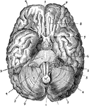
Labels: 1, longitudinal fissure separating the hemispheres; 2 and 3, front and posterior lobes of the cerebrum; 4 cerebellum; 7, optic nerve; 8, olfactory nerve; 9, medulla.

Brain Seen from Below

Labels: 1, longitudinal fissure separating the hemispheres; 2 and 3, front and posterior lobes of the…
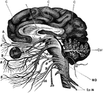
The brain and the cranial nerves seen partly in section and partly in side view. Labels: C, convolutions of the cerebrum; Cbl, cerebellum in section; MD medulla oblongata: Sp N, spinal nerve. The numbers indicate the twelve pairs of cranial nerves in order.

Brain and Cranial Nerves

The brain and the cranial nerves seen partly in section and partly in side view. Labels: C, convolutions…

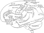
A diagrammatic view displaying the functions of the brain.

Lateral Surface of the Brain

A diagrammatic view displaying the functions of the brain.

A section of the brain and spinal column. Labels: 1, The cerebrum (large brain). 2, The cerebellum (small brain). 3, The Medulla Oblongata. 4, The spinal cord in its canal.

The Spinal Column and Brain

A section of the brain and spinal column. Labels: 1, The cerebrum (large brain). 2, The cerebellum (small…
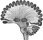
Diagram of human brain in vertical section, showing the situation of the different ganglia and the course of fibers. Labels: 1, Olfactory ganglion. 2, Hemisphere. 3, Corpus striatum. 4, Optic thalamus. 5, Tubercula quadrigemina. 6, Cerebellum. 7, Ganglion of tuber annulare. 8, Ganglion of medulla oblongata.

Diagram of Human Brain in Vertical Section

Diagram of human brain in vertical section, showing the situation of the different ganglia and the course…
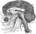
A vertical section of the cerebrum, cerebellum, and the medulla oblongata, showing the relation of the cranial nerves at their origin. Labels: 1, The cerebrum. 2, The cerebellum with its arbor vitae represented. 3, The medulla oblongata. 4, The spinal cord. 5, The corpus callosum. 6, The first pair of nerves. 7, The second pair. 8, The eye. 9, The third pair of nerves. 10, The fourth pair. 11, The fifth pair. 12, The sixth pair. 13, The seventh pair. 14, The eighth pair. 15, The ninth pair. 16, The tenth pair. 17, The eleventh pair. 18, The twelfth pair. 19, Spinal nerves. 21, The tentorium.

Vertical Section of the Brain

A vertical section of the cerebrum, cerebellum, and the medulla oblongata, showing the relation of the…

A back view of the brain and spinal cord. Labels: 1, The cerebrum. 2, The cerebellum. 3, The spinal cord. 4, Nerves of the face. 5, The brachial plexus of nerves. 6, 7, 8, 9, Nerves of the arm. 10, Nerves that pass under the ribs. 11, The lumbar plexus of nerves. 12, The sacral plexus of nerves. 13, 14, 15, 16, Nerves of the lower limbs.

A Back View of the Brain and Spinal Cord

A back view of the brain and spinal cord. Labels: 1, The cerebrum. 2, The cerebellum. 3, The spinal…
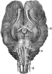
The base of brain of a horse. Labels: l, Cerebrum. 2, Ganglion of sight. 3, Cerebellum. 4, Medulla Oblongata and Spinal Cord.

Base of Brain of a Horse

The base of brain of a horse. Labels: l, Cerebrum. 2, Ganglion of sight. 3, Cerebellum. 4, Medulla Oblongata…

In Amphibians the nervous system is but slightly developed. The cerebrum is small; the cerebellum is scarcely visible. Shown is the brain of an alligator. Labels: 1, Olfactory ganglions. 2, Cerebrum. 3, Optic ganglions. 4, Cerebellum. 5, Medulla Oblongata and Spinal Cord.

Brain of an Alligator

In Amphibians the nervous system is but slightly developed. The cerebrum is small; the cerebellum is…
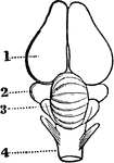
In birds the hemispheres are not united as in humans; the cerebellum is proportionately larger than the medulla oblongata, and the comparative weight of the brain to the body is less than in mammals. Labels: 1, Cerebrum. 2, Optic ganglion. 3, Cerebellum. 4, Medulla oblongata.

Brain of a Bird

In birds the hemispheres are not united as in humans; the cerebellum is proportionately larger than…

The brain of fish are small, it does not fill the whole cranial cavity, there being found within it a spongy fatty mass. Labels: 1, Olfactory ganglions. 2, Cerebrum. 3, Optic ganglions 4, Cerebellum. 5, Medulla Oblongata and Spinal Cord.

Brain of a Fish

The brain of fish are small, it does not fill the whole cranial cavity, there being found within it…

In the nervous system of the centipede the ganglions are arranged in pairs of nearly equal size, except the ganglion that answers to the brain, which is larger, along the surface of the alimentary canal.

Diagram of the Nervous System of a Centipede

In the nervous system of the centipede the ganglions are arranged in pairs of nearly equal size, except…
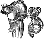
A view of the auditory nerve. Labels: 1, The spinal cord. 2, The medulla oblongata. 3, The lower part of the brain. 4, The auditory nerve. 5, A branch to the semicircular canals. 6, A branch to the cochlea.

View of the Auditory Nerve

A view of the auditory nerve. Labels: 1, The spinal cord. 2, The medulla oblongata. 3, The lower part…

A representation of the brain, spinal cord and spinal nerve. Labels: 1, The cerebrum. 2. The cerebellum. 3, Spinal cord. 4, The sciatic nerve. A. Distribution of the olfactory nerve. Labels: 1, 2, Nerve of smell. B. Optic Nerve. Labels: 15, Nerve of vision. C. The gustatory nerve. Labels: 1, 2, 3, 4, Branches of the nerve of taste. D. Auditory nerve. Labels: 13, Nerve of hearing.

Nervous System

A representation of the brain, spinal cord and spinal nerve. Labels: 1, The cerebrum. 2. The cerebellum.…