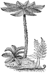
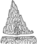
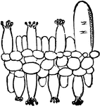
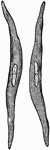
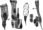
Tree-Fern
A Tree-Fern, Dicksonia arborescens, with a young one near its base. In front a common herbaceous Fern…
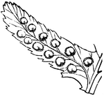
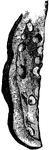
Shield-Fern
A small piece (pinnule) of a Shield-Fern: a row of fruit-dots on each side of the midrib, each covered…
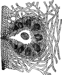
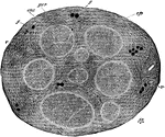
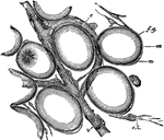
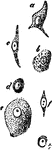
Fertile Conceptacle of Rockweed
Magnified section through a fertile conceptacle of Rockweed, showing the large spores in the midst of…
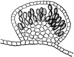
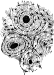
Delesseria Lepriuerei
Section through a conceptacle of Delesseria Leprieurei, showing the spores, which are single specialized…
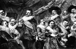
Banquet of the Officers of the Civic Guard
This painting was done by Frans Hals. It is an important piece from the military collection by the artist.…
Trillium
Diagram of flower of Trillium. In this, as in all such diagrams of cross-section of blossoms, the parts…
Anther
Diagram of the lower part of an anther, cut across above, and the upper part of a leaf, to show how…
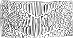
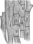
Buttonwood
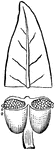
Some wood-cells from Buttonwood, Platanus, a whole cell and lower end of another on the left; a cell…
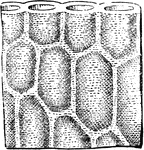
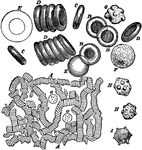
Chara
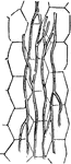
Outlines of a portion of the stem in section, showing the central cell and the outer or cortical cells.
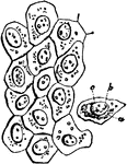
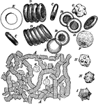
Delesseria Lepreiurei
A piece of the rose-red Delesseria Lepreiurei, showing that it is composed of a layer of cells.
Delesseria Lepreiurei
A piece of the rose-red Delesseria Lepreiurei, showing the cells are gelatinous and thick-walled.
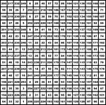
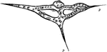
Magic Square
A square divided into equal squares, like a chessboard, in each of which is placed one of a series of…
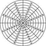
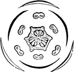
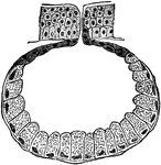
Magic Circle of Circles
The magic circle of circles, first developed by Benjamin Franklin, consists of eight annular rings and…
Section of the Internal Saphenous Nerve
Section of the internal saphenous nerve. Stained in osmic acid and subsequently hardened in alcohol.…
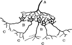
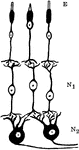
Diagram of the Relation between Cerebrospinal and Sympathetic Neurons
Diagram showing the relation of the cerebrospinal to the sympathetic neurons. Labels: A, a medullated…
Red and White Blood Cells
Red and white corpuscles (cells) of the blood, magnified. Labels: A, moderately magnified, the red corpuscles…
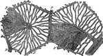
Capillary Network
Isolated capillary network formed by the junction of several hallowed-out cells, and containing colored…
Diagram Showing Various Forms of Secreting Glands
Diagram showing various forms of secreting glands. Labels: 1, general plan of a secreating membrane;…
Hepatic Lobules of the Liver
Diagram of two hepatic lobules of the liver. "The left hand lobule is represented with the intralobular…
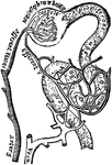
Plan of the Blood Vessels Connected with the Tubules
Plan of the blood vessels connected with the tubules. "The blood passes downwards in straight vessels…
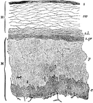
Section of Epidermis (Skin)
Section of epidermis. Labels: H, horny layer, consisting of s, superficial horny scales; sw, swollen-out…
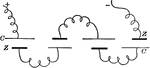
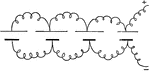
Parallel Connection
"Diagram of a multiple or parallel connection. When connected in this manner the voltage of the battery…
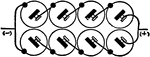
Series Multiple Connection
"Diagram of a series multiple connction. Two sets of cells are connected in series, and the two batteries…
Neurons and Sensory Epithelium in the Retina
Diagram showing relations of the neurons and sensory epithelium in the retina. labels: E, epithelial…
Epithelium Cells
Section of stratified epithelium. Labels: c, lowermost columnar cells; P, polygonal cells above these;…
Epithelium Cells Lining the Bladder
Section of the transitional epithelium lining the bladder, highly magnified. Labels: a, superficial;…
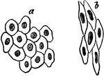
Simple Pavement Epithelium Cells
Simple pavement epithelium. Labels: a, from a serious membrane; b, from a blood vessel. "In simple pavement…
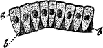
Simple Columnar Epithelium
Simple columnar epithelium. Labels: a, the cells; b, intercellular substance between the lower end of…
Glandular Epithelium Cells
Glandular epithelium with the cells set round a simple saccular gland, highly magnified. "Glandular…
Ciliated Epithelium Cells
Ciliated epithelium from the human trachea, highly magnified. Labels: a, large ciliated cell; d, cell,…
Subcutaneous Areolar Tissue from a Young Rabbit
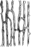
Subcutaneous areolar tissue from a young rabbit, highly magnified. The white fibers are in wavy bundles,…
Fat Cells
A few cat cells from the margin of a fat lobule in adipose tissue, very highly magnified. Labels: f.g.…
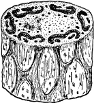
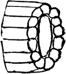
Bone Tissue of Humerus
Transverse section of compact tissue of humerus, magnified about 150 diameters. Three of the Haversian…
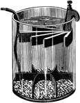
Battery
"The term battery is applied either to a single jar, or cell, containing the generating materials, or…
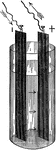
Gravity Cell
"The electric resistance of the porous cup employed in the Daniel cell, and the local action produced…
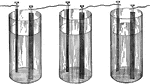
Series Connection
Figure showing a series type of connection between multiple battery cells, where the negative end of…
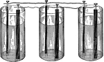
Parallel Connection
Figure showing battery cells connected in parallel, with all of the positive ends wired together, and…
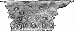
Cells from the Abdominal Lining
Flat cells from the surface of the lining membrane of the abdomen (peritoneum). Labels: a, cell-body;…
Cartilage Tissue
A thin slice of cartilage highly magnified, showing the cartilage cells (a,b) scattered through an almost…
Series Connection
"When several cells are conected so that the positive plate of one is joined to the negative plate of…
Parallel Connection
"When all of the positive plates are connected on one side, and all of the negative plates are connected…
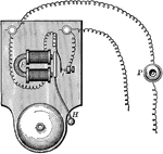
Electric Bell
"An electric bell consists mainly of an electromagnet, E, and a vibrating armature that carries a hammer,…
Muscle Fibers
Muscle fibers from the heart showing the striations and the junctions of the cells, highly magnified.

Blood Cells
Blood corpuscles. Labels: A, magnified about 400 diameters. The red corpuscles have arranged themselves…