Part of the central nervous system found in the skull.

Brain

Part of the central nervous system found in the skull.

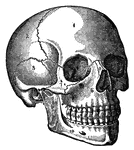
The head is the part of the body that contains the brain and the organs of the special senses.

Head

The head is the part of the body that contains the brain and the organs of the special senses.

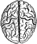
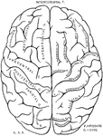
Top view of the human brain.

Human Brain

Top view of the human brain.

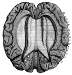
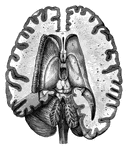
A top view of a dissection of the human brain showing the lateral fourth and fifth ventricles.

Human Brain

A top view of a dissection of the human brain showing the lateral fourth and fifth ventricles.

A view of the Corpus Callosum from above.

Human Brain

A view of the Corpus Callosum from above.

Side diagram of the human brain showing which parts of the brain control hearing, speech, vision, legs, head, arm, and face.

Human Brain

Side diagram of the human brain showing which parts of the brain control hearing, speech, vision, legs,…

Side diagram of the human brain showing which areas perform the sense of taste, smell, and vision.
Human Brain

Side diagram of the human brain showing which areas perform the sense of taste, smell, and vision.

Coral that has branched out and looks like a brain.

Astraea Pallida

Coral that has branched out and looks like a brain.

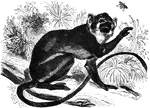
"<em>Genus</em> Saimiri. The animals of this genus are but about ten inches in length and are the most slender, graceful, and interesting of the whole monkey family. The large development of the brain is one of their most striking characteristics. The tail is slightly prehensile, yet used in climbing trees." &mdash;Goodrich, 1885

Saimiri

"Genus Saimiri. The animals of this genus are but about ten inches in length and are the most…
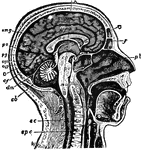
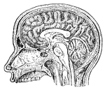
"The Brain is the encephalon, or center of the nervous system and the seat of consciousness and volition in man and the higher animals. Median Longitudinal Section through Head and Upper Part of Neck, to Show relation of Brain to Cranium and the Spinal Cord. <em>c,</em> cerebrum; <em>cb,</em> cerebellum; <em>sc,</em> spinal cord; <em>spc,</em> spinal column; <em>mo,</em> medulla oblongata passing, through foramen magnum, into the spinal cord; <em>pv,</em> pons Varolii; <em>cp,</em> cerebral peduncles or crura cerebri; <em>cqa,</em> anterior corpora quadrigemina; <em>cqp,</em> posterior corpora quadrigemina; <em>pg,</em> pineal gland; <em>pb,</em> pituitary body; <em>cc,</em> corpus collosum, divided transversely; <em>f,</em> fornix; <em>mg,</em> marginal gyrus; <em>gf,</em> gyrus fornicatus; <em>cmg,</em> calloso-marginal suleus; <em>O,</em> occipital lobe; <em>po,</em> parieto-occipital fissure; <em>cf,</em> calcarine fissure; <em>dm,</em>dura mater, separating cerebrum from cerebellum."&mdash;(Charles Leonard-Stuart, 1911)

Human Brain

"The Brain is the encephalon, or center of the nervous system and the seat of consciousness and volition…
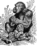
"The Gorilla is a celebrated anthropoid ape, generally belived to come nearer than any known one to man. The height is about 5 feet, almost the same as man. On the other hand, the greatest capacity of the gorilla's brain is only 34 and one half cubic inches, the least 23 against 62 in the least capacious human skull, and 114 in the greatest. The low facial angle also, and the abundant hair, with the extraordinary breadth of the chest, diminish the resemblance. The last mentioned characteristic imparts to the animal a colossal strength. It is a native of Lower Guinea and the interior of equinoctial Africa."—(Charles Leonard-Stuart, 1911)

Female Gorilla

"The Gorilla is a celebrated anthropoid ape, generally belived to come nearer than any known one to…

"Tinoceras, or tinotherium, is a genus of mammals now extinct, found in the Eocene, and representing the order Dinocerata. The individuals were all large, some of them nearly equaling the elephants, while the brain was smaller than that of any living or fossil mammal."&mdash;(Charles Leonard-Stuart, 1911)

Tinoceras

"Tinoceras, or tinotherium, is a genus of mammals now extinct, found in the Eocene, and representing…
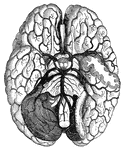
"Arteries and their Branches at the Base of the Brain." &mdash; Blaisedell, 1904

Blood vessels of the brain

"Arteries and their Branches at the Base of the Brain." — Blaisedell, 1904
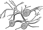
"Wherever nerve cells are abundant, the nerve tissue has a gray color; in other places, it looks white. Most of the gray matter of the brain is on the surface." — Blaisedell, 1904

Nerve Cells of the Brain

"Wherever nerve cells are abundant, the nerve tissue has a gray color; in other places, it looks white.…

"Diagram illustrating the General Arrangement of the Nervous System. (posterior view.)" — Blaisedell, 1904

Nervous System

"Diagram illustrating the General Arrangement of the Nervous System. (posterior view.)" — Blaisedell,…
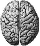
"The Upper Surface of the Cerebrum. Showing its division into two hemispheres, and also of the convolutions." &mdash; Blaisedell, 1904

Cerebrum

"The Upper Surface of the Cerebrum. Showing its division into two hemispheres, and also of the convolutions."…
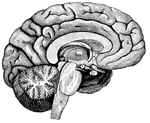
"<em>A</em>,frontal love of the cerebrum; <em>B</em>, parietal lobe; <em>C</em>, parieto-occipital lobe; <em>D</em>, occcipital lobe; <em>E</em>, cerebellum; <em>F</em>; arbor vitae; <em>H</em>, pons Varolii; <em>K</em>, medulla oblongata." — Blaisedell, 1904

Left Half of the Brain

"A,frontal love of the cerebrum; B, parietal lobe; C, parieto-occipital lobe;…
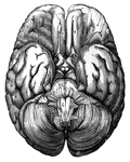
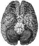
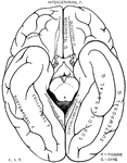
<em>A</em>, anterior lobe of the cerebellum; <em>B</em>, olfactory nerve; <em>C</em>, portion of the posterior lobe; <em>D</em>, optic chiasm; <em>E</em>, optic tract; <em>H, M,</em> hemispheres of the cerebellum; <em>K</em>, portion of the occipital lobe; <em>N</em>, medulla oblongata; <em>R</em>, pons Varolii."

Base of the brain

A, anterior lobe of the cerebellum; B, olfactory nerve; C, portion of the…

Part of a human brain.

Cerebellum

Part of a human brain.

"A nerve cell from the brain." &mdash; Ritchie, 1918

Neuron

"A nerve cell from the brain." — Ritchie, 1918

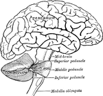
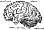
"The brain seen from the side, showing the three principal divisions." &mdash; Ritchie, 1918

Side view of the brain

"The brain seen from the side, showing the three principal divisions." — Ritchie, 1918

"The cerebrum seen from above, showing the hemispheres." — Ritchie, 1918

Top View of the Cerebrum

"The cerebrum seen from above, showing the hemispheres." — Ritchie, 1918

"Brain and spinal cord, with the thirty-one pairs of spinal nerves." — Tracy, 1888

Central Nervous System

"Brain and spinal cord, with the thirty-one pairs of spinal nerves." — Tracy, 1888

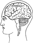
"Diagram showing the position of the nervous centers in the head." &mdash; Tracy, 1888

Cerebellum

"Diagram showing the position of the nervous centers in the head." — Tracy, 1888

"Part of the nervous system viewed in front." —Davison, 1910

Nervous System

"Part of the nervous system viewed in front." —Davison, 1910

A side view of the two great cavities of the body and their organs. 1: The mouth. 2: The thorax. 3: The abdomen. 4: The brain. 5: The spinal cord. 6: A salivary gland. 7: The aesophagus. 8: The trachea and lungs. 9: The heart. 10: The liver. 11: The stomach. 12: The colon. 13: The small intestine. 14: The lacteals. 15: The thoracic duct. 16: The diaphragm. 17, 18, and 20: The walls of the lower cavity, composed of bones, muscles and skin. 19 and 20: The walls of the upper cavity.

Side View of the Body

A side view of the two great cavities of the body and their organs. 1: The mouth. 2: The thorax. 3:…

The cord-like structures composed of delicate filaments by which sensation or stimulative impulses are transmitted to and from the brain and other organs.

Nerves

The cord-like structures composed of delicate filaments by which sensation or stimulative impulses are…

In this Turbellarian the digestive tract (d.t.) is a blind sac. st., boundary of stomodaeum and mesenteron; c, cilia; g, ganglion (brain); g', ganglion of a new individual which is being formed by fission; o, mouth; o', mouth of new individual in process of formation; w, excretory system.

Stenostoma

In this Turbellarian the digestive tract (d.t.) is a blind sac. st., boundary of stomodaeum and mesenteron;…

This illustration shows the arrangement of the nervous material in the anterior end of an Oligochete Worm, seen in profile. That part of the body wall nearest the observer is supposed to be removed. a, anterior; b.w., body wall; g, dorsal ganglia (brain); g', ventral chain of ganglia; n, nerve ring around the pharynx; o, mouth; p, pharynx.

Oligochete Worm

This illustration shows the arrangement of the nervous material in the anterior end of an Oligochete…
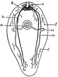
This is a diagram of a Turbellarian, showing the general arrangement of the nervous structures and one of the modes of occurrence of the excretory tubules, which in this case open seperately into the pharynx, on the ventral side of the animal. b., brain; e, eye-spots; ex, excretory canals consisting of a transverse portion passing from the mouth toward the dorsal side, and longitudinal tubes which branch into the capillary vessels terminating in f, the flame cells; lc., lateral nerve cords; m, mouth.

Turbellarian

This is a diagram of a Turbellarian, showing the general arrangement of the nervous structures and one…

This diagram shows a sagittal section of a Rotifer. b, brain; bl., excretory bladder; c, cloaca, the common opening of digestive and reproductive organs; co, caelom; e, eyespot; ex, excretory canal; f, flame cells; f.g., foot gland; ft., foot; g, gut; m, mouth; m.f., longitudinal muscle fibres; mx, mastax; o, ovary; pf., pharynx; s.g., salivary gland; t, tentacle; tr, trochus, or cilia-bearing disc.

Rotifer

This diagram shows a sagittal section of a Rotifer. b, brain; bl., excretory bladder; c, cloaca, the…

This diagram shows a fresh-water annelid. a, appendages; br., brain; d, dissepiments; i, intestine; m, mouth; nph, nephridium; oe, aesophagus; p, pavilion, line with ciliated entoderm; ph., pharynx; pr., processes from the anal segment; z, zone immediately in front of the anal segment where new segments are continually being formed; z', the zone of fission or budding. This takes place in the middle of a segment. The anterior half-segment of z' will produce a region like z for the anterior half of the worm.

Annelid

This diagram shows a fresh-water annelid. a, appendages; br., brain; d, dissepiments; i, intestine;…

This diagram shows the longitudinal section of the anterior end of the annelid. A, sagittal section; B, frontal section to show anterior portion of nervous system. b, brain; co., nervous collar about the mouth; c.v., contractile blood vessels; d, dissepiment; d.m., dermo-muscular wall; d.v., dorsal blood vessel; m, mouth; n.c., nerve cells; n.f., nerve fibers; np., nephridia; p, prostomium; ph., pharynx; s, setae; sn., segmental nerves; v.g, ventral chain of fanglia; v.v., ventral blood vessel. Only a portion of the blood vascular system is shown, and this appears unsectioned in the figure.

Annelid

This diagram shows the longitudinal section of the anterior end of the annelid. A, sagittal section;…

Diagram of the mouth of a snail, showing the lingual ribbon. br, brain; c, buccal cavity; co., caelom; g, gullet; j, jaw, against which the radula works; m, mouth; r., radula; r.s., radula sac, in which the radula is renewed as it is worn away in front.

Snail

Diagram of the mouth of a snail, showing the lingual ribbon. br, brain; c, buccal cavity; co., caelom;…
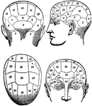
The term applied to the psychological theories of Gall and Spurzheim, founded upon 1, the discovery that the brain, as the organ of the mind, is not so much a single organ as a complex congeries of organs; and 2, observations as to the existence of a certain correspondence between the aptitudes of the individual and the configuration of his skull.

Phrenology

The term applied to the psychological theories of Gall and Spurzheim, founded upon 1, the discovery…
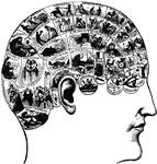
A theory which claims to be able to determine character, personality traits, and criminality on the basis of the shape of the head. This technique was developed in the early 1800's and is currently discredited as a pseudoscience.

Phrenology

A theory which claims to be able to determine character, personality traits, and criminality on the…
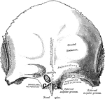
A bone in the human skull that consists of two portions, a vertical portion and a horizontal portion. This bone does a part in protecting your brain from harm.

Frontal Bone

A bone in the human skull that consists of two portions, a vertical portion and a horizontal portion.…
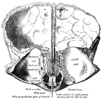
A bone in the human skull that consists of two portions, a vertical portion and a horizontal portion. This bone does a part in protecting your brain from harm.

Frontal Bone

A bone in the human skull that consists of two portions, a vertical portion and a horizontal portion.…

Scheme showing the connection of the several parts to the brain.

Brain

Scheme showing the connection of the several parts to the brain.

A top view of the human brain.

Brain

A top view of the human brain.

Fissures and gyres of the lateral surface of the left hemicerebrum of the brain.

Brain

Fissures and gyres of the lateral surface of the left hemicerebrum of the brain.

Fissures and gyres of the mesal surface of the left hemicerebrum of the brain.

Brain

Fissures and gyres of the mesal surface of the left hemicerebrum of the brain.

Fissures and gyres of the basal surface of the cerebrum portion of the brain.

Brain

Fissures and gyres of the basal surface of the cerebrum portion of the brain.

"Diagrams illustrating two stages in the development of the vertebrate eye. A, showing the relation of the ectoderm, the brain vesicle, and the optic vesicle. The right side of the figure shows a later stage than the left. B, later stage, showing the lens, eye-ball and retina in position. b.v., brain vesicle formed by the invagination oof the ectoderm (ect.); l, lens; mes., mesodermal tissue; o.n., optic nerve; o.s., optic stalk; o.v., optic vesicle, a portion of the brain vesicle; r, retinal layer; v.h., interior of eye-ball which comes to contain the vitreous humor." &mdash; Galloway

Vertebrate Eye

"Diagrams illustrating two stages in the development of the vertebrate eye. A, showing the relation…

"The size of the hemispheres of the brain (A) is so small that they leave exposed the olfactory ganglion (a), the cerebellum (C), and more or less of the optic lobes (B), and they are but partially connected together by the 'fornix' and 'anterior commissure,' the great cerebral commissure known as the 'corpus callosum' being absent." &mdash; Chambers, 1881

Opossum Brain

"The size of the hemispheres of the brain (A) is so small that they leave exposed the olfactory ganglion…

"Profile and vertex views of cerebrum. Dr, the frontal lobe; Par, parietal; Oc, occipital; Ts, temporo-sphenoidal lobe; SS, Sylvian fissure; RR, fissure of Rolando; PO, parieto-occipital fissure; IP, intra-pariental fissure; PP, Parallel fissure; SF and IF, supero- and infero-frontal fissures; 1, inferior, 2 middle, and 3, superior frontal convolutions; 4, ascending frontal convolution; 5, ascending parietal, 5', postero-parietal, and 6, angular convolutions; A, supra-marginal, or convolution of the parietal eminence; 7, superior, 8, middle, and 9, inferior temporo-sphenoidal convolutions; 10, superior, 11, middle, and 12, inferior occipital convolutions." &mdash; Encyclopedia Britanica, 1893

Brain

"Profile and vertex views of cerebrum. Dr, the frontal lobe; Par, parietal; Oc, occipital; Ts, temporo-sphenoidal…

"Brain of Perch. Upper aspect. a, cereoellum; b, optic lobes; c, hemispheres; e, lobi inferiores; f, hypophysis; g, lobi posteriores; i, olfactory lobes; n, nervus opticus; o, nervus olfactorius; p, nervus olulo-motorius; g, nervus trochlearis; r, nervus trigeminus; s, nervus acousticus; t, nrvus vagus; u, nervus abducens; v, fourth ventricle." &mdash; Encyclopedia Britannica, 1893

Perch Brain

"Brain of Perch. Upper aspect. a, cereoellum; b, optic lobes; c, hemispheres; e, lobi inferiores; f,…

"Brain of Perch. Lower aspect. a, cereoellum; b, optic lobes; c, hemispheres; e, lobi inferiores; f, hypophysis; g, lobi posteriores; i, olfactory lobes; n, nervus opticus; o, nervus olfactorius; p, nervus olulo-motorius; g, nervus trochlearis; r, nervus trigeminus; s, nervus acousticus; t, nrvus vagus; u, nervus abducens; v, fourth ventricle." &mdash; Encyclopedia Britannica, 1893

Perch Brain

"Brain of Perch. Lower aspect. a, cereoellum; b, optic lobes; c, hemispheres; e, lobi inferiores; f,…

"Brain of Polypterus. Upper aspect. a, medulia; b, corpora restiformia; c, cerebellum; d, lobi optici; e, hypophysis; f, fissura cerebri magna; g, nervus opticus; g', chiasma; h, hemispheres; i, lobus olfactorius; k, sinus rhomboidalis (fourth ventricle)." &mdash; Encyclopedia Britannica, 1893

Polypterus Brain

"Brain of Polypterus. Upper aspect. a, medulia; b, corpora restiformia; c, cerebellum; d, lobi optici;…

"Brain of Polypterus. Lateral aspect. a, medulia; b, corpora restiformia; c, cerebellum; d, lobi optici; e, hypophysis; f, fissura cerebri magna; g, nervus opticus; g', chiasma; h, hemispheres; i, lobus olfactorius; k, sinus rhomboidalis (fourth ventricle)." &mdash; Encyclopedia Britannica, 1893

Polypterus Brain

"Brain of Polypterus. Lateral aspect. a, medulia; b, corpora restiformia; c, cerebellum; d, lobi optici;…

"Brain of Polypterus. Lower aspect. a, medulia; b, corpora restiformia; c, cerebellum; d, lobi optici; e, hypophysis; f, fissura cerebri magna; g, nervus opticus; g', chiasma; h, hemispheres; i, lobus olfactorius; k, sinus rhomboidalis (fourth ventricle)." &mdash; Encyclopedia Britannica, 1893

Polypterus Brain

"Brain of Polypterus. Lower aspect. a, medulia; b, corpora restiformia; c, cerebellum; d, lobi optici;…

"Brain of Carcharias. ae, nervus acousticus; b, corpus restiforme; c, cerebellum; d, lobus opticus; e, hypophysis; g, nervus opticus; h, hemisphere; i, lobus olfactorius; i', olfactory pedicle; k, nervus olfactorius; l, epiphysis; m, nervus oculo-motorius; tr, nervus trigeminus; v, nervus vagus." &mdash; Encyclopedia Britannica, 1893

Carcharias Brain

"Brain of Carcharias. ae, nervus acousticus; b, corpus restiforme; c, cerebellum; d, lobus opticus;…
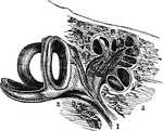
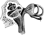
"By anatomists, the auditory nerve is associated with the facial, and is the seventh in order of origin from the brain, counting from before backwards. The seventh pair consists of the portio dura or facial, the portio mollis or auditory, and a small intermediate portion." &mdash; Chambers' Encyclopedia, 1875

Auditory Nerve

"By anatomists, the auditory nerve is associated with the facial, and is the seventh in order of origin…
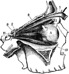
"The external bones of the temple are supposed to be removed in order to render visible the muscular arrangements. The muscle, 1, raises the eye-lid, and is constantly in action while we are awake. During sleep, the muscle being in repose and relaxed, the eye-lid falls and protects the eye from the action of light. The muscle, 4, turns the eye upwards; 5, downwards; 6, outwards; and a corresponding one on the inside, not seen in the figure, turns it inwards. No. 2 and 10 turn the eye round its axis. No. 11 is the great optic nerve, which conveys the sensation to the brain. If this nerve were cut, notwithstanding the eye might be in other respects perfect, the sense of sight would be destroyed." &mdash;Wells, 1857

Eye Muscles

"The external bones of the temple are supposed to be removed in order to render visible the muscular…
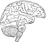
"Diagram illustrating the general relationships of the parts of the brain. A, fore-brain; b, midbrain; B, cerebellum; C, pons Varolii; D, medulla oblongata; B, C, and D together constitute the hind-brain." &mdash;Martin, 1917

Brain

"Diagram illustrating the general relationships of the parts of the brain. A, fore-brain; b, midbrain;…
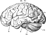
"The brain from the left side. Cb, the cerebral hemispheres forming the main bulkl of the fore-brain; Cbl, the cerebellum; Mo, the medulla oblongata; P, the pons Varolii; *, the fissure of Sylvius; Ro, the fissure of Rolando; Po, the Parieto-occipital fissure." &mdash;Martin, 1917

Brain

"The brain from the left side. Cb, the cerebral hemispheres forming the main bulkl of the fore-brain;…
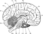
"Diagram of the left half of a vertical median section of the brain. H, H, convoluted inner surface of left cerebral hemisphere; Cc, corpus callosum; Th, optic thalamus; c.q., corpora quadrigemina; Cb, cerebellum; Sp.c, spinal cord; Mo, medulla oblongata; P, pons Varolii; oc, oculo-motor nerve; pt, pituitary body; op, optic nerve; Ro, fissure of Rolando; Po, parieto-occipital fissure; Fr, frontal lobe; Pa, parietal lobe; O, occipital lobe." &mdash;Martin, 1917

Brain

"Diagram of the left half of a vertical median section of the brain. H, H, convoluted inner surface…
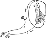
"Diagram of the simple reflex arc. R, receptor; A, afferent (sensory) neuron; E, efferent (motor) meuron; M, muscle." &mdash;Martin, 1917

Reflex Arc

"Diagram of the simple reflex arc. R, receptor; A, afferent (sensory) neuron; E, efferent (motor) meuron;…
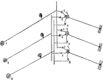
"Diagram to illustrate how a single sensory neuron may communicate with several motor neurons, and a single motor neuron with several sensory neurons." &mdash;Martin, 1917

Sensory Neuron

"Diagram to illustrate how a single sensory neuron may communicate with several motor neurons, and a…