Banner
This banner contains images of several different animals. There are also floral arrangements surrounding…
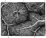
Rabbit Liver Lobule
Lobule of rabbit's liver, vessels and bile ducts injected. Labels: a, central or intralobular vein;…
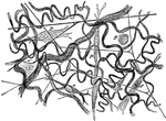
Subcutaneous Areolar Tissue from a Young Rabbit
Subcutaneous areolar tissue from a young rabbit, highly magnified. The white fibers are in wavy bundles,…
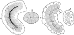
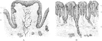
Rabbit's Intestinal Mucous Membrane
Vertical section of the intestinal mucous membrane of the rabbit. Two villi are represented, in one…
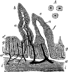
Serous Glands of a Rabbit
Serous glands. Labels: a, rabbit's pancreas "loaded" (resting); c, "discharged" (active) (observed in…
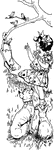
Girl with Animals
Girl outside with a dog and a rabbit, looking and pointing to the birds on a tree branch.
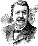
Joel Chandler Harris
(1848-1908) American author, best known as "Uncle Remus," who wrote the famous stories of "Brer Rabbit."
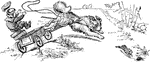
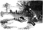
Dog Pulling a Wagon
A Dog pulling a wagon. He chases a rabbit and runs so fast that the boy falls out.
Three Rabbits With Carrot
Illustration of three rabbits and a carrot that can be used to write mathematics story problems involving…
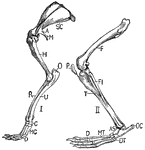
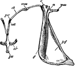
Rabbit Limbs
"Fore-limb and shoulder-girdle (I.) and hind-limb (II.) of rabbit. SC., Scapula; A., acromion; M., metacromion…
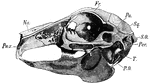
Rabbit Skull
"Side view of rabbit's skull. Pmx., Premaxilla; Na., nasal; Fr., frontal; Pa., parietal; Sq., squamosal;…
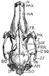
Upper Surface Rabbit Skull
"Upper surface of rabbit's skull. N., Anterior nostril; PMX., premaxilla; NA., nasal; FR., anterior…
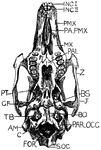
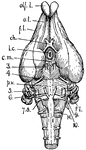
Under Surface Rabbit Skull
"Under surface of rabit's skull. Inc. I., First incisors; Inc. II., second incisors; PMX., premaxilla;…
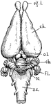
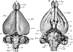
Dorsal View of Rabbit Brain
"Dorsal view of rabbit's brain. olf.l., Olfactory lobes; c.h., cerebral hemispheres; o.l., optic lobes…
Under Surface of Rabbit Brain
"Under surface of rabbit's brain. olf.l., Olfactory lobes; o.t., olfactory tract; f.l., frontal lobe…
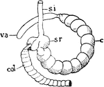
Rabbit Caecum
"Diagram of caecum in rabbit. s.i., Small intestine; s.r., sacculus rotundus; col., sacculated colon;…
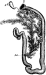
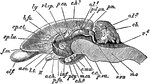
Rabbit Duodenum
"Duodenum of rabbit. P., Pyloric end of stomach; g.b., gall-bladder with bile duct and hepatic ducts;…
Rabbit Shoulder Girdle
"Lepus cuniculus. Shoulder-girdle with anterior end of sternum of young specimen. a, acromion; af, pre-scapular…
Rabbit Brain
"Lepus cuniculus. Brain. A, dorsal view; B, ventral; b. o, olfactory lobe; cb', median lobe of cerebellum…
Rabbit Brain
"Lepus cuniculus. Longitudinal vertical sectioin of the brain. cb, cerebellum, showing arbor vitae;…
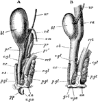
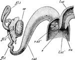
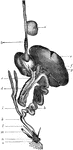
Rabbit Urogenital Organs
"Lepus cuniculus. The urogenital organs. A, of male; B, of female, from the left side. The kidneys and…
Rabbit Vagina
"Lepus cuniculus. The anterior end of the vagina, with the right uterus. Fallopian tube, and ovary.…
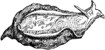
Sea Hare
The name of a genus of gasteropodous mollusca. These animals are slug-like in appearance, and derive…

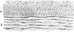
Epithelium of the Rabbit's Cornea
The vertical section of the stratified epithelium of the cornea of a rabbit. Labels: a, Anterior epithelium…
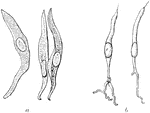
Branched Tendon Cells
The branched character of the cells is seen. Shown is a transverse section from a cross section of the…
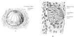
Peyer's Glands
The glands of Peyer occur chiefly but not exclusively in the small intestine. They are found in greatest…
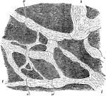
Capillary Network in the Lobules of a Rabbit's Liver
The liver is made up of small roundish or oval portions called lobules, each of which is composed of…
Lymphatics of Rabbit's Diaphragm
Lymphatics of central tendon of rabbit's diaphragm, stained with silver nitrate. The ground substance…
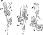
Cells from the Olfactory Region of a Rabbit
Cells from the olfactory region of the rabbit. Labels: st, supporting cells; r, r', varieties of rod-cells;…
Magnified Rabbit's Cornea
Vertical section of rabbit's cornea. Labels: anterior epithelium, showing the different shapes of the…
Section of Rabbit's Cornea
Vertical section of rabbit's cornea, stained with gold chloride. Labels: e, Laminated anterior epithelium.…
Lens of a Rabbit
Meridional section through the lens of a rabbit. Labels: 1, Lens capsule; 2, epithelium of lens; 3,…
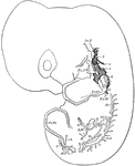
Lymphatics in a Rabbit Embryo
Developing lymphatics in rabbit embryo of 14 days. Lymphatic vessels are heavily shaded; veins are light.…
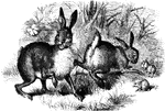
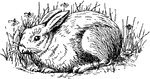
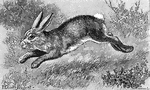
Rabbits
Rabbits are small mammals in the family Leporidae of the order Lagomorpha, found in several parts of…
Tongue of Human and Rabbit
A, Section through papilla vallata of a human tongue. B, Section through part of the papilla foliata…
Taste bud of Rabbit
A, Three-quarter surface view of taste bud from the papilla foliata of a rabbit. B, Vertical section…

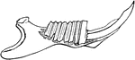
Rabbit Stick
"A throwing-stick, similar to the Australian boomerang, used by the Indians of the southwestern United…
Mandible of a Rabbit
Lateral half of mandible of a rabbi, opened to show the arrangement of rodent teeth.
Alimentary Canal of a Bird
Alimentary canal of a bird. Labels: a, ingluvies; b, proventriculus; c, pancreas; d, duodenum; e, liver;…

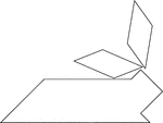
Rabbit Siting on Its Hind Legs
Tangrams, invented by the Chinese, are used to develop geometric thinking and spatial sense. Seven figures…

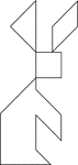
Stylized Rabbit
Tangrams, invented by the Chinese, are used to develop geometric thinking and spatial sense. Seven figures…

Rabbit Lieing Down
Tangrams, invented by the Chinese, are used to develop geometric thinking and spatial sense. Seven figures…
Rabbit Siting on Its Hind Legs
Tangrams, invented by the Chinese, are used to develop geometric thinking and spatial sense. Seven figures…

Stylized Rabbit
Tangrams, invented by the Chinese, are used to develop geometric thinking and spatial sense. Seven figures…
Rabbit Lying Down
Tangrams, invented by the Chinese, are used to develop geometric thinking and spatial sense. Seven figures…

Rabbit Siting on Its Hind Legs
Tangrams, invented by the Chinese, are used to develop geometric thinking and spatial sense. Seven figures…