This human anatomy ClipArt gallery offers 148 illustrations of general dissected or cross-sectional views of the human body that do not focus on a particular system.
Abdomen
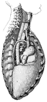
"The Abdominal Viscera in situ, as seen when the abdomin is laid open and the great omentum…
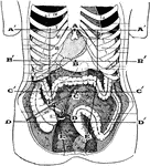
Principal Organs of the Thorax and Abdomen
"The principal muscles are seen on the left, and superficial veins on the right." — Blaisedell, 1904
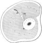
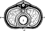
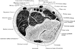
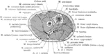
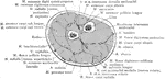
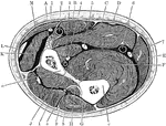
Transverse Section of the Abdomen
A transverse section of the abdomen in the lumbar region showing abdominal muscles.
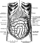
Abdominal Region
Showing the average position of the abdominal viscera with their surface markings. Labels: A, sterno-ensiform…
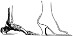
High Heel and Arch of Foot
"Showing the arch of the foot, and how a high-heeled shoe props it up on end." — Ritchie, 1918
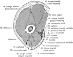
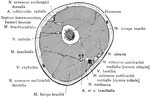
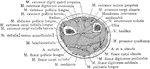
Cross Section Through Upper Arm above Epicondyles
Section passes through the right upper arm one inch above the epicondyles.
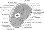
Cross Section Through Arm below Insertion of Deltoid
Section through upper third of arm, immediately below the insertion of the deltoid.
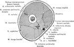
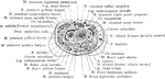
Cross Section Through Lower Third of the Upper Arm
Section through the lower third of the upper arm.
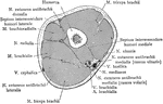
Cross Section Through Middle Thirds of the Upper Arm
Section through the middle thirds of the upper arm.
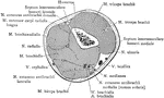
Horizontal Section of Arm
Horizontal section at middle of right arm. Labels: B.V., basilic vein; CEPH. V., cephalic vein; I.C.N.…
Transverse Section Through Arm
Transverse section through the middle of the right forearm, in the position of semipronation.
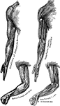
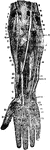
Bones and Muscles of the Arms
Showing relations of the muscles and bones of the arms from the inner side.
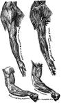
Bones and Muscles of the Arms
Showing relations of the muscles and bones of the arms from the outer side
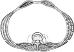
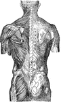
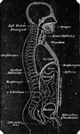
Vertical Section of the Back
"The spinal column below the twelfth dorsal vertebra at A has been removed, as well as the…
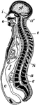
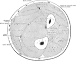
Longitudinal Section of Body
Diagrammatic longitudinal section of the body. Labels: a, the neural tube, with its upper enlargement…
Buttock and Thigh
Buttock and back of thigh. Labels: 1, gluteus maximus; 2, gluteus medius; 3, gluteal artery; 4, gluteus…
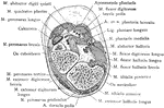
Transverse Section Through the Carpus
Transverse section through the carpus, showing the relative position of the tendons, vessels, and nerves.
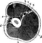
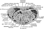
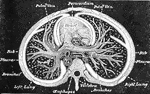
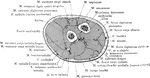
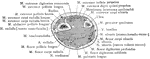
Section Across the Body in the Chest Region
A Diagrammatic section across the body in the chest region. Labels: x, the dorsal tube, which contains…
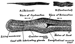
Lateral section of the chest
"A, a muscle which aids in pushing the food down the esophagus; B, esophagus; C,…
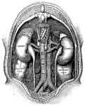
Circulatory
1: Right auricle of the heart. 2 and 3: Large veins that open into the right auricle. 4: Veins of the…
Human Esophagus
An Image of an esophagus showing how a mass of food, or bolus, passes through the esophagus into the…
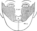
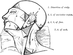
Development of the Face
Showing the development of the face. F.N.P., Part formed from the frontonasal process; L, from its lateral…
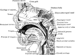
Mesial Sagittal Section of Child's Face
Anterior portion of mesial sagittal section of child's head, probably of about three year.
Cross Section Five Inches Above the Lower End of the Fibula
Section five inches above the lower end of the fibula.
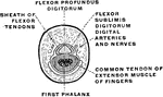
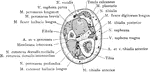
Section Through the Finger
Section passing through the middle third of the first phalanx of the middle finer. The tendon of the…

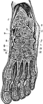
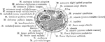
Frontal Section of Foot and Ankle
Frontal section of the right ankle and foot. Viewed from in front.

Oblique Anteroposterior Section of
Oblique anteroposterior section of foot, to show the synovial cavities of the tarsus. Labels: 1, tibia;…
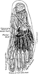
Forearm and Hand
Deep dissection of the front of the forearm and hand, showing the muscles, nerves, blood vessels, etc.…
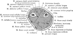
Cross Section Through Forearm One Inch below Elbow
Section through right forearm one inch below the elbow.
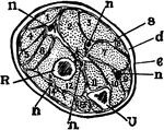
Section Across the Forearm
A section across the forearm a short distance below the elbow-joint. R and U, its two supporting bones,…
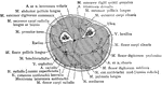
Cross Section Through Forearm Two Inches below Elbow
Section through right forearm, two inches below the elbow.
Cross Section One Inch above the Styloid Process of the Forearm
Section one inch above the styloid process of the right radius.
Cross Section Three Inches above the Styloid Process of the Forearm
Section three inches above the styloid process of the right radius.
Cross Section Through the Styloid Process of the Forearm
Section through the styloid process of the right radius.
Cross Section Through Forearm Two Inches below Elbow
Section through upper third of the right forearm.
Cross Section Two Inches above the Styloid Process of the Forearm
Section two inches above the styloid process of the right radius.
Dissection of Forearm
Deep dissection of front of the forearm. Labels: 1, supinator longus; 2, ulnar nerve; 3, brachialis;…
Dissection of the Forearm
Deep dissection of the front of the forearm. Labels: 1, supinator longus; 2, ulnar nerve; 3, brachialis…
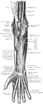
Muscles and Nerves of the Forearm
The muscles and nerves on the front of the forearm and hand. The pronator radii teres, flexor carpi…
Section Across Forearm
Section across the forearm in the middle third. Labels: A, pronator radii teres; B, flexor carpi radialis;…
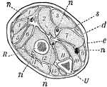
Forearm, Section of
A section across the forearm a short distance below the elbow-joint. R and U, its two supporting bones,…
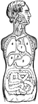
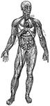
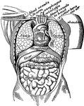
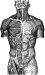
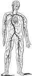
Front View of the Body
The position of the organs of the mouth, thorax, and abdomen. 1, 2, and 3: Salivary glands. 4: The larynx…
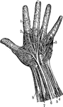
Hand
1. Nerves of the skin 2. Tendons 3. Arteries of the palm of the hand 4. Elbow nerve 5. Elbow artery…