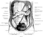
The abdomen viscera after the removal of the jejunum and ileum. The transverse colon is much more regular than usual. Both the liver and caecum extend lower down than normal. The subdivisions of the abdominal cavity are indicated by dark lines.

Abdomen Laid Open After Removal of Jejunum and Ileum

The abdomen viscera after the removal of the jejunum and ileum. The transverse colon is much more regular…
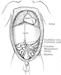
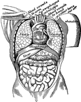
The abdominal and thoracic viscera of a five months fetus. The large liver and large size if its left lobe at this age should be noted.

Abdomen of Fetus

The abdominal and thoracic viscera of a five months fetus. The large liver and large size if its left…
The air passages of the lungs.

Air Passages

The air passages of the lungs.

"The air passages of the lungs." — Richardson, 1906

Air Passages

"The air passages of the lungs." — Richardson, 1906

"Diagram showing the capillary network of the air sacs and origin of the pulmonary veins.. <em>A</em>, small branch of pulmonary artery; <em>B</em>, twigs of pulmonary artery; <em>C</em>, capillary network around the walls of the air sacs; <em>D</em>, branches of network converging to form the veinlets of the pulmonary veins." — Blaisedell, 1904

Capillaries of the Air Sac

"Diagram showing the capillary network of the air sacs and origin of the pulmonary veins.. A,…
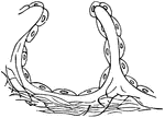
"<em>A</em>, epithelial lining wall; <em>B</em>, partition between two adjacent sacs, in which run capillaries; <em>C</em>, fibers of elastic tissues." &mdash; Blaisedell, 1904

Diagrammatic view of an air sac

"A, epithelial lining wall; B, partition between two adjacent sacs, in which run capillaries;…
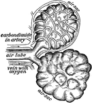
Two of the air sacs from the lungs with the network of blood tubes shown about one.

Air Sacs from the Lungs

Two of the air sacs from the lungs with the network of blood tubes shown about one.
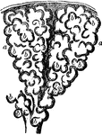
Diagram of the air tubes with the lung tissue removed.

Diagram of Air Tubes in the Lungs

Diagram of the air tubes with the lung tissue removed.

Two alveoli of the lung, highly magnified. Alveoli are cavities which are honeycombed with bulgings in order to increase the surface area exposed to air and covered by capillaries. Labels: b,b, bulgings of the alveoli, a, a.

Two Alveoli of the Lung

Two alveoli of the lung, highly magnified. Alveoli are cavities which are honeycombed with bulgings…
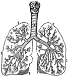
"Organs in the body cavity viewed from the front." —Davison, 1910

Organs of the Body Cavity

"Organs in the body cavity viewed from the front." —Davison, 1910

The bronchia. Labels; 1, Outline of the right lung. 2, Outline of the left lung. 3, Larynx. 4, Trachea. 5, 6, 7, 8, Bronchial tubes. 9, Air-cells.

The Bronchia

The bronchia. Labels; 1, Outline of the right lung. 2, Outline of the left lung. 3, Larynx. 4, Trachea.…
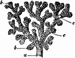
A small bronchial tube. Labels: a, dividing into its terminal branches, c; these have pouched or sacculated walls and end in the sacculated infundibula, b.

A Bronchial Tube

A small bronchial tube. Labels: a, dividing into its terminal branches, c; these have pouched or sacculated…

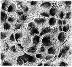
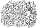
The capillary network of the pulmonary blood vessels in the human lung.

Capillary Network in Lung

The capillary network of the pulmonary blood vessels in the human lung.

"Lung of Ceratodus, opened in its lower half to show its cellular pouches. a, right half; b, left half; c, cellular pouches; e, vena pulmonalis; f, arterial blood-vessel; oe, esophagus, opened to show glottis." &mdash; Encyclopedia Britannica, 1893

Ceratodus Lung

"Lung of Ceratodus, opened in its lower half to show its cellular pouches. a, right half; b, left half;…

Transverse section of the chest.

Transverse Section of the Chest

Transverse section of the chest.

An ideal view of the circulation in the lungs and system.

Circulation

An ideal view of the circulation in the lungs and system.

"<em>R.A.</em>, right auricle; <em>L.A.</em>, left auricle; <em>R.V.</em>, right ventricle; <em>L.V.</em>, left ventricle; <em>P.A.</i., pulmonary artery; <em>A</em>, pulmonary artery and vein of right lung; <em>B</em>, pulmonary artery and vein of left lung; <em>C</em>, carotid artery to head, showing branch of left subclavian artery; <em>D</em>, portal vein; <em>F</em>, hepatic artery; <em>G</em>, superior vena cava, bringing blood from head and upper limbs to right auricle." &mdash; Blaisedell, 1904

Diagram of the circulation of the blood

"R.A., right auricle; L.A., left auricle; R.V., right ventricle; L.V.,…

"The heart and blood-vessels diagrammatically represented. L, lung; M, intestine; P, liver; dotted lines represent lymphatic vessels." &mdash;Martin, 1917

Heart

"The heart and blood-vessels diagrammatically represented. L, lung; M, intestine; P, liver; dotted lines…
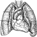
1, The trachea or windpipe; 2 and 3, right and left common carotid arteries; 4 and 5, right and left sub clavian arteries; 6, innominate artery; 7 and 8, right and left innominate veins; 9, Superior vena cava; 10, arch of the aorta; 11, ascending aorta; 12, pulmonary artery; 13, right atrium and auricula; 14 and 15, right and left lung; 16, conus arteriosus; 17, right ventricle; 18, left ventricle.

Heart and Lungs

1, The trachea or windpipe; 2 and 3, right and left common carotid arteries; 4 and 5, right and left…
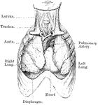
The heart and lungs.

The Heart and Lungs

The heart and lungs.

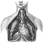
View of heart and lungs in situ. The front portion of the chest wall, and the outer or parietal layers of the pleurae and pericardium have been removed. The lungs are partly collapsed.

Heart and Lungs

View of heart and lungs in situ. The front portion of the chest wall, and the outer or parietal layers…
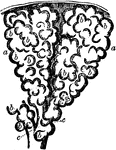
Two infundibula of the lung much magnified. Labels: b, b, hollow protrusions of the alveolus, opening into its central cavity; c, terminal branches of a bronchial tube.

Infundibula of the Lung

Two infundibula of the lung much magnified. Labels: b, b, hollow protrusions of the alveolus, opening…
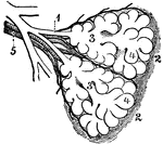
Diagram of the two primary lobules of the lungs, magnified. Labels: 1, Bronchial tube. 2, A pair of primary lobules connected by elastic tissue. 3, Intercellular air-passages. 4, Air-cells. 5, Branches of the pulmonary artery and vein.

Diagram of the Two Primary Lobules of the Lung

Diagram of the two primary lobules of the lungs, magnified. Labels: 1, Bronchial tube. 2, A pair of…

"Showing the structure of a lobule of the lung. The lobule has been injected with mercury, afterwards dried and cut open. A large bronchial tube with its various branches is well shown." &mdash; Blaisedell, 1904

Lobule of a Lung

"Showing the structure of a lobule of the lung. The lobule has been injected with mercury, afterwards…
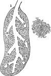
In birds the lungs are confined to the back wall of the chest. They are not separated into lobes, but are oblong and flattened in shape, and connected with a series of air-receptacles scattered through various parts of the body. Shown is a section of a bird lung, magnified. Labels: 1, A bronchial tube that ends in a sac (caeca). 2, Division of the bronchial tubes that branch out among lobules. B, A plexus of capillary vessels.

Section of the Lung of a Bird

In birds the lungs are confined to the back wall of the chest. They are not separated into lobes, but…

In birds the lungs are confined to the back wall of the chest. They are not separated into lobes, but are oblong and flattened in shape, and connected with a series of air-receptacles scattered through various parts of the body. Shown is the right lung of a goose. Labels: 1, A bronchial tube which divides into two tubes that open into the abdominal air-receptacles (2).

The Right Lung of a Goose

In birds the lungs are confined to the back wall of the chest. They are not separated into lobes, but…
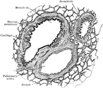
Section of lung, showing small air tubes and branch of pulmonary artery.

Section of Lung Showing Air Tubes

Section of lung, showing small air tubes and branch of pulmonary artery.
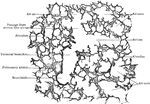
Section of lung, showing general relations of division of air tubes.

Section of Lung Showing General Relations of Divisions of Air Tubes

Section of lung, showing general relations of division of air tubes.
Lung with pleuro-pneumonia.

Lung with Pleuro-Pneumonia

Lung with pleuro-pneumonia.

Lung with croupos of vesicular pneumonia. Stage of consolidation.

Lung with Vesicular Pneumonia

Lung with croupos of vesicular pneumonia. Stage of consolidation.

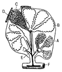
"<em>A</em>, an air sac; <em>B</em>, an air sac cut open; <em>C</em>, capillary network over an air sac; <em>D</em>, branch of pulmonary artery; <em>E</em>, branch of pulmonary vein; <em>F</em>, bronchial tube." &mdash; Blaisedell, 1904

Lobule of the lung

"A, an air sac; B, an air sac cut open; C, capillary network over an air…

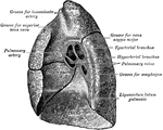
Mediastinal surface of right lung.

Mediastinal Surface of the Lung

Mediastinal surface of right lung.

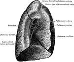
Mediastinal surface of left lung.

Mediastinal Surface of the Lung

Mediastinal surface of left lung.

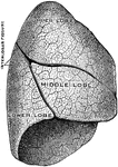
The right lung. The outer or costal surface.

Outer Surface of the Lung

The right lung. The outer or costal surface.

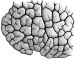
"Lobules of various sizes are well shown." &mdash; Blaisedell, 1904

Portion of a child's lung

"Lobules of various sizes are well shown." — Blaisedell, 1904

Section of lung with distended blood vessels, highly magnified. Labels: c,c, partitions between alveoli; b, small artery giving off capillaries to alveoli.

Section of Lung

Section of lung with distended blood vessels, highly magnified. Labels: c,c, partitions between alveoli;…
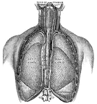
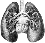
"<em>L.A.</em>, left ventricle; <em>B</em>, right ventricle; <em>F</em>, pulmonary artery; <em>G</em>, aorta; <em>H</em>, arch of the aorta; <em>K</em>, innominate artery; <em>L</em>, right common carotid artery; <em>M</em>, right subclavian artery; <em>N</em>, thyroid cartilage forming upper portions of the larynx; <em>O</em>, trachea." &mdash; Blaisedell, 1904

Lungs

"L.A., left ventricle; B, right ventricle; F, pulmonary artery; G,…

"<em>A</em>, division of left bronchus to upper lobe; <em>B</em>, left branch of the pulmonary artery; <em>C</em>, left bronchus; <em>D</em>, left superior pulmonary vein; <em>E</em>, left inferior pulmonary vein; <em>F</em>, left auricle; <em>K</em>, inferior vena cava; <em>L</em>; division of the right bronchus to lower lobe; <em>M</em>, right inferior pulmonary vein; <em>N</em>, right superior pulmonary vein, <em>O</em>, right branch of the pulmonary artery; <em>P</em>, division of right bronchus to upper lobe; <em>R</em>, left ventricle; <em>S</em>, right ventricle." &mdash; Blaisedell, 1904

Lungs

"A, division of left bronchus to upper lobe; B, left branch of the pulmonary artery;…

"Relative Postion of the Lungs, the Heart, and Some of the Great Vessels belonging to the latter. <em>A</em>, left common carotid artery; <em>B</em>, external carotid artery; <em>C</em>, internal carotid artery; <em>D</em>, left jugular vein; <em>E</em>, sterno-cleido muscle; <em>F</em>, right innominate artery with branches to head and arm; <em>G</em>, left subclavian artery; <em>H</em>, great azygos vein; <em>K</em>, thoracic duct." &mdash; Blaisedell, 1904

Lungs

"Relative Postion of the Lungs, the Heart, and Some of the Great Vessels belonging to the latter. A,…

The lung is the essential organ of respiration in air-breathing vertebrates. Its principal function is to transport oxygen from the atmosphere into the bloodstream, and excrete carbon dioxide from the bloodstream into the atmosphere.

Lungs

The lung is the essential organ of respiration in air-breathing vertebrates. Its principal function…
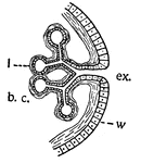
This is a diagram illustrating lungs or tracheae. b.c., the cavity in which the body fluids circulate; l, the walls of the lung, which are much thinned inpockedings of the body wall (w); ex., the external medium-usually the atmosphere-in which the oxygen is found.

Lungs

This is a diagram illustrating lungs or tracheae. b.c., the cavity in which the body fluids circulate;…
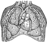
The lungs. Labels: 3, The lobes of the right lung. 4, The lobes of the left lung. 5, 6, 7, The heart. 9, 10, 11, The large blood-vessels. 12, The trachea. 15, The diaphragm.

The Lungs

The lungs. Labels: 3, The lobes of the right lung. 4, The lobes of the left lung. 5, 6, 7, The heart.…

The lungs and air passages seen from the front. On the left of the figure the pulmonary tissue has been dissected away to show the ramifications of the bronchial tubes. Labels: a, larynx; b, trachea; d, right bronchus. The left bronchus is seen entering the root of its lung.

The Lungs and Air Passages

The lungs and air passages seen from the front. On the left of the figure the pulmonary tissue has been…
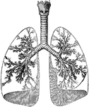
The lungs and windpipe (trachea). Labels: 1, larynx; 2, windpipe (trachea); 3, right lung, showing bronchi and bronchial tube.

Lungs and Trachea

The lungs and windpipe (trachea). Labels: 1, larynx; 2, windpipe (trachea); 3, right lung, showing bronchi…

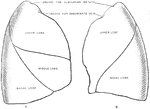
Costal surfaces of the two lungs. A, right lung. B, left lung.

Costal Surfaces of the Lungs

Costal surfaces of the two lungs. A, right lung. B, left lung.

Mediastinal surfaces of the two lungs of a subject hardened by formalin injection. A, right lung. B, left lung. Labels: a, base; 2, fissure; 3, cardiac depression; 4, groove for innominate vein; 5, groove for innominate artery; 6, apex pulmonis; 7, groove for vena azygos major; 8, eparterial bronchus; 9, pulmonary artery (right); 10, fissure; 11, groove for aorta; 12, bronchus; 13, pulmonary artery (left); 14, apex pulmonis; 15, groove for left subclavian artery; 16, groove for left innominate vein; 17, cardiac depression; 18, fissure; 19, base.

Mediastinal Surfaces of the Lungs

Mediastinal surfaces of the two lungs of a subject hardened by formalin injection. A, right lung. B,…
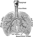
"The lungs fill up most of the cavity of the chest. One lies on either side of the heart which is in the middle of the chest. The lungs in animals are called lights because they are spongy sacs and so light as to float when thrown into water." &mdash;Davison, 1910

The lungs

"The lungs fill up most of the cavity of the chest. One lies on either side of the heart which is in…
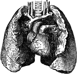
The lung, which are the two essential organs of respiration contained in the cavity of the thorax.

The Lungs

The lung, which are the two essential organs of respiration contained in the cavity of the thorax.

Left pleural sac in a subject hardened by formalin injection, opened into by the removal of the costal part of the parietal pleura. The lung has also been removed so as to display the mediastinal pleura.

Pleural Sac

Left pleural sac in a subject hardened by formalin injection, opened into by the removal of the costal…
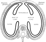
Diagram showing arrangement of pleural sacs, as seen in transverse section.

Arrangement of the Pleural Sacs

Diagram showing arrangement of pleural sacs, as seen in transverse section.
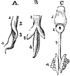
The development of the respiratory organs. A, is the esophagus of a chick on the fourth day of incubation, with the rudiments of the trachea on the lung of the left side, viewed laterally; 1, the inferior wall of the esophagus; 2, the upper portion of the same tube; 3, the rudimentary lung; 4, the stomach. B, is the same object seen from below, so that both lungs are visible. C, shows the tongue and respiratory organs of the embryo of a horse; 1, the tongue; 2, the larynx; 3, the trachea; 4, the lungs, viewed from the upper side.

Development of Respiratory Organs

The development of the respiratory organs. A, is the esophagus of a chick on the fourth day of incubation,…
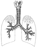
"Larynx, trachea, and bronchi, showing the manner of division, and the rings of cartilage." &mdash; Tracy, 1888

Respiratory system

"Larynx, trachea, and bronchi, showing the manner of division, and the rings of cartilage." —…

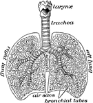
The respiratory system. Labels: 1. the larynx; 2. the trachea; 3. right bronchia; 4. left bronchia; 5. left lung divided into three lobes; 7. large bronchial tubes; 8. small bronchial tubes ending in air cells or vesicles.

Respiratory System

The respiratory system. Labels: 1. the larynx; 2. the trachea; 3. right bronchia; 4. left bronchia;…
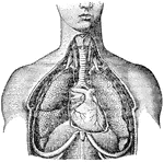
The respiratory system. Labels: 1, larynx; 2, trachea; 3, right lung; 4, left lung; 5, heart.

Respiratory System

The respiratory system. Labels: 1, larynx; 2, trachea; 3, right lung; 4, left lung; 5, heart.
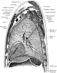
Sagittal section through left shoulder and left lung.

Sagittal Section Through Shoulder and Lung

Sagittal section through left shoulder and left lung.

Sagittal section through left shoulder, lung, and apex of the heart.

Sagittal Section Through Shoulder and Lung

Sagittal section through left shoulder, lung, and apex of the heart.

"Anatomy of the Snail: a, the mouth; bb, foot; c, anus; dd, lung; e, stomach, covered above by the salivary glands; ff, intestine; g, liver; h, heart; i, aorta; j, gastric artery; l, hepatic artery; k, artery of the foot; mm, abdominal cavity, supplying the place of a cenous sinus; nn, irregular canal in communication with the abdominal cavaty, and carrying the blood to the lung; oo, vessel carrying the blood from the lung to the heart." &mdash; Chambers, 1881

Snail Anatomy

"Anatomy of the Snail: a, the mouth; bb, foot; c, anus; dd, lung; e, stomach, covered above by the salivary…

"A contrivance for measuring the extreme differential capacity of the human lungs. The instrument most commonly employed consists on an inverted chamber submerged in a water bath. The breath is conducted by a flexible pipe and internal tube so as to collect in the chamber, which rises in the water, and is fitted with an index which marks the cubic inches of air expired after a forced inspiration...a, is a small gas-holder containing an inverted vessel a'; b, index which shows on the scale c the number of cubic inches expired; d, manometer, which, when a' is held down, shows the pressure which the lungs can exert; e, plug-vent for outlet of the expired air; f, cock for outlet of water; g, tube through which the expiration is made." &mdash;Whitney, 1889

Spirometer

"A contrivance for measuring the extreme differential capacity of the human lungs. The instrument most…
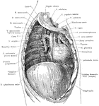
Deep structures of the right thoracic cavity, after removal of the right lung.

Thoracic Cavity after Removal of Lung

Deep structures of the right thoracic cavity, after removal of the right lung.
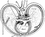
"Cross-section of thorax. A, bronchus, entering the lung; B, the aorta cut at its origin and again at the descending part of its arch; C, the pericardial space; D, the pleural cavity; E, the alimentary canal; PA, the pulmonary artery; X, the neural canal." &mdash;Martin, 1917

Thorax

"Cross-section of thorax. A, bronchus, entering the lung; B, the aorta cut at its origin and again at…