This human anatomy ClipArt gallery offers 242 illustrations the human muscular system. This includes both human skeletal muscle and human smooth and cardiac muscle. Human skeletal muscles (also known as striped, striated, or voluntary muscle) are those muscles that attach to bones and are involved in voluntary movements. Smooth muscles (also known as non-striated or involuntary muscle) are found lining the surface of internal organs such as the stomach, and cardiac muscles are those muscles specific to the heart. Both smooth and cardiac muscles are involuntarily controlled by the autonomic nervous system.
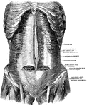
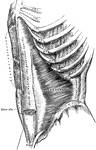
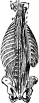
Muscles of the Abdomen
The muscles of the abdomen, showing the semilunar fold of Douglas. Viewed from in front.
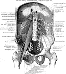
Muscles of the Abdominal Wall
View of the posterior abdominal wall to show the muscles and the nerves of the lumbo sacral plexus.
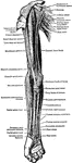
Muscles on the Front of the Arm and Forearm
Superficial muscles on the front of the arm and forearm.
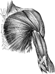
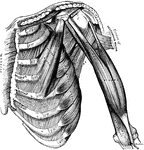
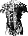
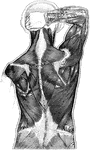
Muscles of the Arm and Thorax
Muscles of the thorax and front of the arm, showing some of the boundaries of the axilla.
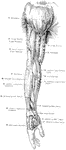
Arm Muscles
"abc, deltoid muscle; d, coraco branchfalls muacle; r, r, triceps; e, i, extensors of wrist and long…
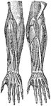
Muscles of the Forearm
Muscles in front of forearm. Labels: 62, pronator teres; 63, 65, 66, 67, flexors; 70, supinator longus;…
Arm Muscles
The muscles on the back of the hand, forearm, and lower half of the arm, as exposed on dissecting away…
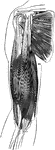
Arm Muscles
Two of the principal muscles f the arm (4 and 7). Between these is the bone of the arm (1) and the bones…
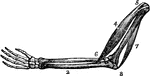
Arm Muscles in Action
Diagram of the muscles of the arm in action. Labels: 1, humerus; 2, ulna, 3, biceps muscle; 4, attachment…
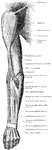
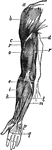
Front View of the Superficial Muscles of the Arm
Superficial muscles of the right arm, viewed from in front.
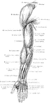
Lateral View of the Superficial Muscles of the Arm
Superficial muscles of the right arm, lateral view.
Muscles of the Arm
"Muscles on the front of the arm. Note the while cords, the tendons at the wrist." —Davison, 1910
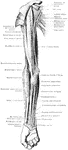
Posterior View of the Superficial Muscles of the Arm
Superficial muscles of the right arm, posterior view.
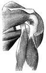
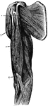
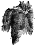
Muscles of the Back and Shoulder
"Some of the Larger Muscles on the Back of the Shoulder and the Arm." — Blaisedell, 1904
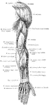
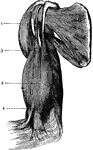
Back Forearm Muscles
The muscles of the back forearm. 1, biceps; 2, brachialis internus; 3, triceps; 4, pronator radii teres;…
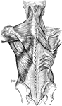
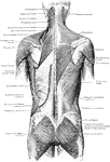
Back Muscles
Muscles of the back. 1, trapezius; 2, its origin; 3, spine of scapula; 4, latissimus dorsi; 5, delltoid;…
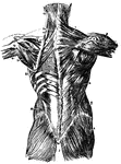
Muscles of the Human Back
Muscles of the back. Labels: 50, latissimus dorsi; 51, trapezius; 52, deltoid. The muscles of the back…
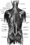
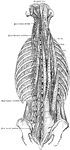
Muscles of the Back
Second layer of muscles of the back. Labels: 1, trapezius; 2, a portion of the ten dinous ellipse formed…
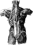
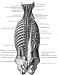
Back Muscles
Muscles of the back. On the left side is exposed the first layer; on the right side, the second layer…
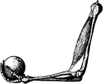
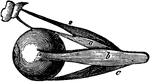
Biceps
Biceps muscle relaxed. 1 and 3: Two heads of the muscle; 2: Muscular portion; 3: Tendon fastening to…
Biceps
Biceps muscle contracted to raise the fore-arm. 1 and 3: Two heads fastened at the shoulder; 2: Contracting…
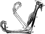
Biceps
The bicep muscle and the arm bones, to illustrate how, under ordinary circumstances, the elbow joint…
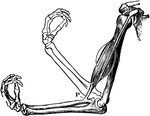
The Biceps Muscle and Arm Bones
The biceps muscle and arm bones, to illustrate how, under ordinary circumstances, the elbow-joint is…
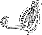
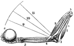
Muscular Cycle of the Biceps
A diagram showing the muscular cycle form by the biceps or flexor muscle, and the triceps or extensor…
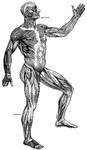
Superficial Muscles of the Body
"A single muscle rarely or never contracts alone, but always in harmony with a number of other muscles.…
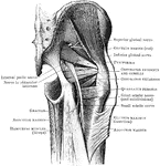
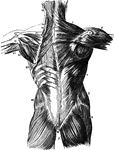
Muscles and Nerves of the Buttock
The muscles and nerves of the buttock. The gluteus maximus is reflected; and the gluteus medius is cut…
Muscles of the Chest and Abdomen
Muscles of the back. Labels: 50, latissimus dorsi; 51, trapezius; 52, deltoid. The muscles of the back…
Chest Muscles
A frontal view of the chest muscles. 1, sterno-hyoid; 2, sterno-mastoid; 3, sterno-thyroid; 4, sterno-mastoid;…
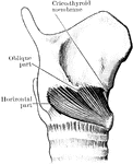
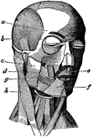
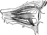
Eye Muscles
"The external bones of the temple are supposed to be removed in order to render visible the muscular…
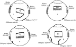
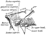
Muscles of the Eye
The plan of the insertions of the eight ocular muscles as seen: a, from Above; b, from the inner side;…
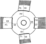
Muscles of the Eye
The plan of the insertions of the recti (muscles) into the right eyeball. The muscles have been shaded.…
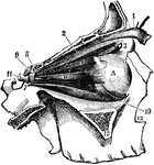
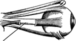
Eyeball Muscles
Muscles of the eyeball. There are muscles that move the eyeball, 5 of which are shown here.
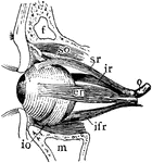
Muscles of Left Eyeball
"Muscles of Left Human Eyeball. so, superior oblique, passing through a trochlea or pulley; io, inferior…
Muscles of the Eyes
"The eye is moved about by six muscles. The back ends of these muscles are attached to the eye sockets.…