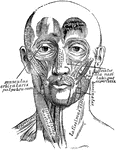
This human anatomy ClipArt gallery offers 242 illustrations the human muscular system. This includes both human skeletal muscle and human smooth and cardiac muscle. Human skeletal muscles (also known as striped, striated, or voluntary muscle) are those muscles that attach to bones and are involved in voluntary movements. Smooth muscles (also known as non-striated or involuntary muscle) are found lining the surface of internal organs such as the stomach, and cardiac muscles are those muscles specific to the heart. Both smooth and cardiac muscles are involuntarily controlled by the autonomic nervous system.
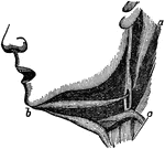
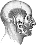
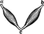
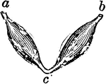
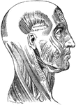
Muscles of the Lower Jaw
Muscles of the lower jaw. There are 2 muscles that work to draw down the lower jaw (a and b).
Muscle Diagram
"Diagram illustrating the muscles (drawn in thick black lines) which pass before and behind the joints…
Muscle Fiber
Diagram of muscle fiber with sarcolemma attached. Muscular tissue is the tissue by means of which the…

Muscle Fiber
Diagram of the appearance in fresh muscle fiber. Labels: A, At low focus (B) the muscle columns appear…
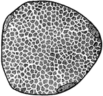
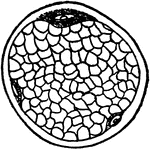
Section of a Muscle Fiber
Section of a muscle fiber, showing areas of Cohnheim. three nuclei are seen lying close to the sarcolemma.

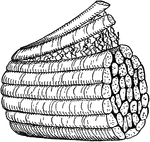
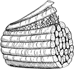
Muscle Fibers
A small bit of muscle composed of five primary fasciculi (bundles). Labels: A, natural size; B, the…
Muscle Fibers
A small piece of muscular fiber highly magnified. At a the fiber has been crushed and twisted so as…
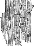
Muscle Fibers
Muscle fibers from the heart showing the striations and the junctions of the cells, highly magnified.
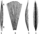
Representation of the Direction and Arrangement of the Muscle Fibers
Representation of the direction and arrangement of the fibers in a spindle-shaped muscle (1), a radiated…
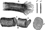
Fragments of Striped (Striated) Muscle Fibers
Fragments of striped (striated) muscle fibers, showing a cleavage in opposite directions, magnified…

Arm Muscle
A,b,c, deltoid muscle; d, coracobrachialis muscle; r,r, triceps;e,i, extensors of the hand; km, flexor…
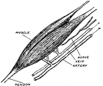
Bicep Muscle
The upper bicep of the right arm. Included are the tendons, blood vessels, and its nerve.

Microscopic View of a Muscle
A microscopic view of a muscle showing, at one end, the fibrillae; and at the other, the disk, or cells…
Striated Muscle
Striped, or striated, muscle which quickly contracts causing the alternating black and white lines.…
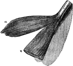
Two Portions of Muscle
Two portion of muscle; one of which, a, is covered with membrane; the other, b, is uncovered; c, the…

Muscles
A frontal view of the human muscles. The right half shows superficial muscles and the left half shows…
Muscles
Diagram illustrating the muscles (drawn in thick black lines) which pass before and behind the joints,…
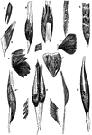
Forms of Muscles and Tendons
Forms of muscles and tendons. Labels: A, adductor of thigh; B, biceps of arm; D, deltoid, G, gastrocnemius;…

Muscles of Different Forms
Diagrams illustrating, A, typical muscle with a central belly and two terminal tendons; B, a penniform…

Back View of the Muscles of the Body
Muscle of the body, back view: The fascia is left upon the left limbs, removed from the right.

Front View of the Muscles of the Body
Muscle of the body, front view: On the right half, the superficial muscles; left half, deep-seated muscles.

Front View of the Superficial Muscles of the Body
A front view of the superficial muscles of the body. Labels: 1, The frontal swells of the occipito-frontalis.…
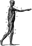
Side View of the Muscles of the Body
Side view of the muscles of the body, showing those that lie directly under the skin. Other deeper muscles…
Muscles of the forearm
"The extensor muscles on the back of the forearm. Note the tendons at the wrist." —Davison, 1910
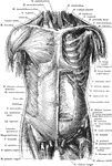
Anterior View of the Muscles of the Trunk
Superficial and deep muscles of the trunk. The sternocleidomastoid, pectoralis major, anterior portion…
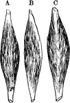
Typical Muscles
Diagrams of typical muscles with a central belly and two terminal tendons. Labels: b, a penniform muscle;…

Recording of a Muscular Contraction
Diagram to illustrate the method of obtaining a graphic record of a muscular contraction.
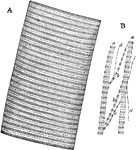
Muscular Fiber
A. Portion of a medium sized human muscular fiber. B. Separated bundles of fibrils equally magnified.…
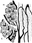
Muscular Fiber
Three muscular fibers running longitudinally, and two bundles of fibers in transverse section, M, from…

Muscular Fiber Contracting
Wave of contraction passing over a muscular fiber of dytiscus, very highly magnified. When a muscle…

Torn Muscular Fiber
Muscular fiber torn across; the sarcolemma still connecting the two parts of the fiber.

The Development of Muscular Fibers from Cells
The development of muscular fibers from cells. Labels: a, simple cell. b, a pair of cells fused together.…

Muscular Tissue
This illustration shows a diagram of nervous and cross-striate muscular tissue, showing the mode of…
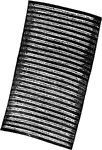
Fiber of Muscular Tissue Showing Alternating Bands
A fiber of cross-striped muscular tissue, showing the alternating bands.

Muscular Tissue Showing Transverse Cleavage
Fragment of a fiber of cross-striped muscular tissue, hardened, showing transverse cleavage.
