The Comparative Anatomy ClipArt gallery offers 228 images of anatomy comparing numerous parts of humans and animals, showing similarities and differences among species and between different taxonomic orders.
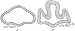
Excretory System Stage 3
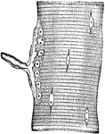
"The origin of the mesonephric tubules is seen. They arise from the upper part of the lateral plate,…
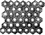
Eye of the Butterfly
Eye of the phalaena or butterfly, magnified, consisting of 11,300 square sections. The eye of the mordella…
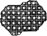
Eye of the Yellow Beetle
Eye of the yellow beetle magnified, composed of 8,820 hexagonal cylinders, the interior of each tube…
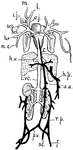
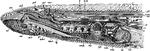
Fish Circulation Vessels
"Diagram of the principal vessels in the circulation of a Fish, lateral view. a, aorta; au., auricle;…
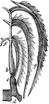
Respiration in Fishes
This figure shows the mode of respiration in fishes. The gills are seen bent over in the form of a feather.…
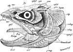
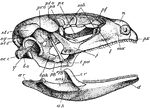
Fish Skull
"Salmo fario, the entire skull, from the left side. art, articular; branchiost, branchiostegal rays;…
Fish Vertebrae
"Diagram of vertebrae of a bony fish. A, caudal; B, trunk. c, centrum or body of the vertebra; ch.,…
Evolution of Horse Foot
Generic development of the horse's foot. A, Foot of Eohippus; B, That of Orohippus; C, That of Hipparion;…
Section Through Forebrain of Human and Lepidosteus Embryos
Two cross sections through the forebrain. A. Through the forebrain of the early human embryo. B. Through…
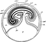
Fowl Embryo
"Diagram of a longitudinal section through the embryo of a fowl, showing formation of amnion and allantois…
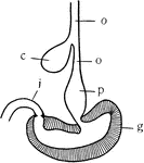
Fowl Stomach
"Diagram of the stomach and esophagus of the fowl. o, esophagus; c, crop; p, proventriculus or glandular…
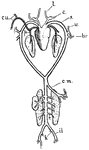
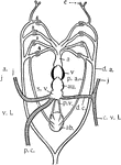
Frog Arterial System
"Arterial system of frog. l., Lingual; c., carotid; s., systemic; cu., cutaneous; p., pulmonary; v.,…
Frog Blood Corpuscles
Blood corpuscles of a frog. Highly magnified. The appearance of blood cells in different animals varies…
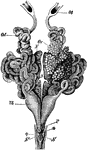
Edible Frog Brain
"Rana esculenta. The brain. A, from above; B, from below. ch. opt, optic chiasma; HH, cerebellum; Hyp,…
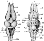
Female Edible Frog Genital Organs
"Rana esculenta. Urinogenital organs of the female. N, kidneys; Od, oviduct; Ot, its coelomic aperture;…
Male Edible Frog Genital Organs
"Rana esculenta. Urinogenital organs of the male. Ao, dorsal aorta; Cl, cloaca; Cv, post-caval vein;…
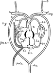
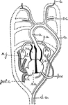
Frog Heart
"Diagram of the heart and branchial arches in the Frog. c.g., carotid gland; l., lungs; l.a., left auricle;…
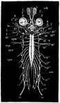
Frog Nervous System
"Nervous system of frog. 1-10, The cranial nerves; oc., eyes; crb., in front of optic chiasma; to.,…
Frog Pectoral Girdle
"Pectoral girdle of Rana esculenta. The cartilaginous parts are dotted. Ep., Episternum; om., omosternum;…
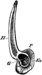
Edible Frog Pelvic Girdle
"Rana esculenta. Pelvic girdle from the right side. G, acetabulum; Il, P, ilium; Is, ischium; Kn, pubis."…
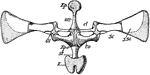
Edible Frog Shoulder Girdle
"Rana esculenta. The shoulder girdle from the ventral aspect. Co, coracoid; Co', epicoracoid; Cl, clavicle;…
Frog Skeleton
"Skeleton of frog. The half of the pectoral girdle, and fore- and hind-limb of the right side are not…
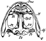
Lower Surface of Frog Skull
"Skull of frog. Lower surface-- Pmx., premaxilla; M., maxilla; Q.j., quadrato-jugal; Q., quadrate; Pt.,…
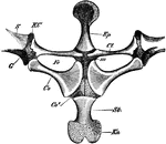
Upper Surface of Frog Skull
"Skull of frog. Upper surface-- Pmx., premaxilla; N., nasal; M., maxilla; Sq., squamosal; Q.j., quadrato-jugal;…
Frog Venous System
"Venous system of frog. m., l., Mandibular and lingual; e.j., external jugular; i.j., internal jugular;…
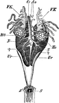
Frog Viscera
"General view of the viscera of a male frog, from the right side. a, stomach; b, urinary bladder; c,…
Circulation of Blood in a Frog's Foot
The circulation of the blood in the web of a frog's foot. A, an artery; B, capillaries crowded with…
Gorilla Skeleton
"Skeleton of male gorilla. cl., Clavicle; sc., tip of scapula; S., praesternum; H., humerus; r., radius;…
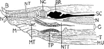
Hagfish Anterior
"Median longitudinal section of anterior region of Myxine. B., Barbule; N., nasal aperture; NT., nasal…
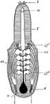
Hagfish Respiratory System
"Respiratory system of hag, from ventral surface. b., Barbules; m., mouth opening on ventral surface;…
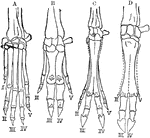
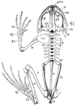
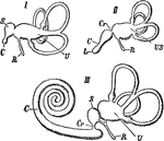
Hands of Vertebrates
A comparison of vertebrate hands. A, hand or anterior foot of the dog; B, that of the hog; C, that of…
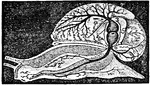
The Heart and Arteries of a Snail
In high orders of Mollusca the circulation resembles that of fish. Shown is the heart and arteries of…
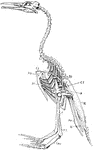
Hesperornis Skeleton
"Hesperornis. ST., Sternum; CO., coracoid; CL., clavicle; H., rudimentary humerus; SC., scapula; P.,…
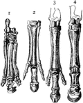
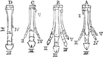
Horse Feet
"Feet of horse and its predecessors. 1, Palaeotherium; 2, Anchitherium; 3, Hippotherium; 4, Equus."…
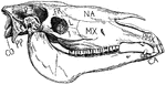
Horse Skull
"Side view of horse's skull. P., Parietal; FR., frontal; NA., nasal; PMX., premaxilla; MX., maxilla;…
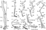
Human Leg (Front View), and Comparative Diagrams showing Modifications of the Leg
This illustration shows a human leg (front view), and comparative diagrams showing modifications of…
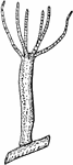
Freshwater Hydra
A freshwater hydra, magnified. Example of an animal with no skeleton. There are animals without a skeleton…
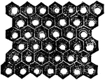
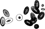
Eye of an Insect
A magnified view of a small portion of the surface of an insect's compound eye which is composed of…
Internal Ear of Vertebrates
Internal ear of different vertebrates. I, fish; II, bird; III, mammal. Labels: U, utriculus with semicircular…
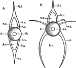
Invertebrate Simple Eye
"Diagrams showing some of the stages in the increasing complexity of the simple eye in Invertebrates.…
Kangaroo Foot
"Foot of young kangaroo. 2, 3, Small syndactylous toes; 4, large fourth toe; 5, fifth toe." -Thomson,…
Lamprey Anatomy
The dissection of a female sea lamprey or Petromyzon marinus. ad, anterior dorsal cartilage; an, annular…
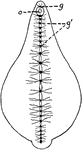
Leech Nervous System
"The central nervous system in a leech. g, dorsal ganglia (brain); g', ventral chain of ganglia; o,…
Liver Lobule of a Pig
A lobule of a pig liver, magnified, showing the hepatic cells radially arranged around the central intralobular…
Monitor Lizard Heart
"Heart of monitor (Varanus) dissected to show the cavity of the ventricle and the vessels leading out…
Nerve Ending in Muscle Fiber of Lizard
Nerve ending in muscular fiber of a lizard (Kühne). The end-plate, or motorial ending of the axone,…
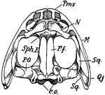
Lizard Skull
"Side view of skull of Lacerta. px., Premaxilla; mx., maxilla; l., lachrymal; j., jugal; t.pa., transpalatine;…
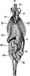
Sand Lizard Viscera
"Lacerta agilis. General view of the viscera in their naturaal relations. Bl, urinary bladder; Ci, post-caval…
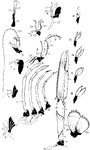
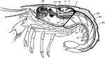
Norway Lobster Appendages
"Appendages of Norway lobster. Ex., Exopodite: En., endopodite; protopodite dark throughout; Ep., epipodite.…
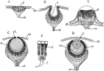
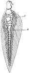
Lobster Ommatidium
"An ommatidium or eye-element from the eye of the Lobster. c, cornea (cuticle); c.h., corneal hypodermis,…
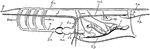
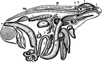
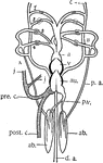
Lobster Organs
"Longitudinal section of lobster, showing some of the organs. H., Heart; AO., ophthalmic artery; SA.,…
Queensland Lungfish Fin
"Skeleton of Ceratodus fin. a., Central axis; r., radials; h., basal piece." -Thomson, 1916
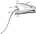
African Lungfish Head
"Head region of Protopterus. sn.t., Sensory tubes; l.l., lateral line; e.br., external gills; pc.l.,…
African Lungfish Heart
"Diagram of the heart and branchial arches in Protopterus (one of the Dipno). pre.c., precaval vein,…
Queensland Lungfish Heart
"Diagram of heart and branchial arches in Ceratodus (one of the Dipnoi). a.b., air bladder (lung); p.a.,…
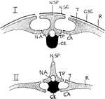
Mammal and Chelonian Backbones
"Vertical section through backbone and ribs of Chelonian (I.) and Mammal (II.). N.SP., neural spine;…
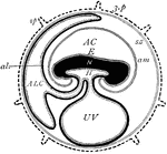
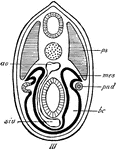
Mammal Fetus
"Diagram of foetal membranes. E., Embryo; H., gut lined by hypoblast, dotted-- the dark is mesoblast;…
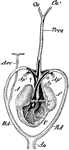
Mammal Heart
"Diagram of the heart and the branchial arches in Mammals. A dotted outline of the arches of the Fish…
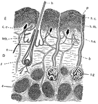
Mammal Skin
This is a diagram of the skin of mammals, showing the multiple layered condition, together with outgrowths…
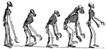
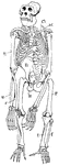
Man and Ape Skeletons
This illustration shows the skeletons of Anthropid Apes compared with that of Man.