The Comparative Anatomy ClipArt gallery offers 228 images of anatomy comparing numerous parts of humans and animals, showing similarities and differences among species and between different taxonomic orders.
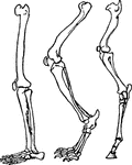
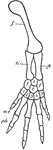
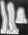
"Spiral valve of skate." -Thomson, 1916

Skate Spiral Valve

"Spiral valve of skate." -Thomson, 1916

The part of the limb upon which an animal stands when resting. A comparison of the foot in various animals brings out many interesting modifications.

Skeletal Feet

The part of the limb upon which an animal stands when resting. A comparison of the foot in various animals…
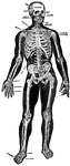
The framework of animals, which in vertebrates is composed of bone and cartilage. It serves to support the fleshy parts and the nervous system, and forms levers for the muscles.

Skeleton

The framework of animals, which in vertebrates is composed of bone and cartilage. It serves to support…

This diagram shows the skeletons of an Orang, Chimpanzee, and a Man.

Skeletons

This diagram shows the skeletons of an Orang, Chimpanzee, and a Man.

"Reproductive organs of Helix pomatia. O.T., Ovotestis; H.D., hermaphrodite duct; A.G., albumen gland; F.D., female side of common duct; M.D., male side of common duct; O., oviduct; R.S., receptaculum seminis; M.G., mucus glands; D.S., dart-sac; V.D., vas deferens; FL., flagellum; P., penis; M., retractor muscle of penis; AP., genital aperture." -Thomson, 1916

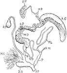
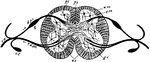
Snail Reproductive Organs

"Reproductive organs of Helix pomatia. O.T., Ovotestis; H.D., hermaphrodite duct; A.G., albumen gland;…
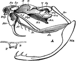
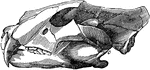
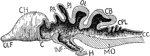
"A, lateral view of skull of rattlesnake (Crotalus). B. O, basi-occipital; B. S, basi-sphenoid; E. O, exoccipital; F. O, fossa ovalis; La, conjoined lacrymal and pre-fontal; L. f, articulation between lacrymal and frontal; Mn, mandible; Mx, maxilla; Na, nasal; Pl, palatine; Pmx, pre-maxilla; P. Sph, pre-sphenoid; Pt, pterygoid; Qu, quadrate; Sq, squamosal; II. V, foramina of exit of the second and fifth cranial nerves. B, transverse section at point lettered B in Fig. .A; T, trabeculae." -Parker, 1900

Rattlesnake Skull

"A, lateral view of skull of rattlesnake (Crotalus). B. O, basi-occipital; B. S, basi-sphenoid; E. O,…

"Forms of spermatozoa (not drawn to scale). 1 and 2. Immature and mature spermatozoa of snail; 3. of bird; 4. of man (h., head; m., middle portion; t., tail); 5. of salamander, with vibratile fringe (f.); 6. of Ascaris, slightly amoeboid with cap (c); 7. of crayfish." -Thomson, 1916

Spermatozoa

"Forms of spermatozoa (not drawn to scale). 1 and 2. Immature and mature spermatozoa of snail; 3. of…

Spermatozoa of the salamander (1) and human (2). Labels: a, long pointed head; b, elliptical structure ; c, filiform body; d, fine filament; e, homogeneous membrane.

Spermatozoa of a Salamander and Human

Spermatozoa of the salamander (1) and human (2). Labels: a, long pointed head; b, elliptical structure…

The backward and forward movement of the jaws and the great size and strength of the lower jaw, adapt rodents and gnawers to their mode of life, as in the squirrel. Labels: 1, The enamel of the gnawing tooth. 2, The ivory. 3, The lateral furrows of the molar teeth.

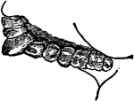
The Lower Jaw of a Squirrel

The backward and forward movement of the jaws and the great size and strength of the lower jaw, adapt…

"Diagram showing arrangement of the nervous matter in Starfish. c, ganglionated ring about the mouth; o, mouth; r.n., radial nerve in each arm." -Galloway, 1915

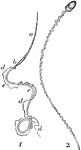
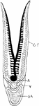
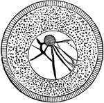
Starfish Nervous System

"Diagram showing arrangement of the nervous matter in Starfish. c, ganglionated ring about the mouth;…

Ruminants (those animals that chew the cud), as the sheep, have a stomach with four cavities. Labels: 1, esophagus; 2, rumen; 3, reticulum; 4, omasum; 5, abomasum or rennet; 6, intestine.

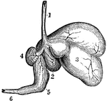
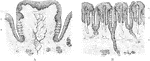
The Stomach of a Sheep

Ruminants (those animals that chew the cud), as the sheep, have a stomach with four cavities. Labels:…

Teeth of a carnivorous animal that lives on flesh alone. The front teeth are designed for tearing, while the back teeth have sharp edges for cutting.

Teeth of a Carnivorous Animal

Teeth of a carnivorous animal that lives on flesh alone. The front teeth are designed for tearing, while…

Teeth of a frugivore (fruit-eating animal). Animals that live on soft fruits do not need such grinders as grass-eating animals do, instead they have rounded teeth which serve to crush their food.

Teeth of a Frugivore

Teeth of a frugivore (fruit-eating animal). Animals that live on soft fruits do not need such grinders…

Teeth of an herbivore, showing the rough surface of some of these teeth. Herbivores have no tearing teeth. Instead they have two kinds of teeth, cutting teeth in the front and grinding teeth in the back. This figure shows a peculiar arrangement of the enamel, which admirably fits them to grind up the fibers of the grass in the back grinding teeth of an herbivore. The enamel is not only on the outside as it is in human teeth, rather there are ridges on it standing up in the middle of each tooth.

Teeth of an Herbivore

Teeth of an herbivore, showing the rough surface of some of these teeth. Herbivores have no tearing…

The image on the left are the teeth of a carnivora or flesh-eating animal. The teeth on the right belong t a insectivora or insect-eating animal. Notice the projecting jaw, the wide mouth, and sharp teeth of the carnivore animal in contrast with the elongated, tapering muzzle and cone-pointed molars of the insectivorous animal.

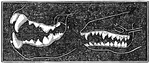
Comparing Teeth of Carnivora and Insectivora Animals

The image on the left are the teeth of a carnivora or flesh-eating animal. The teeth on the right belong…

1. Dentition (teeth) of man. 2. Dentition of hyena. 3. Dentition of pig. 4. Dentition of Patagonian cavy (type of rodent). 5. Section of skull of Indian elephant, showing dentition of right side. 6. Crown of upper molar of horse, showing enamal folds. 7. Grinding surface of molar of African elephant, with enamal folds. 8. Single tooth of blue shark. 9. Longitudinal section of human tooth. 1. Incisors (human); C, canines; P, premolars; M, molars. a, enamel; b, dentine; c, cement (crusta petrosa); d, pulp cavity.

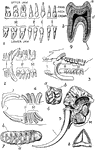
Teeth of Man and Several Animal Species

1. Dentition (teeth) of man. 2. Dentition of hyena. 3. Dentition of pig. 4. Dentition of Patagonian…

The first step in the development of teeth consists in a downward growth from the Rete Malpighi or the deeper layer of stratified epithelium of the mucous membrane of the mouth, which first becomes thickened in the neighborhood of the maxillae now in the course of formation. This development is shown in a section of the upper jaw of a fetal sheep. A- 1, common enamel germ dipping down into the mucous membrane; 2, palantine process of jaw; 3, Reta Malpighi. B- Section similar to A, but passing through one of the special enamel germs here becoming flask-shaped; c, d, epithelium of mouth; f, neck; f', body of special enamel organ; p, papilla; s, dental sac forming; f p, the enamel germ of permanent tooth; m, bone of jaw; v, vessel cut across.

Development of Teeth

The first step in the development of teeth consists in a downward growth from the Rete Malpighi or the…

"Diagram of the heart, the branchial arches, and the principal veins in the Teleosts. Ventral view. The heart is represented without the sigmoid flexure; that is, with the auricle posterior. a, aorta; au., auricle; br.a., branchial arches of the aorta (1-4, numbering from the front); c, carotid; c.v., cardinal veins (right and left); d.a., dorsal arteries; j, jugular veins; d.c., ductus Cuvieri; s.v., sinus venosus; v, ventricle. Only four arterial arches are shown." -Galloway, 1915

Teleost Heart

"Diagram of the heart, the branchial arches, and the principal veins in the Teleosts. Ventral view.…

"Diagram of Teleostean circulation. A., auricle; V., ventricle; v.a., ventral aorta; a.br., afferent branchials; e.br., efferent branchials; c.c., cephalic circle; e., carotids; A.c.v., anterior cardinal veins; P.C.V., posterior cardinal veins; d.c., ductuss Cuvieri; d.a., dorsal aortaa; c.v., caudal vein; c.a., caudal artery; K., kidney." -Thomson, 1916

Teleostean Circulation

"Diagram of Teleostean circulation. A., auricle; V., ventricle; v.a., ventral aorta; a.br., afferent…

"Section of a Teleostean gill. G.F., Gill-filament; A., artery (venous blood); V., vein (pure blood); G.A., gill-arch." -Thomson, 1916

Teleostean Gill

"Section of a Teleostean gill. G.F., Gill-filament; A., artery (venous blood); V., vein (pure blood);…
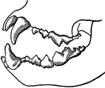
Jaws of a tiger, showing that when the jaws are closed the ends of the teeth do not press upon the ends of the teeth that are opposite to them, but instead the teeth pass by each other. This arrangement of these long tearing teeth gives them great power in tearing flesh.

Jaws of a Tiger

Jaws of a tiger, showing that when the jaws are closed the ends of the teeth do not press upon the ends…

A, Section through papilla vallata of a human tongue. B, Section through part of the papilla foliata of a rabbit. Labels: 1, papilla; 2, vallum; 3, taste buds; 4, papillae; 5, taste buds; 6, ducts of serous gland.

Tongue of Human and Rabbit

A, Section through papilla vallata of a human tongue. B, Section through part of the papilla foliata…

Section through a premolar tooth of a cat still embedded in its socket. Labels: 1, enamel; 2, dentine; 3, cement; 4, the gum; , the bone of the lower jaw; c, the pulp-cavity.

Premolar Tooth

Section through a premolar tooth of a cat still embedded in its socket. Labels: 1, enamel; 2, dentine;…
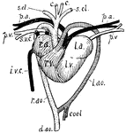
"Heart and associated vessels of tortoise. r.a., Right auricle; superior venae cavae (s.v.c.) and inferior vena cava (i.v.c.) enter it. r.v., Right half of ventricle; pulmonary arteries (p.a.) and left aortic arch (l.ao.) leave it; coel., coeliac; d.ao., dorsal aorta. l.a., Left auricle; p.v., pulmonary veins enter it. l.v., Left half of ventricle; right aortic arch (r.ao.), giving off carotids (c.) and subclavians (s.cl.)." -Thomson, 1916

Tortoise Heart

"Heart and associated vessels of tortoise. r.a., Right auricle; superior venae cavae (s.v.c.) and inferior…
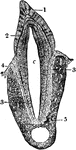
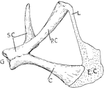
"Internal view of tortoise skeleton. H., humerus; SC., scapula running dorsally; PC., precoracoid; C., coracoid; EC., epicoracoid cartilage; P., pubis; IL., ilium running dorsally to sacral vertebrae; IS., ischium; DV., dorsal vertebrae fused in carapace; R., head of a rib; CEV., cervical vertebrae free; CAV., caudal vertebrae free." -Thomson, 1916

Tortoise Skeleton

"Internal view of tortoise skeleton. H., humerus; SC., scapula running dorsally; PC., precoracoid; C.,…
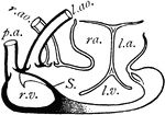
"Dissection of Chelonian heart. r.v., Right half of ventricle; S., septum; l.v., left half of ventricle; r.a., right auricle; l.a., left auricle; l.ao., left aortic arch; r.ao., right aortic arch; p.a., pulmonary arch." -Thomson, 1916

Turtle Heart

"Dissection of Chelonian heart. r.v., Right half of ventricle; S., septum; l.v., left half of ventricle;…
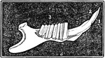
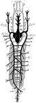
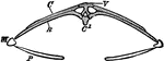
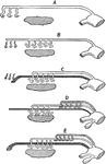
"Hyoid apparatus of a Chelonian. BH., Body of the hyoid (basihyal); H., representing another part of the hyoid arch; A.C., anterior cornu, representing the first branchial arch; P.C., posterior cornu, representing the second branchial arch." -Thomson, 1916

Turtle Hyoid

"Hyoid apparatus of a Chelonian. BH., Body of the hyoid (basihyal); H., representing another part of…

"Pectoral girdle of a Chelonian. G., Glenoid cavity; SC., scapula; P.C., procoracoid fused to the scapula; C., coracoid; E.C., epicoracoid cartilage; L., ligament." -Thomson, 1916

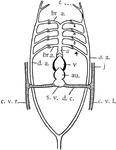
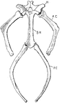
Turtle Pectoral Girdle

"Pectoral girdle of a Chelonian. G., Glenoid cavity; SC., scapula; P.C., procoracoid fused to the scapula;…

"Chelone midas. Transverse section of skeleton. C, costal plate; C', centrum; M, marginal plate; P, lateral element of plastron; R, rib; V, expanded neural plate." -Parker, 1900

Green Turtle Skeleton

"Chelone midas. Transverse section of skeleton. C, costal plate; C', centrum; M, marginal plate; P,…

"Cistudo lutaria. Skeleton seen from below; the plastron has been removed and is represented on one side. C, costal plate; Co, coracoid; e, entoplastron; Ep, epiplastron; F, fibula; Fe, femur; H, humerus; Il, ilium; Is, ischium; M, marginal plates; Nu, nuchal plate; Pb, pubis; Pro, pro-coracoid; Py, pygal plates; R, radius; Sc, scapula; T, tibia; U, ulna." -Parker, 1900

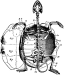
Marsh Turtle Skeleton

"Cistudo lutaria. Skeleton seen from below; the plastron has been removed and is represented on one…
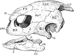
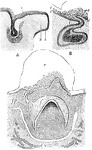
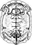
"Skull of turtle. "S.O., supra-occipital; PAR., parietal; FR., frontal; P.F., pre-frontal; PO.F., post-frontal; SQ., squamosal; PMX., pre-maxilla; MX., maxilla; J., jugal; Q.J., quadrato-jugal; Q., quadrate; D., dentary; AN., angular; AR., articular; S., surangular." -Thomson, 1916

Turtle Skull

"Skull of turtle. "S.O., supra-occipital; PAR., parietal; FR., frontal; P.F., pre-frontal; PO.F., post-frontal;…
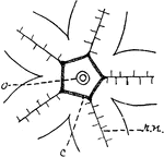
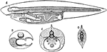
"Ovum of a Sea-Urchin, showing the radially striated cell-membrane, the protoplasm, containing yolk-granules, the large nucleus (germinal vescicle), with its network of chromatin and a large nucleolus (germinal spot)." -Parker, 1900

Sea Urchin Ovum

"Ovum of a Sea-Urchin, showing the radially striated cell-membrane, the protoplasm, containing yolk-granules,…

Diagrams illustrating the development of the urinogenital organs of a vertebrate. A, pronephros and segmental duct; B, atrophy of pronephros, development of mesonephros; C, appearance of Mullerian duct; D, development of metanephros, male type; E, the same, female type. The sex gland, ovary and testis is obliquely shaded; phonephros and mesonephros unshaded, metanephros stippled; Mullerian duct heavily shaded. The large chamber to the right, into which these ducts as well as the intestine open, is the cloaca.

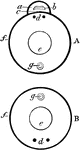
Urinogenital Organs of a Vertebrate

Diagrams illustrating the development of the urinogenital organs of a vertebrate. A, pronephros and…

Transverse section through the middle of the ventricles (right and left) of a dog's heart in diastole (top) and in systole (bottom).

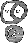
Transverse Section through the Ventricle of a Dog's Heart

Transverse section through the middle of the ventricles (right and left) of a dog's heart in diastole…

The vertebra of a fish, which is very different from that of a human. It has but two processes, <em> f f </em>. In humans there is a single short spinous process behind, while the vertebra is round in front. But in the fish there are two quite long spinous processes, one in front and the other in the rear (which is above and below, respectively, when the fish is in water).

Vertebra of a Fish

The vertebra of a fish, which is very different from that of a human. It has but two processes,

A, Diagrammatic transverse section through the body of a vertebrate. B, A similar section through a highly organized invertebrate animal. Labels: a, neural canal; b, cerebrospinal nerve center; c, notochord; d, sympathetic nerve center; e, alimentary canal; f, visceral canal wall; g, haemal system.

Vertebrate and Invertebrate

A, Diagrammatic transverse section through the body of a vertebrate. B, A similar section through a…

"Diagrams of the girdles and appendages in a typical Vertebrate. A, anterior; B, posterior. ac., acetabulum, articulation of the humerus with its girdle; c, coracoid; ca., carpals; c.e., centralia; d.c., distal carpals; d.t., distal tarsals; el., elbow joint; f, fibula; fe., femur; fi., fibulare; gc., glenoid cavity, articulation of arm with girdle; h, humerus; il., ilium; in., intermediale; is. ischium; kn., knee joint; m.c., metacarpals (1-5); m.t., metatarsals (1-5); p, pubis; ph., phalanges (1-5); pr.c., precoracoid; r, radius; ra., radiale; sc., scapula; t, tibia; ta., tarsals; ti., tibiale; u., ulna; ul., ulnare." -Galloway, 1915

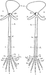
Vertebrate Appendages

"Diagrams of the girdles and appendages in a typical Vertebrate. A, anterior; B, posterior. ac., acetabulum,…

"Partial section of a Vertebrate brain (diagrammatic). OLF., Olfactory lobe; CH., cerebral hemispheres; C., wall of cerebrum cut to show ventricle, behind this the figure is that of a median section; PA., parietal organ arising from thalamencephalon; Pl., pineal organ; INF., infundibulum descending from thalamencephalon; H., hypophysis; OL., optic lobes; CB., cerebellum; CPL., choroid plexus on roof of fourth ventricle; MO., floor of the medulla oblongata; CC., central canal of spinal cord." -Thomson, 1916

Vertebrate Brain

"Partial section of a Vertebrate brain (diagrammatic). OLF., Olfactory lobe; CH., cerebral hemispheres;…

"Ideal fore limb. H., Humerus; R., radius; U., ulna; r'., radiale; u'., ulnare; i., intermedium; c., centrale; 1-5, carpalia bearing the corresponding digits with metacarpals (mc.) and phalanges (ph)." -Thomson, 1916

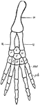
Vertebrate Fore Limb

"Ideal fore limb. H., Humerus; R., radius; U., ulna; r'., radiale; u'., ulnare; i., intermedium; c.,…
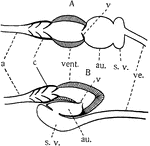
"Diagrams of the structure of the heart in the lower Vertebrates. A, primitive condition; B, the position of the parts in the fishes. a, artery; au., auricle; c, conus arteriosus with valves; s.v., sinus venosus; v, valves; ve., vein; vent., ventricle. The dorsal portion of the heart is toward the bottom of the figure." -Galloway, 1915

Vertebrate Heart

"Diagrams of the structure of the heart in the lower Vertebrates. A, primitive condition; B, the position…

"f., Femur; ti., tibia; fi., fibula; i., intermedium; t., tibiale (astragalus); f., fibulare (os calcis); c., centrale; 1-5, tarsalia bearing the corresponding digits with metatarsals (mt.) and phalanges (ph.)." -Thomson, 1916

Vertebrate Hind Limb

"f., Femur; ti., tibia; fi., fibula; i., intermedium; t., tibiale (astragalus); f., fibulare (os calcis);…

"Diagrammatic section of spinal cord. p.f., Posterior fissure; p.c., posterior column of white matter; d.p.s., dorsal, posterior, sensory or afferent root; g., ganglion; v.a.m., ventral, anterior, motor or efferent root; c.n., compound spinal nerve with branches; s.g., sympathetic ganglion; a.c., anterior column--the anterior fissure is exaggerated; g.c., ganglion cells; g.m., grey matter; w.m., white matter." -Thomson, 1916

Vertebrate Spinal Cord

"Diagrammatic section of spinal cord. p.f., Posterior fissure; p.c., posterior column of white matter;…

Diagrammatic sections of the ideal vertebrate. A, sagittal section showing the brain and spinal cord on the dorsal side of the notochord, and the alimentary canal and viscera on the ventral side of it. B, transverse section of the head, showing a gill arch and filaments of the left and a gill cleft on the right. C, transverse section of the trunk, showing the gut, the genital glands, and the excretory organs in the body cavity. D, transverse section of the tail.

Section of Vertebrate

Diagrammatic sections of the ideal vertebrate. A, sagittal section showing the brain and spinal cord…

"Fore limbs of vertebrates showing similarity of structure. <em>A</em>, salamander; <em>B</em>, turtle; <em>C</em>, very young bird; <em>D</em>, adult bird; <em>E</em>, dog; <em>F</em>, man." &mdash; Davison, 1906

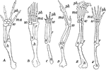
Forearms of vertebrates

"Fore limbs of vertebrates showing similarity of structure. A, salamander; B, turtle;…
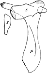
A, Villus of sheep. B, Villi of human.

Villus of Humans and Sheep

A, Villus of sheep. B, Villi of human.

"Left fore-limb of Balaenoptera. Sc., Sca pula with spine (sp.); H., humerus; R., radius; U., ulna; C., carpals embedded in matrix; Mc., metacarpals; Ph., phalanges." -Thomson, 1916

Whale Forelimb

"Left fore-limb of Balaenoptera. Sc., Sca pula with spine (sp.); H., humerus; R., radius; U., ulna;…

"Fore-limb of whale (Megaptera longimana)." -Thomson, 1916

Whale Forelimb

"Fore-limb of whale (Megaptera longimana)." -Thomson, 1916

"Pelvis and hind-limb of Greenland whale (Balaena). P., Pelvis; F., femur; T., tibia." -Thomson, 1916

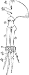
Greenland Whale Pelvis

"Pelvis and hind-limb of Greenland whale (Balaena). P., Pelvis; F., femur; T., tibia." -Thomson, 1916