Types of vascular bundles: "B, the collateral type, with phloem, h, standing in front of the xylem, k." -Stevens, 1916

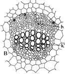
Collateral Vascular Bundle

Types of vascular bundles: "B, the collateral type, with phloem, h, standing in front of the xylem,…
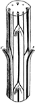
"B, diagram of vascular bundles in external view and in cross section of the stem of Cerastium. The leaves are shown cut off at 1, 2, 2' and 3." -Stevens, 1916

Cerastium Stem

"B, diagram of vascular bundles in external view and in cross section of the stem of Cerastium. The…
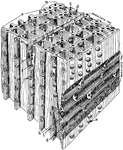
"Diagrammatic representation of a block of pine wood highly magnified. a, Early growth; b, late growth; c, intercellular space; d, bordered pit in tangential wall of late growth; m, f and e, bordered pit in radial wall of early growth from different points of view; h, row of medullary cells for carrying food; g, row of medullary ray cells for carrying water; k, thin place in radial wall of ray cells that carry food." -Stevens, 1916

Magnified Pine

"Diagrammatic representation of a block of pine wood highly magnified. a, Early growth; b, late growth;…
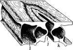
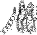
"A typical stoma in cross section and surface view combined. k, guard cell; j, the gap or stoma between the guard cells; l, epidermal cell bordering a guard cell." -Stevens, 1916

Stoma

"A typical stoma in cross section and surface view combined. k, guard cell; j, the gap or stoma between…

"Portion of leaf of Sphagnum, in cross section on the left, and surface view on the right. h, hole through the wall; i, chlorophyll-bearing cells; j, water-storage and water-conducting cells, with annular thickenings at k." -Stevens, 1916

Sphagnum Leaf

"Portion of leaf of Sphagnum, in cross section on the left, and surface view on the right. h, hole through…

"Cross section, A, and surface view, B, of a leaf of common moss, showing chloroplasts, c." -Stevens, 1916

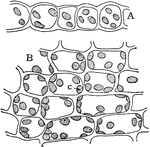
Moss Leaf Chloroplasts

"Cross section, A, and surface view, B, of a leaf of common moss, showing chloroplasts, c." -Stevens,…
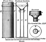
"Microscopic Structure of white Statuary marble." -Geikie, 1893

Marble

"Microscopic Structure of white Statuary marble." -Geikie, 1893

"Rain gauges are usually vertically placed sheet-metal hollow cylinders of from 5 to 8 inches in diameter. (The figure) shows a rain gauge with a funnel-shaped mouth, A, and an inside receiver, C, of less diameter than the outer cylinder." -Waldo, 1896

Rain Gauge

"Rain gauges are usually vertically placed sheet-metal hollow cylinders of from 5 to 8 inches in diameter.…
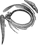
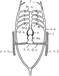
Side view of the muscles of the eye in their natural positions. Labels: a,b,c,d, the four straight muscles. a. is turned up to prevent the others from being hidden. e. the superior oblique muscle. f. the optic nerve. The other oblique muscle is not shown, but it lies on the other side of the ball, and comes round, and is inserted near the superior oblique; so that they pull opposite ways, like two men sawing timber. By the combined action of all these muscles, an infinite variety of motions of the eye can be produced.

Muscle of the Eye

Side view of the muscles of the eye in their natural positions. Labels: a,b,c,d, the four straight muscles.…

Side view of the eyeball. Labels: a, the eyeball, and b,b, are the upper and lower sides. Now in order to prevent dust and other bodies from working their way between the ball and the lid, through passages at c,c, into the socket of the eye where they would excite great inconvenience and pain, we find the common skin of the eye lids d,d, after covering their edges turn in a little way between the lid and the ball, and then turn back and are reflected over the surface of the cornea; where, to prevent the obstruction of vision, it becomes perfectly transparent.

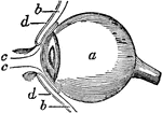
Side View of the Eyeball

Side view of the eyeball. Labels: a, the eyeball, and b,b, are the upper and lower sides. Now in order…

The eyelids viewed from before; a,a, the lachrymal canals; b, the lachrymal sack. The lachrymal sac is a bag of an oval shape, fixed to the end of the double canal, and lies in a depression of the nasal bones. It terminates in the tube called the duct, which passes through a hole made for it, in the bones of the nose, and opens into the nostril.

Eyelids, Viewed from the Front

The eyelids viewed from before; a,a, the lachrymal canals; b, the lachrymal sack. The lachrymal sac…

General sectional view of the structure of the ear. Labels: a, the meatus auditorius externus; b, the tympanum; c, the malleus; d, the incus, e, the os orbiculare; f, the stapes; g, the semicircular canals; h, the cochlea; i, the meatus auditorius internus; k, the eustachian tube.

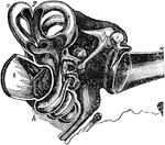
Sectional View of the Ear

General sectional view of the structure of the ear. Labels: a, the meatus auditorius externus; b, the…
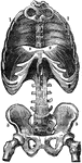
View of the diaphragm; 1, cavity of the thorax; 2, diaphragm separating the cavity of the thorax from that of the abdomen; 3, cavity of the pelvis.

Diaphragm

View of the diaphragm; 1, cavity of the thorax; 2, diaphragm separating the cavity of the thorax from…
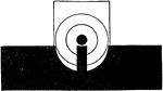
"Look through the rear-sight notch at the bull's-eye or mark and bring the top of the front sight on a line with the top of and in the center of the rear-sight notch and aligned upon the point of aim." -Moss, 1914

Normal Sight

"Look through the rear-sight notch at the bull's-eye or mark and bring the top of the front sight on…
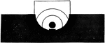
"Look through the peep hole at the bull's-eye or mark and bring the top of the front sight to the center of the aperture and aligned upon the point of aim." -Moss, 1914

Peep Sight

"Look through the peep hole at the bull's-eye or mark and bring the top of the front sight to the center…

"Although men will be found occasionally who can get excellent results by using the fine sight, the average man can not, and this form of sighting is not recommended." -Moss, 1914

Fine Sight

"Although men will be found occasionally who can get excellent results by using the fine sight, the…

"Fracture of the arm: Apply two splints, one in front, the other behind, if the lower part of the bone is broke; or to the inner and outer sides, if the fracture is in the middle or upper part; support by sling." -Moss, 1914

Splints and Sling

"Fracture of the arm: Apply two splints, one in front, the other behind, if the lower part of the bone…
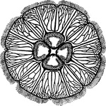
"Surface view of Aurelia. Showing four genital pockets in centre, much branched radial canals, eight peripheral niches for sense organs, and peripheral tentacles." -Thomson, 1916

Aurelia

"Surface view of Aurelia. Showing four genital pockets in centre, much branched radial canals, eight…
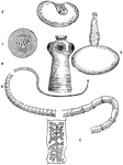
"Life history of Taenia solium. 1. Six-hooked embryo in egg-case; 2. proscolex or bladder-worm stage, with invaginated head; 3. bladder-worm with evaginated head; 4. enlarged head of adult, showing suckers and hooks; 5. general view of the tape-worm, from small head and thin neck to the rip joints; 6. a rip joint or proglottis with branched uterus; all other organs are now lost." -Thomson, 1916

Pork Tapeworm

"Life history of Taenia solium. 1. Six-hooked embryo in egg-case; 2. proscolex or bladder-worm stage,…

"Diagrammatic longitudinal section of a Nemertean (Amphiporum lactifloreus), dorsal view. p.p., Proboscis pore; b., brain giving off the lateral nerve-cords (n.); po., oesophageal pocket; p., proboscis lying within its sheath; st., stilet of proboscis; m., retractor muscles of proboscis; g., gut shown in outline at the sides of proboscis; e., the three main, longitudinal blood vessels, which unite both anteriorly and posteriorly." -Thomson, 1916

Nemertea

"Diagrammatic longitudinal section of a Nemertean (Amphiporum lactifloreus), dorsal view. p.p., Proboscis…
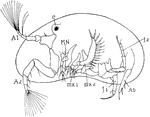
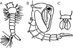
"Cypris, side view, after removal of one valve. e., Eye; A.1, first antennae; A.2, second antennae; MN., mandibles; mx.1, first maxilla; mx.2, second maxilla; f.1, f.2, thoracic legs; Ab., rudimentary abdomen." -Thomson, 1916

Cypris

"Cypris, side view, after removal of one valve. e., Eye; A.1, first antennae; A.2, second antennae;…
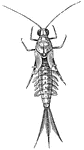
"Young may-fly or ephemerid. Showing tracheal gills, and wings appearing in front of them." -Thomson, 1916

Mayfly

"Young may-fly or ephemerid. Showing tracheal gills, and wings appearing in front of them." -Thomson,…
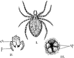
"Garden spider. I., Female garden spider; II., end view of head of the same showing the simple eyes, the poison fangs (ch.), and the pedipalps (p.); III., posterior end of body showing two pairs of spinnerets (sp.), with anus above." -Thomson, 1916

Garden Spider

"Garden spider. I., Female garden spider; II., end view of head of the same showing the simple eyes,…

The side view of Sepia officinalis, or the common cuttlefish.

Cuttlefish

The side view of Sepia officinalis, or the common cuttlefish.

View of the bronchial tubes, terminating in air vesicles. On the left is the external view (1, bronchial tube; 2, air vesicles). On the right is the bronchial tube and air vesicles laid open.

Bronchial Tubes Terminating in Air Vesicles

View of the bronchial tubes, terminating in air vesicles. On the left is the external view (1, bronchial…
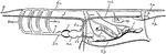
"Lateral view of Amphioxus. The notochord runs from tip to tip. t., Tentacular cirri; G., reproductive organs; a.p., atriopore; a., position of anus; 40 and 62, indicate number of myotomes." -Thomson, 1916

Lancelet

"Lateral view of Amphioxus. The notochord runs from tip to tip. t., Tentacular cirri; G., reproductive…
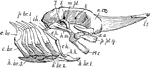
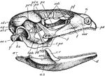
"Side view of skate's skull. l1., First labial cartilage; n.c., nasal capsule; a.o., antorbital; p.pt.q., palato-pterygo-quadrate; M.c., Meckel's cartilage; h.m., hyo-mandibular; e.h., epi-hyal; c.h., cerato-hyal; h.h., hypo-hyal; h.br.1-5, hypobranchials; c.br., ceratobranchial; e.br., epibranchial; p.br.1., first prebranchial; i.h., inter-hyal; m.pt., meta-pterygoid; 2, 5, 7, foramina of exit of the corresponding nerves." -Thomson, 1916

Skate Skull

"Side view of skate's skull. l1., First labial cartilage; n.c., nasal capsule; a.o., antorbital; p.pt.q.,…
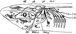
"Side view of chief cranial nerves of Elasmobranchs." -Thomson, 1916

Shark Nerves

"Side view of chief cranial nerves of Elasmobranchs." -Thomson, 1916

"The haddock. n.a., Nasal apertures (double on each side); d.f.1, d.f.2, d.f.3, dorsal unpaired fins; c.f., the caudal fin of the homocercal tail. b., Barbule; op., operculum covering the four gills; br.m., continuation of the gill-cover forming the branchiostegal membrane; pv.f., pelvic fin (=hind-limb)--note its jugular position in front of p.f., the pectoral fin (=fore-limb)." -Thomson, 1916

Haddock

"The haddock. n.a., Nasal apertures (double on each side); d.f.1, d.f.2, d.f.3, dorsal unpaired fins;…
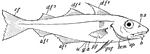
"Lateral view of dogfish (Scyllium catulus). Note ventral mouth with naso-buccal groove, heterocercal tail, and unpaired fins. gs., Gill-slits; pc., pectoral fins; pv., pelvic fins." -Thomson, 1916

Dogfish

"Lateral view of dogfish (Scyllium catulus). Note ventral mouth with naso-buccal groove, heterocercal…

"Nervous system of frog. 1-10, The cranial nerves; oc., eyes; crb., in front of optic chiasma; to., optic tract; sym., sympathetic system; msp., spinal cord; sp., spinal nerves." -Thomson, 1916

Frog Nervous System

"Nervous system of frog. 1-10, The cranial nerves; oc., eyes; crb., in front of optic chiasma; to.,…

"Internal view of the plastron of the Greek tortoise. EP., Epiplastron (clavicle?); ENT., entoplastron (interclavicle?); HYO., hyoplastron; HYPO., hypoplastron; XIPHI., xiphiplastron." -Thomson, 1916

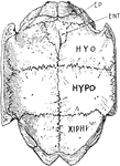
Greek Tortoise Plastron

"Internal view of the plastron of the Greek tortoise. EP., Epiplastron (clavicle?); ENT., entoplastron…
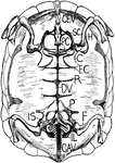
"Internal view of tortoise skeleton. H., humerus; SC., scapula running dorsally; PC., precoracoid; C., coracoid; EC., epicoracoid cartilage; P., pubis; IL., ilium running dorsally to sacral vertebrae; IS., ischium; DV., dorsal vertebrae fused in carapace; R., head of a rib; CEV., cervical vertebrae free; CAV., caudal vertebrae free." -Thomson, 1916

Tortoise Skeleton

"Internal view of tortoise skeleton. H., humerus; SC., scapula running dorsally; PC., precoracoid; C.,…
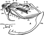
"Side view of skull of Lacerta. px., Premaxilla; mx., maxilla; l., lachrymal; j., jugal; t.pa., transpalatine; epg., epipterygoid; pg., pterygoid; bpg., basipterygoid; b.o., basioccipital; q., quadrate; oc.c., occipital condyle; sq., squamosal; pr.o., pro-otic; pt.o., postorbital; st.1, st.2, supratemporals; ps., presphenoid (the optic nerve is seen issuing in front of the end of the reference line); p.e., mesethmoid; s.ob., supraorbitals; pf., prefrontal; n., nasal; ar., articular; ag., angular; sag., surangular; cr., coranary; d., dentary." -Thomson, 1916

Lizard Skull

"Side view of skull of Lacerta. px., Premaxilla; mx., maxilla; l., lachrymal; j., jugal; t.pa., transpalatine;…
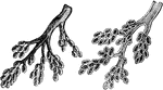
"A., Filoplume. B., very young feather within its sheath (sh.); c., the core of dermis; b., the barbs. C., the same, external view." -Thomson, 1916

Feathers

"A., Filoplume. B., very young feather within its sheath (sh.); c., the core of dermis; b., the barbs.…
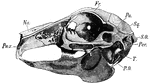
"Side view of rabbit's skull. Pmx., Premaxilla; Na., nasal; Fr., frontal; Pa., parietal; Sq., squamosal; S.O., supraoccipital; Per., periotic; T., tympanic (the reference line points to the bony external auditory meatus, beneath it lies the inflated bulla); PO., paroccipital process." -Thomson, 1916

Rabbit Skull

"Side view of rabbit's skull. Pmx., Premaxilla; Na., nasal; Fr., frontal; Pa., parietal; Sq., squamosal;…

"Dorsal view of rabbit's brain. olf.l., Olfactory lobes; c.h., cerebral hemispheres; o.l., optic lobes (corpora quadrigemina); cb., median part of cerrebellum; Fl., flocculus of cerebellum; 10, root of the tenth or vagus nerve; s.c., spinal cord." -Thomson, 1916

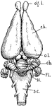
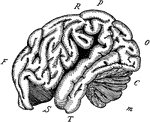
Dorsal View of Rabbit Brain

"Dorsal view of rabbit's brain. olf.l., Olfactory lobes; c.h., cerebral hemispheres; o.l., optic lobes…
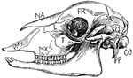
"Side view of sheep's skull. PMX., Premaxilla; MX., maxilla; NA., nasal; J., Jugal; L., lachrymal; FR., frontal; PA., parietal; SQ., squamosal; CO., condyle; PP., paroccipital process." -Thomson, 1916

Sheep Skull

"Side view of sheep's skull. PMX., Premaxilla; MX., maxilla; NA., nasal; J., Jugal; L., lachrymal; FR.,…
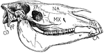
"Side view of horse's skull. P., Parietal; FR., frontal; NA., nasal; PMX., premaxilla; MX., maxilla; J., jugal; L., lachrymal; SQ., squamosal; PP., paroccipital process; CO., condyle CA., canine." -Thomson, 1916

Horse Skull

"Side view of horse's skull. P., Parietal; FR., frontal; NA., nasal; PMX., premaxilla; MX., maxilla;…

View of the heart enclosed in its bag, or pericardium, which is a serious membrane. It is here laid open and turned back.

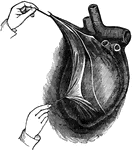
Heart in the Pericardium

View of the heart enclosed in its bag, or pericardium, which is a serious membrane. It is here laid…

View of the heart with its several chambers exposed and the vessels in connection with them. Labels: 1, the superior vena cava. 2, the inferior vena cava. 3. the chamber called the auricle. 4. the right ventricle. 5. the line marking the passage between the two chambers, and the points of attachment of one margin of the valve. 6. the septum between the two ventricles. 7. the pulmonary artery, arising from the right ventricle, and dividing at 8 into right and left, of the corresponding lungs. 9. the four pulmonary veins, bringing the blood from the lungs into 10, the left auricle. 11. the left ventricle. 12. the aorta, arising from the left ventricle, and passing down behind the heart, to distribute blood to every part of the system. Thus the blood moves in a double circle, one from the heart to the body, and from the body back to the heart, called the systemic circle; the other, from the heart to the lung, and from the lung back to the heart, called the pulmonic circle.

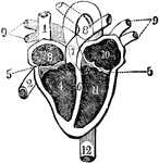
Heart and its Chambers

View of the heart with its several chambers exposed and the vessels in connection with them. Labels:…

"Diagram showing some stages in the life history of the Tapeworm (taenia). A, Cysticercus or Bladderworm stage, before the "head" protrudes from the bladder; B, same, later stage; C, Strobila, or chain of proglottides, many being omitted; D, embro, such as fill the uterus of the mature proglottides. It is protected by a shell. b, bladder; ex., excretory canals; g, genital pore; h, head or scolex provided with hooks and suckers (s); u, uterus in a mature posterior proglottis; z, zone of budding or segment formation. The numerals show the approximate number of the segments, reckoning from the front." -Galloway, 1915

Tapeworm

"Diagram showing some stages in the life history of the Tapeworm (taenia). A, Cysticercus or Bladderworm…

"Two stages in the metamorphosis of the Mosquito. A, larva; B, pupa; C, ventral view of the oar-like appendages of the last segment of the pupa." -Galloway, 1915

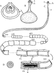
Mosquito Metamorphosis

"Two stages in the metamorphosis of the Mosquito. A, larva; B, pupa; C, ventral view of the oar-like…

"Diagram of the heart, the branchial arches, and the principal veins in the Teleosts. Ventral view. The heart is represented without the sigmoid flexure; that is, with the auricle posterior. a, aorta; au., auricle; br.a., branchial arches of the aorta (1-4, numbering from the front); c, carotid; c.v., cardinal veins (right and left); d.a., dorsal arteries; j, jugular veins; d.c., ductus Cuvieri; s.v., sinus venosus; v, ventricle. Only four arterial arches are shown." -Galloway, 1915

Teleost Heart

"Diagram of the heart, the branchial arches, and the principal veins in the Teleosts. Ventral view.…

"Diagram of the principal vessels in the circulation of a Fish, lateral view. a, aorta; au., auricle; br.a., afferent branchial arteries; br.e., efferent branchial arteries; bu., bulbus (or conus) arteriosus; c, carotid; c.a., caudal artery; c.v.r., right cardinal vein; c.v.l., left cardinal vein; d.a., dorsal artery; d.c., ductus Cuvieri; g, gills; h.v., hepatic vein; h.p., hepatic portal vein; i, intestine; j, jugular vein; k, kidney; l, liver; r.p., renal portal vein; s.v., sinus venosus; v, ventricle." -Galloway, 1915

Fish Circulation Vessels

"Diagram of the principal vessels in the circulation of a Fish, lateral view. a, aorta; au., auricle;…

"The outline of the brain of an orang outang. Front portion F to O, cerebrum; C, cerebellum; M, medulla and spinal cord; F, the frontal lobe; P, the parietal lobe; O, the occipital lobe; T, the temporal lobe; R, the fissure of Rolando; S, the fissure of Silvius." -Galloway, 1915

Orangoutang Brain

"The outline of the brain of an orang outang. Front portion F to O, cerebrum; C, cerebellum; M, medulla…
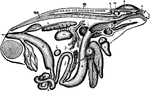
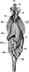
A spindle root.

Spindle Root

A spindle root.

A suitor in front of the mirror practicing and admiring himself.

Beau

A suitor in front of the mirror practicing and admiring himself.

"General view of the viscera of a male frog, from the right side. a, stomach; b, urinary bladder; c, small intestine; cl, cloacal aperture; d, large intestine; e, liver; f, bile duct; g, gall bladder; h, spleen; i, lung; k, larynx; l, fat body; m, testis; n, ureter; o, kidney; p, pancreas; s, cerebral hemisphere; sp, spinal cord; t, tongue; u, auricle; ur, urostyle; v, ventricle; v.s, vesicula seminalis; w, optic lobe; x, cerebellum; y, Eustachian recess; z, nasal sac." -Parker, 1900

Frog Viscera

"General view of the viscera of a male frog, from the right side. a, stomach; b, urinary bladder; c,…

"Transverse section through the wall of a cylinder (parallel with the course of the canals), showing one incurrent canal (IC), and one radial (R) throughout their length; sp, triradiate spicules; sp', oxeote spicules of dermal cortex (dc); sp'', tetraradiate spicules of gastral cortex (gc); ec, ectoderm; en, endoderm; pm, pore membrane; pp, prosopyles; ap, apopyle; di, diaphragm; exc, excurrent passage; PG, paragastric cavity; em, early embryo; em, late embryo. The arrows indicate the course of the water through the sponge." -Parker, 1900

Sycon Gelatinosum

"Transverse section through the wall of a cylinder (parallel with the course of the canals), showing…

"Ophioglypha lacertosa. A, outline, of the natural size; B, central disc, dorsal view; C, the disc, ventral view showing the mouth and genital fissures." -Parker, 1900

O. Lacertosa

"Ophioglypha lacertosa. A, outline, of the natural size; B, central disc, dorsal view; C, the disc,…
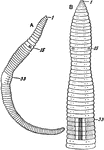
"Lumbricus agricola. A, entire specimen, lateral view; B, ventral view of anterior portion of the body, magnified. 1, 15, 33, first, fifteenth, and thirty-third segments. The black dots represent the setae." -Parker, 1900

Lumbricus Agricola

"Lumbricus agricola. A, entire specimen, lateral view; B, ventral view of anterior portion of the body,…
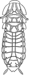
"Periplaneta. View of the arrangement of the principal trunks of the tracheal system." -Parker, 1900

Cockroach

"Periplaneta. View of the arrangement of the principal trunks of the tracheal system." -Parker, 1900

"Periplaneta. General view of the nervous system. abd. 6, sixth abdominal ganglion; ant, antennary nerve; br, brain; conn, oesophageal connective; inf, sub-oesophageal ganglion; opt, optic nerve; thor. 1, thor. 2, thor. 3, first, second, and third thoracic ganglia." -Parker, 1900

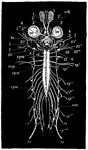
Cockroach Nervous System

"Periplaneta. General view of the nervous system. abd. 6, sixth abdominal ganglion; ant, antennary nerve;…
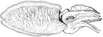
"Cattle Tick. Above, nat. size, side view; below, dorsal view, enlarged." -Parker, 1900

Cattle Tick

"Cattle Tick. Above, nat. size, side view; below, dorsal view, enlarged." -Parker, 1900

"Spirula peronii, lateral view. d, terminal sucker; f, funnel; s1, s2, projecting portions of the shell, the internal part of which is indicated by dotted lines." -Parker, 1900

Spirula Peronii

"Spirula peronii, lateral view. d, terminal sucker; f, funnel; s1, s2, projecting portions of the shell,…
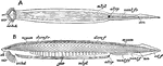
"Amphioxus lanceolatus. A, ventral; B, side view of the entire animal. an, anus; atrp, atriopore; cd. f, caudal fin; cir, cirri; dors. f, dorsal fin; dors. f. r. dorsal fin-rays; gon, gonads; mtpl, metapleure; myom, myomeres; nch, notochord; or. hd, oral hood; vent. f, ventral fin; vent. f. r, ventral fin-rays." -Parker, 1900

Lancelet

"Amphioxus lanceolatus. A, ventral; B, side view of the entire animal. an, anus; atrp, atriopore; cd.…

"Lacerta agilis. General view of the viscera in their naturaal relations. Bl, urinary bladder; Ci, post-caval vein; ED, rectum; GB, gall-bladder; H, heart; Lg, Lg', the lungs; M, stomach; MD, small intestine; Oe, oesophagus; Pn, pancreas; Tr, trachea." -Parker, 1900

Sand Lizard Viscera

"Lacerta agilis. General view of the viscera in their naturaal relations. Bl, urinary bladder; Ci, post-caval…

"A, lateral view of skull of rattlesnake (Crotalus). B. O, basi-occipital; B. S, basi-sphenoid; E. O, exoccipital; F. O, fossa ovalis; La, conjoined lacrymal and pre-fontal; L. f, articulation between lacrymal and frontal; Mn, mandible; Mx, maxilla; Na, nasal; Pl, palatine; Pmx, pre-maxilla; P. Sph, pre-sphenoid; Pt, pterygoid; Qu, quadrate; Sq, squamosal; II. V, foramina of exit of the second and fifth cranial nerves. B, transverse section at point lettered B in Fig. .A; T, trabeculae." -Parker, 1900

Rattlesnake Skull

"A, lateral view of skull of rattlesnake (Crotalus). B. O, basi-occipital; B. S, basi-sphenoid; E. O,…
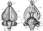
"Lepus cuniculus. Brain. A, dorsal view; B, ventral; b. o, olfactory lobe; cb', median lobe of cerebellum (vermis); cb'', lateral lobe of cerebellum; cr, cauro-cerebri; ep, epiphysis; f. b, parencephala; f. p, longitudinal fissure; h. b, hind-brain; hp, hypophysis; m. b, mid-brain (corpora quadrigemina); md, medulla oblongata; p.v, pons Varolii; i-xii, cranial nerves." -Parker, 1900

Rabbit Brain

"Lepus cuniculus. Brain. A, dorsal view; B, ventral; b. o, olfactory lobe; cb', median lobe of cerebellum…