"An evergreen of the laurel family, having glossy leaves and bearing clusters of yellowish flowers, which are succeeded by branches of fruit resembling black currants." -Lupton

Camphor Tree

"An evergreen of the laurel family, having glossy leaves and bearing clusters of yellowish flowers,…

"This tree, a native of India, is remarkable for its vast branches. It is a species of fig; has ovate, heart-shaped, entire leaves, about five or six inches long, and produces a fruit of a rich scarlet, not larger than a cherry, growing in pairs from the axils of the leaves." -Lupton

Banyan Tree

"This tree, a native of India, is remarkable for its vast branches. It is a species of fig; has ovate,…
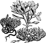
A small cruciferous plant, growing in arid Arabia and Palestine. When full grown and ripe its leaves drop and it becomes rolled up like a ball in the dry season, but opens its branches and seed vessels when it comes in contact with moisture.

Illustration of Roots and Jericho Rose

A small cruciferous plant, growing in arid Arabia and Palestine. When full grown and ripe its leaves…
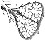
Diagram of the two primary lobules of the lungs, magnified. Labels: 1, Bronchial tube. 2, A pair of primary lobules connected by elastic tissue. 3, Intercellular air-passages. 4, Air-cells. 5, Branches of the pulmonary artery and vein.

Diagram of the Two Primary Lobules of the Lung

Diagram of the two primary lobules of the lungs, magnified. Labels: 1, Bronchial tube. 2, A pair of…
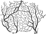
The arteries and veins of a section of the skin. A, arterial branches. B, capillary, or hair-like vessels, in which the large branches terminate. C, the venous trunk, collecting the blood from the capillaries.

The Arteries and Veins of a Section of the Skin

The arteries and veins of a section of the skin. A, arterial branches. B, capillary, or hair-like vessels,…
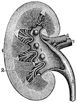
A longitudinal section of a kidney. Labels: 1, 2, 3, Parts of the Kidney. 4, Pelvis. 5, Ureter. 6, Renal artery. 7, Renal vein. 8, Branches of the latter vessels in the kidney.

Longitudinal Section of a Kidney

A longitudinal section of a kidney. Labels: 1, 2, 3, Parts of the Kidney. 4, Pelvis. 5, Ureter. 6, Renal…
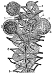
A diagram of the structure of the kidney. Labels: 1, Tubules or minute tubes. 2, Enlargement of a tubule at its extremity. 3, Branches of the real artery ending in vessels which enter the enlargements as seen at 4, 5. 6, Knot of blood-vessels freed from its investment. 7, Veins emerging from the knots. 8, Plexus formed by the latter veins among the tubules, from which plexus originate the branches of the renal vein.

Diagram of the Structure of the Kidney

A diagram of the structure of the kidney. Labels: 1, Tubules or minute tubes. 2, Enlargement of a tubule…

The sympathetic ganglions and their connection with other nerves. Labels: A, The semilunar ganglion and solar plexus. D, The thoracic (chest) ganglions. E, The external and internal branched of the thoracic ganglions. G, H, The right and left coronary (heart) plexus. I, N, Q , The inferior, middle and superior cervical (neck) ganglions. 1, The renal plexus of nerves. 2, The lumbar (loin) ganglions. 3, Their internal branches. 4, Their external branches. 5, The aortic plexus of nerves.

The Sympathetic Ganglions and their Connection to other Nerves

The sympathetic ganglions and their connection with other nerves. Labels: A, The semilunar ganglion…
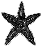
In the Radiata, the starfish manifests one of the simplest forms of nervous systems. it consists of a central mass, with five arms radiating from it. In the center is the mouth, and beneath it the stomach or gastric cavity, which sends branches to each limb. The nervous system consists of five similar ganglions situated in the central portion at the base of the arms. These ganglions are connected and each sends off nerve-filaments to corresponding limbs.

Diagram of a Radiata

In the Radiata, the starfish manifests one of the simplest forms of nervous systems. it consists of…
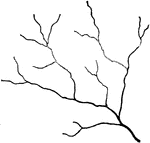
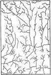
"Normal twig-like branching of streamlets—the type produced in homogenous rock material when uninfluenced by geological structure planes." -Walcott, 1901

Streamlet Branching

"Normal twig-like branching of streamlets—the type produced in homogenous rock material when uninfluenced…

"Streamlet branchings of the abnormal type found in the area in and about the Pomperaug Valley. a and i, streamlets controlled by two sets or airs of fault planes. b and c, streamlets preponderantly controlled by a single series of parallel planes; d, f, and h, by a nearly rectangular system of fault planes. e and g, streamlets indicating that neighboring streamlet branchings have been controlled by the same fault plane. The side streamlets of g indicate control by a second series of faults." -Walcott, 1901

Streamlet Branchings

"Streamlet branchings of the abnormal type found in the area in and about the Pomperaug Valley. a and…

"Drainage which has been influenced by joints." -Walcott, 1901

Drainage Streamlets

"Drainage which has been influenced by joints." -Walcott, 1901

A representation of the brain, spinal cord and spinal nerve. Labels: 1, The cerebrum. 2. The cerebellum. 3, Spinal cord. 4, The sciatic nerve. A. Distribution of the olfactory nerve. Labels: 1, 2, Nerve of smell. B. Optic Nerve. Labels: 15, Nerve of vision. C. The gustatory nerve. Labels: 1, 2, 3, 4, Branches of the nerve of taste. D. Auditory nerve. Labels: 13, Nerve of hearing.

Nervous System

A representation of the brain, spinal cord and spinal nerve. Labels: 1, The cerebrum. 2. The cerebellum.…
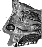
The olfactory system. Labels: a, b, c, d, interior of the nose, which is lined by a mucous membrane; n, the nose; e, the wing of the nose; q, the nose bones; o, the upper lip; g, section of the upper jawbone; h, the upper part of the mouth, or hard palate; m, frontal bone of the skull; k, the ganglion or bulb of the olfactory nerve in the skull, from which are seen the branches of the nerve passing in all direction.

Olfactory System

The olfactory system. Labels: a, b, c, d, interior of the nose, which is lined by a mucous membrane;…
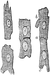
Muscular fiber cells from the heart. The fibers which lie side by side are united at frequent intervals by short branches.

Muscular Fiber Cells from the Heart

Muscular fiber cells from the heart. The fibers which lie side by side are united at frequent intervals…

Two striped muscle fibers of the hyoglossus of frog. Labels: a, Nerve end plate; b, nerve fibers leaving the end plate; c, nerve fibers, terminating after diving into branches d, a nucleus in which two nerve fibers anastomose.

Striped Muscle Fibers of a Frog

Two striped muscle fibers of the hyoglossus of frog. Labels: a, Nerve end plate; b, nerve fibers leaving…

The Pacinian bodies or corpuscles are elongated oval bodies situated on some of the cerebrospinal and sympathetic nerves, especially the cutaneous nerves of the hands and feet; and on branches of the large sympathetic plexus about the abdominal aorta. Shown are the extremities of a nerve of the finger with Pacinian corpuscles attached.

Pacinian Corpuscles in a Human

The Pacinian bodies or corpuscles are elongated oval bodies situated on some of the cerebrospinal and…

The Pacinian bodies or corpuscles are elongated oval bodies situated on some of the cerebrospinal and sympathetic nerves, especially the cutaneous nerves of the hands and feet; and on branches of the large sympathetic plexus about the abdominal aorta. Shown is a Pacinian corpuscle of the cat's mesentery. The stalk consist of a nerve fiber (N) wit its think outer sheath. The peripheral capsules of the Pacinian corpuscle are narrower near the entrance of the axis cylinder into the clear central mass. A hook-shaped termination with the end bulb; it possesses a sheath which is the continuation of the peripheral capsules of the Pacinian corpuscle.

Pacinian Corpuscle of a Cat

The Pacinian bodies or corpuscles are elongated oval bodies situated on some of the cerebrospinal and…

The Pacinian bodies or corpuscles are elongated oval bodies situated on some of the cerebrospinal and sympathetic nerves, especially the cutaneous nerves of the hands and feet; and on branches of the large sympathetic plexus about the abdominal aorta. Shown is the summit of a Pacinian corpuscle of a human finger, showing the endothelial membranes linking the capsules.

Pacinian Corpuscle of a Human

The Pacinian bodies or corpuscles are elongated oval bodies situated on some of the cerebrospinal and…
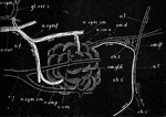
Diagrammatic representation of the submaxillary gland of the dog with its nerves and blood vessels. Labels: sm. gld., the submaxillary gland into the duct (sm. d.) of which a cannula has been tied. The sublingual gland and duct are not proceeding from the facial nerve, becoming conjoined with the lingual at n.l'., and afterwards diverging and passing to the gland along the duct; sm. gl., submaxillary ganglion with its roots; n. l., the lingual nerve proceeding to the tongue; a. car., the carotid artery. two branches of which, a. sm. a. and r. sm. p. pass to the anterior and posterior parts of the gland; v. sym., the conjoined vagus and sympathetic trunks; gl. cer. s., the superior cervical ganglion, two branches of which forming a plexus, a.f., over the facial artery are distributed (n.sym.sm.) along the two glandular arteries to the anterior and posterior portion of the gland. The arrows indicate the direction taken by the nervous impulses; during reflex stimulations of the gland they ascend to the brain by the lingual and descend by the chorda tympani.

Submaxillary Gland of Dog with Nerves and Blood Vessels

Diagrammatic representation of the submaxillary gland of the dog with its nerves and blood vessels.…

Section of a mucous gland from the tongue. Labels: A, opening of the duct on the free surface; C, basement membrane with nuclei; B, flattened epithelial cells lining duct. The duct divides into several branches, which are convoluted and end blindly, being lined throughout by columnar epithelium. D, lumen of one of the tubuli of the gland.

Mucous Gland from Tongue

Section of a mucous gland from the tongue. Labels: A, opening of the duct on the free surface; C, basement…
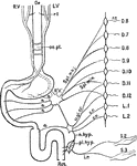
Diagrammatic representation of the nerves of the alimentary canal. Oe to Rct, the various parts of the alimentary canal from esophagus to rectum: L.V, left vagus, ending in front of stomach; rl, recurrent laryngeal nerve, supplying upper part of esophagus; R.V, right vagus, joining left vagus in esophageal plexus; oe.pl., supplying the posterior part of the stomach , and continues at R'V' to join the solar plexus, here represented by a single ganglion, and connected with the inferior mesenteric ganglion m.gl.: a, branches from the solar plexus to stomach and small intestine , and from the mesenteric ganglia and rami communicantes; r.c., belonging to dorsal nerves from the 6th to the 9th (or 10th); Spl.min., small splanchnic nerve similarly from the 10th and 11th dorsal nerves. These both join the solar plexus, and thence make their way to the alimentary canal; c.r., nerves from the ganglia, &c., belonging to 11th and 12th dorsal and 1st and 2nd lumbar nerves, proceeding to the inferior mesenteric ganglia (or plexus), m.gl., and thence by the hypogastic nerve, n.hyp., and the hypogastric nerve, n.hyp., and the hypogastric plexus, pl.hyp., to the circular muscles of the rectum; 1.r., nerves from the 2nd and 3rd sacral nerves, S.2, S.3 (nervi erigentes) proceeding by the hypogastric plexus to the longitudinal muscles of the rectum.

Nerves of the Alimentary Canal

Diagrammatic representation of the nerves of the alimentary canal. Oe to Rct, the various parts of the…
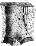
Longitudinal section of a portal canal, containing a portal vein, hepatic artery and hepatic duct from the pig. Labels: P, branch of vena portae, situate in a portal canal formed amongst the lobule of the liver; l, and giving off vaginal branches; there are also seen within the larger portal vein numerous orifices of the small interlobular veins arising directly from it; a, hepatic artery; d, hepatic duct.

Portal Canal

Longitudinal section of a portal canal, containing a portal vein, hepatic artery and hepatic duct from…
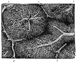
The liver is made up of small roundish or oval portions called lobules, each of which is composed of minute branches of the portal vein, hepatic artery, hepatic duct, and hepatic vein. Shown is the capillary network of the lobules of the rabbit's liver. The figure is taken from a very successful injection of the hepatic veins showing nearly the whole of two lobules, and parts of three others. Labels: p, portal branches running in the interlobular spaces; h, hepatic veins penetrating and radiating from the center of the lobules.

Capillary Network in the Lobules of a Rabbit's Liver

The liver is made up of small roundish or oval portions called lobules, each of which is composed of…

Diagrammatic view of the Sympathetic cord of the right side, showing its connections with the principal cerebrospinal nerves and the main prae-aortic plexuses. Cerebrospinal nerves. VI, a portion of the sixth cranial as it passes through the cavernous sinus, receiving two twigs from the carotid plexus of the sympathetic nerve; O, opthalmic ganglion connected by a twig with the carotid plexus; M, connection of the spheno-palatine ganglion by the Vidian nerve with the carotid plexus; C, cervical plexus; Br, brachial plexus; D 6, sixth intercostal nerve; D 12, twelfth; L 3, third lumbar nerve; S 5, fifth; Cr, anterior crural nerve winding round the subclavian artery. Sympathetic Cord. c, superior cervical ganglion; c', second or middle; c", inferior; from each of these ganglia cardiac nerves (all deep on this side) are seen descending to the cardiac plexus; d 1, placed immediately below the first dorsal sympathetic ganglion; d 6, is opposite he sixth; l 1, first lumbar ganglion. Preaortic and Visceral Plexuses. p, pharyngeal, and, lower down, laryngeal plexus; pl, post-pulmonary plexus spreading from the vagus on the back of the right bronchus; c a,on the aorta , the cardiac plexus, towards which , in addition to the cardiac nerves from the three cervical sympathetic ganglia, other branches are seen descending from the vagus and recurrent nerves; co, right or posterior and co1, left or an. coronary plexus; o, esophageal plexus in long meshes on the gullet; sp, great splanchnic nerve formed by branched from the fifth, sixth, seventh, eight, and ninth dorsal ganglia; +, small splanchnic from the ninth and tenth; ++, smallest or third splanchnic from the eleventh: the first and second of theses are shown joining the solar plexus, s o; the third descending to the renal plexus, r e; connecting branches between the solar plexus and the vagi are also represented; pn', above the place where the right vagus passes to the lower or posterior surface of the stomach; pn", the left distribution on the anterior side or upper surface of the cardiac portion of the organ: from the solar plexus large branches are seen surrounding the arteries of the coeliac axis, and descending from the solar and sup. mesenteric plexuses and from the lumbar ganglia; mi, the inf. mesenteric plexus surrounding the corresponding artery; hy, hypogastric plexus; pl, the right pelvic or inf. hypogastric plexuses; pl, the right pelvic plexus; from this the nerves descending are joined by those from the plexus on the sup. hemorrhoidal vessels, mi', by nerves from the third and fourth sacral spinal nerves, and there are thus formed the rectal, vesical, and other plexuses, which ramify upon the viscera, ,as towards ir, and v, the rectum and bladder.

Sympathetic System

Diagrammatic view of the Sympathetic cord of the right side, showing its connections with the principal…
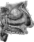
Nerves of the outer walls of the nasal fossae. 1, network of the branches of the olfactory nerve, descending upon the region of the superior and middle turbinated region of the superior and middle turbinated bones; 2, external twig of the ethmoidal branch of the nasal nerves; 3, spheno-palatine ganglion; 4, ramification of the anterior palatine nerves; 5, posterior, and 6, middle division of the palatine nerves; 7, branch to the region of the superior and middle turbinated bones; 9, naso-palatine branch to the septum cut short.

Nerves of the Nasal Fossae

Nerves of the outer walls of the nasal fossae. 1, network of the branches of the olfactory nerve, descending…
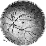
The posterior half of the retina of the left eye, viewed from before; s, the cut edge of the sclerotic coat; ch, the choroid; r, the retina; in the interior at the middle the macula lutea with the depression of the fovea centralis is represented by a slight oval shade; towards the left side the light spot indicates the colliculus or eminence at the entrance of the optic nerve, from the center of which the arteria centralis is seen spreading its branches into the retina, leaving the part occupied by the macula comparatively free.

Posterior Half of the Retina

The posterior half of the retina of the left eye, viewed from before; s, the cut edge of the sclerotic…

Capillary blood vessel of the tail of a young larval frog. a, capillaries permeable to blood; b, fat granules attached to the walls of the vessels, and concealing the nuclei; c, hollow prolongation of a capillary ending in a point; d, a branching cell with nucleus and fat granules; it communicates by three branches with prolongation of capillaries already formed; e, blood corpuscles still containing granules of fat.

Capillary Blood Vessels of Larval Frog

Capillary blood vessel of the tail of a young larval frog. a, capillaries permeable to blood; b, fat…
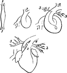
Fetal heart in successive stages of development. 1, venous extremity; 2, arterial extremity; 3, pulmonary branches; 4, ductus arteriosus.

Fetal Heart in Successive Stages of Development

Fetal heart in successive stages of development. 1, venous extremity; 2, arterial extremity; 3, pulmonary…
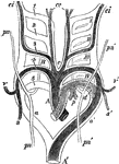
Diagram of the aortic arches in a mammal, showing transformations which give rise to the permanent arterial vessels. A, primitive arterial stem or aortic bulb, now divided into A, the ascending part of the aortic arch, and p, the pulmonary; a, a', right and left aortic roots; A', descending aorta; 1, 2, 3, 4, 5, the five primitive aortic or branchial arches; I, II, III, IV, the four branchial clefts which, for the sake of clearness, have been omitted on the right side. The permanent systemic vessels are deeply, the pulmonary arteries lightly, shaded; the parts of the primitive arches which are transitory are simply outlined; c, placed between the permanent common carotid arteries; c e, external carotid arteries; c i, internal carotid arteries; s, right subclavian, rising from the right aortic root beyond the fifth arch; v, right vertebral from the same, opposite the fourth arch; v' s', left vertebral and subclavian arteries rising together from the left, or permanent aortic root, opposite the forth arch; p, pulmonary arteries rising together from the left fifth arch, forming ductus arteriosus; p n, p n', right and left pneumogastric nerves descending in from of aortic arch, with their recurrent branches represented diagrammatically as passing behind, to illustrate the relations of these nerves respectively to the right subclavian artery (4) and the arch of the aorta and ductus arteriosus (d).

Aortic Arches in a Mammal

Diagram of the aortic arches in a mammal, showing transformations which give rise to the permanent arterial…
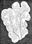
In mammalia, each salivary gland first appears as a simple canal with bud-like processes, lying in a gelatinous nidus or blastema, and communicating with the cavity of the mouth. As the development of the gland advances, the canal becomes more and more ramified, increasing at the expense of the blastema in which it is still enclosed. the branches or salivary ducts constitute an independent system of closed tubes. Shown are lobules of the parotid, with salivary ducts, in the embryo of the sheep at a more advanced stage.

Lobules of Parotid of a Sheep

In mammalia, each salivary gland first appears as a simple canal with bud-like processes, lying in a…
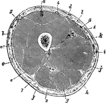
Transverse section through the middle of the thigh. Labels: a, Rectus femoris; b, vastus externus; c, crureus; d, vastus internus; e, short head of biceps; f, long head of biceps; g, semitendinosus; h, semimembranosus; i, adductor magnus; j, gracilis; k, adductor longus; l, sartorius; 1, femoral artery; 2, great sciatic nerve; 3, great saphenous vein; 6, perforating branches from profunda; 7, small sciatic nerve.

Transverse Section Through the Thigh

Transverse section through the middle of the thigh. Labels: a, Rectus femoris; b, vastus externus; c,…

A famous road with many branches which connected Rome with Southern Italy

Construction of a Portion of Appian Way

A famous road with many branches which connected Rome with Southern Italy

Cutaneous nerve supply of legs. Anterior aspect. 1, ilioinguinal; 2, genitocrural; 3, external cutaneous; 4, middle cutaneous; 5, internal cutaneous; 6, patellar plexus; 7, branches of external popliteal; 8, internal saphenous; 9, musculocutaneous; 10, external saphenous; 11, anterior tibial. Posterior aspect. 1, 2, and 3, Small sciatic; 4, external cutaneous; 5, internal cutaneous; 6, internal saphenous; 7, branches of external popliteal; 8, short saphenous; 9, posterior tibial; 10, internal saphenous; 11, internal plantar; 12, external plantar.

Nerve Supply of Legs

Cutaneous nerve supply of legs. Anterior aspect. 1, ilioinguinal; 2, genitocrural; 3, external cutaneous;…
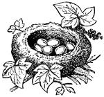
"The Kinglet, or Golden-Crested Wren, builds its nest among ivy or dependent from fir branches, generally near some brook."

Nest of Kinglet

"The Kinglet, or Golden-Crested Wren, builds its nest among ivy or dependent from fir branches, generally…

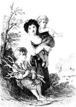
Rustic Children, a painting by Thomas Gainsborough.

Rustic Children

Rustic Children, a painting by Thomas Gainsborough.

"Female Cicada Laying her Eggs in the Groove She has Bored in the Branch of a Tree. While the female does not sing, she is given an organ which is of more practical value. This is a sort of an auger, destined to penetrate the back of the branches of trees."

Female Cicada Laying her Eggs

"Female Cicada Laying her Eggs in the Groove She has Bored in the Branch of a Tree. While the female…
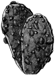
"The larvae are changed into perfect insects, which take up their abode permanently on the branches of the cacti."

Cochineal Insects on the Branches of the Cactus

"The larvae are changed into perfect insects, which take up their abode permanently on the branches…

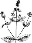
An annual plant , 6 to 15 inches high, with many slender branches of a brownish or straw color, more or less hairy, parasitic upon the roots of other plants.

Broomrape (orobanche ramosa).

An annual plant , 6 to 15 inches high, with many slender branches of a brownish or straw color, more…
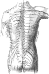
The distribution of cutaneous nerves n the back of the trunk. On the left side the distribution of the several nerves is represented, the letters indicating their nomenclature. ACR.; Posterior supraclavicular branches from cervical plexus; CIRC: Cutaneous branches of axillary nerve; E.C.: Lateral femoral cutaneous nerve, L.1: Lateral cutaneous branch of iliohypogastric nerve; S.1; Lateral branches of sacral nerves; T: T.1-12: Cutaneous branches of the posterior rami of the thoracic nerves. On the right side is a schematic representation of the areas supplied by the above nerves, the numeral and letter s indicate the spinal origins of the nerves distributed to each area.

Posterior View of the Cutaneous Nerves of Trunk

The distribution of cutaneous nerves n the back of the trunk. On the left side the distribution of the…

"They resemble so exactly dry sticks that it is impossible to tell the difference. It is an inoffensive insect, walks gently along the branches of trees, and likes to repose in the sun."

Stick Insect (Phasma Rossia)

"They resemble so exactly dry sticks that it is impossible to tell the difference. It is an inoffensive…
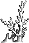
Oculina ranges from Cape Hatteras, North Carolina through the Gulf of Mexico and Caribbean, though the main population of concern is off east-central Florida where it forms unique thicket-type structures in 70-100 meters (230 to 330 ft) depth known as the "Oculina Banks". Colonies are arborescent, with highly clumped, irregular bushy branches.

Oculina Virginea (Lamarck)

Oculina ranges from Cape Hatteras, North Carolina through the Gulf of Mexico and Caribbean, though the…
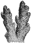
"The Plantain Madrepore is an interesting species, the polyps presenting themselves in tufts, with slender and prolific branches."

Madrepora Plantaginea (Lamarck)

"The Plantain Madrepore is an interesting species, the polyps presenting themselves in tufts, with slender…
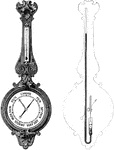
The wheel barometer consists of a siphon barometer, the two branches of which have usually the same diameter.

Wheel Barometer

The wheel barometer consists of a siphon barometer, the two branches of which have usually the same…

"A species of <em>Isis</em> has numerous slender branches, furnished with cylindrical knows at intervals, contracted toward the middle, finely striated; it has a singular resemblance to the Common Mare's Tail."

Isis Hippuris

"A species of Isis has numerous slender branches, furnished with cylindrical knows at intervals,…
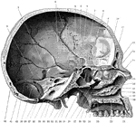
Shown is the inner aspect of the left half of the skull sagittally divided. Labels: 1, suture between parietal and temporal bone; 2, remains of subarcuate fossa; 3, grooves for branches of the middle meningeal artery; 4, dorsum sellae; 5, pituitary fossa; 6, anterior clinoid fossa; 7, optic foramen; 8, sphenoidal suture; 9, nasal surface of superior turbinated bone; 10, cribriform plate of ethmoid; 11, nasal surface of middle turbinated bone; 12, frontal sinus; 13, nasal spine; 14, nasal bone; 15, nasal process of superior maxilla; 16, middle meatus of nose; 17, directed towards opening of antrum; 18, nasal surface of inferior turbinated bone; 19, inferior meatus of nose; 20, anterior nasal spine; 21, anterior palatine canal; 22, palatal process of superior maxilla; 23, palatal process of palate bone; 24, posterior nasal spine; 25, hamular process of internal pterygoid plate; 26, external pterygoid plate; 27, superior meatus of nose; 28,sphenopalatine foramen; 29, pterygospinous ligament almost completely ossified to enclose a foramen; 30, styloid process of temporal bone; 31, alar spine of sphenoid; 32, mastoid process; 33, basion (midpoint of posterior border of foramen magnum); 34, internal auditory meatus; 35, anterior condylic foramen; 36, leading into jugular foramen; 37, opisthion (midpoint of posterior border of foramen magnum); 38, groove for sigmoid sinus; 39, opening of mastoid foramen; 40, for lateral sinus and attachment of tentorium cerebelli; 41, fossa for lodgment of cerebellar hemisphere; 42, internal occipital protuberance.

Skull Seen From Side

Shown is the inner aspect of the left half of the skull sagittally divided. Labels: 1, suture between…

Yule is a winter festival historically celebrated primarily in northern Europe but now celebrated in many other countries in various forms. Yule celebrations often coincide with Christmas. Modern Yule traditions include decorating a fir or spruce tree, burning a Yule log, hanging mistletoe and holly branches, giving gifts, and general celebration and merriment.

Yule Celebration

Yule is a winter festival historically celebrated primarily in northern Europe but now celebrated in…

The developmental stages exhibited by a pyramidal cell of the brain. Labels: a, neuroblast with rudimentary axon, but no dendrites; b and c, the dendrites beginning to sprout out; d and e, further development of the dendrites and appearance of collateral branches on the axon.

Pyramidal Cells of the Brain

The developmental stages exhibited by a pyramidal cell of the brain. Labels: a, neuroblast with rudimentary…

The distribution of the cutaneous nerves on the back of the trunk. On one side the distribution of the several nerves is represented, the letters indicating their nomenclature. Labels: G.O (C.2), great occipital; C.3, least occipital; T.1, et seq., posterior primary division of thoracic nerves; L.1, et seq., posterior primary divisions of first three lumbar nerves; S.1, et seq., posterior primary division of sacral nerves; Acr, acromial branches from cervical plexus; T.2-12, lateral branches of nerve; E.C, external cutaneous nerve; S.Sc, small sciatic nerve. One the other side a schematic representation is given of the areas supplied by the above nerves, the numeral indicating the spinal origin of the branches of distribution to each area.

Distribution of Cutaneous Nerves on the Back

The distribution of the cutaneous nerves on the back of the trunk. On one side the distribution of the…

Distribution of cutaneous nerves on the back of the lower legs. On the one side the distribution of the several nerves is represented the letters indicating their nomenclature. Labels: L.1, 2, 3, S.1, 2, 3, posterior primary division of lumbar and sacral nerves: I.H, iliohypgastric; T.12, lateral and posterior branches of the last thoracic nerve; A.Co1, posterior sacrococcygeal nerve; A.Co2, anterior sacro-coccygeal nerve; PERF., perforating cutaneous nerve; S.Sc, small sciatic; E.C, external cutaneous; OBT, obturator; I.C, internal cutaneous; E.P.S, sural branches of peroneal; I.S., internal saphenous; E.S, external saphenous; M.C, musculocutaneous; CALC, calcanean branch of posterior tibial; I.P, internal plantar; E.P. external plantar nerve. On the other side a schematic representation is given of the areas supplied by the above nerves, the letters indicating the spinal origin of the branches of distribution to each area.

Cutaneous Nerves of the Back Legs

Distribution of cutaneous nerves on the back of the lower legs. On the one side the distribution of…

Scheme of distribution of the plantar nerves. Labels: I.P1, internal plantar nerve, and its cutaneous and muscular branches; F.B.D, flexor brevis digitorum; Abd.H, abductor hallucis; F.B.H., flexor brevis hallucis; L.I., first lumbricalis; E.Pl., external plantar nerve, and its cutaneous and muscular branches; Acc., accessorius, Abd.m.d, abductor minimi digiti; F.B.M.D, flexor brevis minimi digiti; R.P., ramus profundus.

Plantar Nerves of the Foot

Scheme of distribution of the plantar nerves. Labels: I.P1, internal plantar nerve, and its cutaneous…
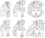
Development of the spinal nerves. A, Formation of nerve roots. B, Formation of nerve trunk (N).C, Formation of nerves. D, E, Formation of subordinate branches. F, Formation of nerve trunks in relation to the limb: dorsal and ventral trunks corresponding to lateral and anterior trunks in D and E. Labels: D.R., dorsal root; A1.C., alimentary canal; V.R., ventral root; Ao, aorta; N.T., neural tube; V., cardinal vein; No., notochord, M.P., muscle plate; D.G., dorsal ganglion; Sy., sympathetic cord; W.D., wolffian duct; Co., coelom; So., somatic division; V1, visceral branch; P, posterior primary division; Lat, lateral branches; Ant., anterior branches.

Development of the Spinal Nerve

Development of the spinal nerves. A, Formation of nerve roots. B, Formation of nerve trunk (N).C, Formation…
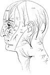
Distribution of sensory nerves to the head and neck. Labels: Ophth, ophthalmic division of the fifth nerve; St, supratrochlear branch; S.O, supra-orbital branch; I.T, infratrochlear branch; L, lachrymal branch; N, external nasal branch; Sup.Max, superior maxillary division; T, temporal branch; M, malar branch; I.O, infra-orbital branch; Inf.Max, inferior maxillary branch; M, mental branch; C2, 3, branches of the second and third cervical nervs; G.O, great occipital nerve; S.O, small occipital nerve; G.A, great auricle nerve; S.C, superficial cervical nerve; C3, least occipital nerve; 4, 5, 6, posterior primary division of 4th, 5th, 6th cervical nerve.

Sensory Nerves to the Head and Neck

Distribution of sensory nerves to the head and neck. Labels: Ophth, ophthalmic division of the fifth…
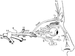
Scheme of the distribution of the ophthalmic nerve. Labels: Vs, trigeminal nerve, afferent root; Mo, efferent root; G.G, gasserian ganglion; M, meningeal branch; I.C, branch to internal carotid artery; Oph, opthalmic nerve; S.M, superior maxillary nerve; I.M, inferior maxillary nerve; III, communication to oculomotor nerve; IV, to trochlear nerve; L, branches to upper eyelid; L.G, long root to lenticular ganglion; Sy, root from sympathetic (on carotid artery); III, short root from motor oculi nerve; C, short ciliary branches; L.C, long ciliary nerves; I.T, infra-trochlear nerve; E.N, external nasal nerve; I.N, internal nasal nerve; O, orbital branch of superior maxillary nerve; L.Gl, lachrymal gland; C, conjuctival branch; L, branch to eyelids and face.

Ophthalmic Nerve

Scheme of the distribution of the ophthalmic nerve. Labels: Vs, trigeminal nerve, afferent root; Mo,…
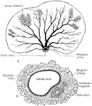
A diagram to illustrate the structure of the liver. A, arrangement of liver lobules around the sublobular branches of the hepatic vein; B, Section of portal canal, showing its contained branches of the portal vein, hepatic artery, and bile duct, surrounded by prolongation of Glisson's capsule.

Structure of the Liver

A diagram to illustrate the structure of the liver. A, arrangement of liver lobules around the sublobular…
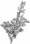
Junipers are coniferous plants in the genus Juniperus of the cypress family Cupressaceae. Depending on taxonomic viewpoint, there are between 50-67 species of juniper, widely distributed throughout the northern hemisphere, from the Arctic, south to tropical Africa in the Old World, and to the mountains of Central America. Junipers vary in size and shape from tall trees, 20-40 m tall, to columnar or low spreading shrubs with long trailing branches. They are evergreen with needle-like and/or scale-like leaves. They can be either monoecious or dioecious

Juniper

Junipers are coniferous plants in the genus Juniperus of the cypress family Cupressaceae. Depending…
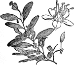
Coca is a plant in the family Erythroxylaceae, native to north-western South America. The plant plays a significant role in traditional Andean culture. Coca leaves contain cocaine alkaloids, a basis for the drug cocaine, which is a powerful stimulant. Coca should not be confused with the similarly named South American cocoa bean from which chocolate is made. The plant resembles a blackthorn bush, and grows to a height of 2&ndash;3 m (7&ndash;10 ft). The branches are straight, and the leaves, which have a green tint, are thin, opaque, oval, and taper at the extremities. A marked characteristic of the leaf is an areolated portion bounded by two longitudinal curved lines, one line on each side of the midrib, and more conspicuous on the under face of the leaf. The flowers are small, and disposed in little clusters on short stalks; the corolla is composed of five yellowish-white petals, the anthers are heart-shaped, and the pistil consists of three carpels united to form a three-chambered ovary. The flowers mature into red berries.

Coca Flower

Coca is a plant in the family Erythroxylaceae, native to north-western South America. The plant plays…
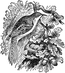
The Brown Creeper (Mohoua novaeseelandiae), also known by its M&#x0101;ori name, Pipipi, is a small passerine bird endemic to the South Island of New Zealand. They are specialist insectivores, gleaning insects from branches and leaves. They have strong legs and toes for hanging upside down while feeding.

Brown Creeper

The Brown Creeper (Mohoua novaeseelandiae), also known by its Māori name, Pipipi, is a small…
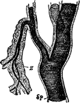
Part of a tracheal stem and branches of an insect. Labels: Z, cellular outer wall; SP, cuticular inner wall with spiral fiber.

Tracheal Stem and Branches

Part of a tracheal stem and branches of an insect. Labels: Z, cellular outer wall; SP, cuticular inner…
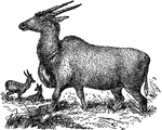
The common eland (Taurotragus oryx, also known as the southern eland) is a savannah and plains antelope found in East and Southern Africa. Common eland live on the open plains of Southern Africa and along the foothills of the great South African plateau. They eat grass, branches and leaves and are diurnal but tend to be inactive during the heat of day. Herds usually have 30 to 80 individuals, but are known to exceed 400. The common eland has an unusual social life, leaving or joining herds as necessary without forming close ties. Predators in the wild are predominantly lions and the endangered African wild dog.

Eland

The common eland (Taurotragus oryx, also known as the southern eland) is a savannah and plains antelope…