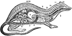
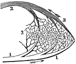
The Respiratory System of a Small Mammal
The respiratory apparatus of other mammals is similar to humans in both structure and function. The…
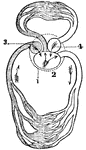
A Diagram of the Heart
A diagram of the heart. Labels: 1, Right and left auricle. 2, Right and left ventricle. 3, 4, The pericardium.…
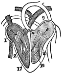
A Diagram of the Heart
A diagram of the heart. Labels: 1, Right auricle. 2, Right ventricle. 9, Left auricle. 10, Left ventricle.…
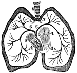
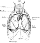
The Pulmonary Artery
The pulmonary artery. Labels: t, The trachea. h, The heart. a, The aorta. p, The pulmonary artery. 1,…
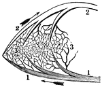
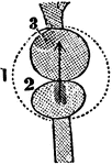
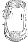
A Portion of the Pulmonic Circulation
A portion of the pulmonic circulation. 1, A branch of the artery that carries the impure blood to the…
A Portion of the Systemic Circulation
A portion of the systemic circulation. 1, A branch of the aorta. This terminates in the capillaries…
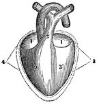
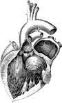
The Heart
A diagram of the heart. Labels: 1. Left auricle. 2, Right auricle. 3, Left ventricle. 4, Right ventricle.…
The Heart
A diagram of the heart. Labels: 1. Right auricle. 2, Left auricle. 3, Right ventricle. 4, Left ventricle.…
A Diagram of the Heart of a Reptile
A diagram of the heart of a reptile. Labels: 1, Pericardium. 2, Single ventricle. 3, Left auricle. 4,…
A Diagram of the Heart of a Fish
A diagram of the heart of a fish. Labels: 1, Pericardium. 2, The ventricle that receives the blood from…
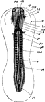
The Heart and Arteries of a Lobster
In the class of Crustacea there is a single ventricle, which receives the blood from the gills and propels…
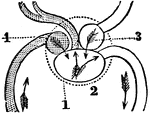
Diagram of the Circulation of an Insect.
Insects have neither arteries nor veins. The circulation, such as it is, is animated by the action of…
The Heart and Arteries of a Snail
In high orders of Mollusca the circulation resembles that of fish. Shown is the heart and arteries of…
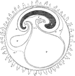
Circulation of a Frog
A diagram of the circulation of a frog. Labels: 1, The pericardium. 2, The single ventricle. 3, The…
Circulation of a Fish
A diagram of the circulation of a fish. Labels: 1, The pericardium. 2, The single auricle. 3, The single…
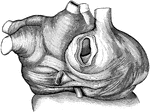
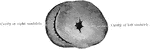
The Circulatory Organs
The circulatory organs. Labels: 1, The left auricle. 2, The right auricle. 3, The left ventricle. 4,…
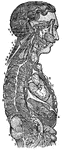
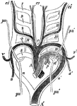
The Sympathetic Ganglions and their Connection to other Nerves
The sympathetic ganglions and their connection with other nerves. Labels: A, The semilunar ganglion…
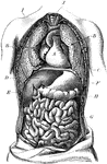
Torso
The human torso. Labels: A, the heart; B, the lungs drawn aside to show the internal organs; C, the…
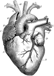
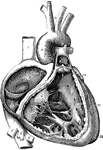
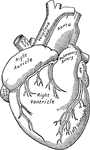
Heart
The heart. Labels: A, the right ventricle; B, the left ventricle; C, the right auricle; D, the left…
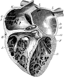
Chambers of the Heart
The chambers of the heart. Labels: A, right ventricle; B, left ventricle; C, right auricle; D, left…
Heart Valves
A diagram showing the peculiar fibrous structure of the heart and the shape of the valves. A, triscupid…
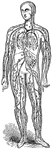
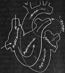
Circulation of Blood
A diagram illustrating the circulation of the blood. Labels: A, vena cava descending (superior); Z,…
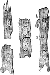
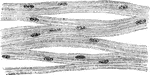
Muscular Fiber Cells from the Heart
Muscular fiber cells from the heart. The fibers which lie side by side are united at frequent intervals…
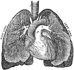
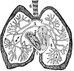
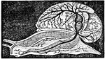
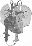
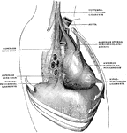
Heart and Lungs
View of heart and lungs in situ. The front portion of the chest wall, and the outer or parietal layers…
A Diagram of the Heart
The right auricle and ventricle opened, and a part of their right and anterior walls removed, so as…
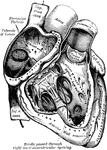
A Diagram of the Heart
The left auricle and ventricle opened and a part of their anterior and left walls removed. The pulmonary…
Muscular Fiber Showing the Nuclei of the Muscle Corpuscles
Network of muscular fibers from the heart of a pig. The nuclei of the muscle corpuscles are well shown
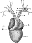
Heart of a Frog
The heart of a frog (Rana esculenta) from the front. Labels: V, ventricle, Ad, right auricle; As, left…
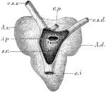
Heart of a Frog
The heart of a frog (Rana esculenta) from the back. Labels: s.v., sinus venosus opened; c.s.s., left…
Nerves of the Heart of a Frog
Course of the nerves in the auricular partition wall of the heart of a frog. Labels: d, dorsal branch;…
Nerve Cells
The nerve cells that compose the ganglia are generally unipolar, and seldom bipolar; sometimes two cells…
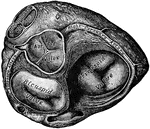
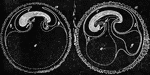
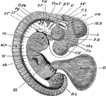
Embryo Chick
Embryo chick (36 hours), viewed from beneath as a transparent object (magnified). Labels:pl, outline…
Membranes of the Ovum
Diagrammatic section showing the relation in a mammal between the primitive alimentary canal and the…
Chorion Villi
Very soon after the entrance of the ovum into the uterus, in the human subject, the outer surface of…
Embryo Chick at Fourth Day
Embryo chick at fourth day, viewed as a transparent object, lying on its left side. CH, cerebral hemispheres;…
Embryo at Fourth Week
A human embryo of the fourth week. I, the chorion; 3, part of the amnion; 4, umbilical vesicle with…
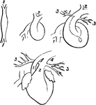
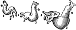
Fetal Heart in Successive Stages of Development
Fetal heart in successive stages of development. 1, venous extremity; 2, arterial extremity; 3, pulmonary…
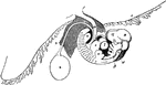
Development of the Heart of a Chick
Heart of a chick at the 45th, 65th, and 85th hours of incubation. I, the venous trunks; 2, the auricle;…
Aortic Arches in a Mammal
Diagram of the aortic arches in a mammal, showing transformations which give rise to the permanent arterial…
Intestine of a Chick
Rudiments of the liver on the intestines of a chick at the fifth day of incubation. Labels: 1, heart;…
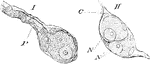
Aplysia Depilans (Linn.)
"The Molluscous Gasteropoda have the organs of respiration formed for aerial respiration or for respiration…
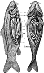
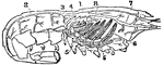
Anatomy of the Carp
br: the branchiae, or gill-openings c: the heart f: the liver vn: swimming bladders ci: intestinal canal…
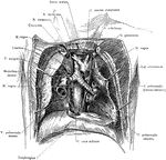
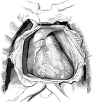
Dissection of the Thorax
Topography of the retrocardiac structures of the mediastinum, after the removal of the heart and pericardium.
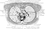
Cross Section of the Trunk at the Level of Junction of the Manubrium
Section at the level of the junction of the manubrium and corpus sterni, exposing the great vessels…
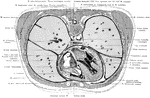
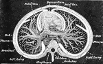
Cross Section of the Trunk Exposing the Ventricles of the Heart
Section exposing the ventricles of the heart.
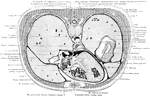
Cross Section of the Trunk Through the Inferior Portion of the Heart
Section through the inferior portion of the heart, exposing the dome of the diaphragm on the right side.
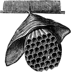
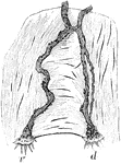
Hanging Hornet's Nest
Hornets "make their nests in trunks of old trees, perforating the sound wood to arrive at the heart,…
Muscle Fibers of the Heart
Anastomosing muscle fibers of the heart, seen in longitudinal section. On the right the limits of the…
Pericardium Ligaments
The pericardium is a conical serofibrous sac in which the hear and the commencement of the great vessels…
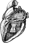
Heart with Right Auricle and Ventricle Laid Open
The right auricle and ventricle laid open, the anterior walls of both being removed.
Heart with Left Auricle and Ventricle Laid Open
The left auricle and ventricle laid open, the posterior walls of both being removed.