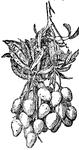
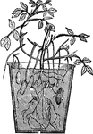
Mango
The fruit is kidney-shaped, four or five inches long, with smooth, pale green to reddish skin.
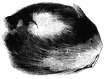
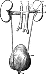
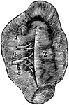
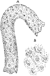
Musk bag
"A small kidney shaped, pendulous bag of the size of a hen's egg, situated below the abdomen and peculiar…
Vertical Section of the Back
"The spinal column below the twelfth dorsal vertebra at A has been removed, as well as the…
Kidney
Two glands having the function of secreting urine from the system, situated at the back of the abdominal…
Fish Circulation
"Diagram of the principal vessels in the circulation of a fish, ventral view. a, aorta; au., auricle;…
Arachis
"A genus of plants of the natural order Leguminosae, sub-order Papilionaceae, natives of the warm parts…
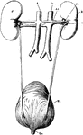
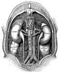
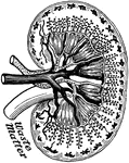
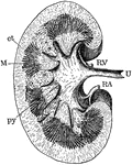
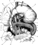
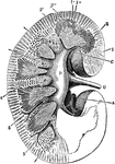
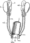
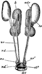
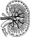
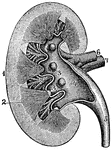
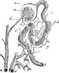
Renal Organs
"The renal organs, viewed from behind. R, right kidney; A, aorta; Ar, right renal artery; Vc, inferior…
Ailmentary Canal
"ailmentary canal of a honey bee. at, honey stomach; s, true stomach; nt, intestine; o, esophagus; sg,…
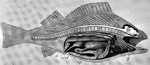
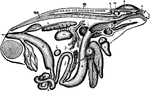
Dissected Fish
"Dissected fish. a, air bladder; b, urinary bladder; b, urinary bladder; br, brain; c, spinal cord;…

Dissected Frog
"Frog with the left side cut away and some of the organs pulled downward. a, aorta leading from the…
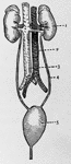
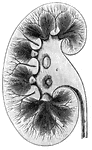
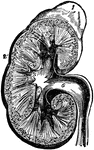
Kidneys
Kidneys and their vessels. 1: Left kidney; 2: Ascending vein; 3: Aorta; 4: Left ureter; 5: Bladder.
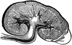
Kidney
Section of Kidney. 1: Body of Kidney; 2: Internal vessels; 3: Ureter, leading to the bladder.
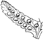
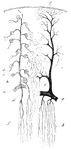
Shield-Fern
A small piece (pinnule) of a Shield-Fern: a row of fruit-dots on each side of the midrib, each covered…
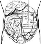
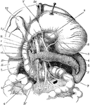
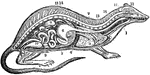
Regions of the Abdomen and their Contents
Regions of the abdomen and their contents (edge of costal cartilages in dotted outline). "For convenience…
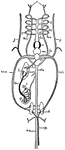
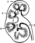
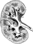
The Renal Organs Viewed from Behind
The renal organs viewed from behind. labels: R, right kidney; A, aorta; Ar, right erenal artery; Vc,…
Section through the Kidney
Section through the kidney showing the medullary and cortical portions, and the beginning of the ureter.…
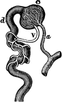
Vascular Supply of Kidney
Vascular supply of kidney. Labels: a, partof arterial arch; b, arterial branch passing upwards through…
Plan of the Blood Vessels Connected with the Tubules
Plan of the blood vessels connected with the tubules. "The blood passes downwards in straight vessels…
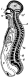
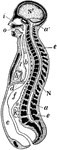
Longitudinal Section of Body
Diagrammatic longitudinal section of the body. Labels: a, the neural tube, with its upper enlargement…
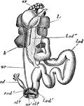
The Stomach, Pancreas, Liver, and Duodenum
The stomach, pancreas, liver, and duodenum, with part of the rest of the small intestine and the mesentery;…
Kidney Section
Section through the right kidney from its outer to inner border. Labels: 1, cortex; 2, medulla; 2',…
Kidney Circulation
Circulation in the kidney. Labels: ai, small branch of renal artery giving off the branch va, which…
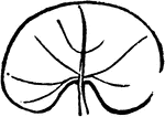
The Spleen
The spleen, a soft, brittle, highly vascular organ, of dark purplish color, in size about 5 x 3 x 1.5…
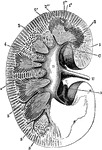
The Kidney
The basic structure of the kidney, which consists of the cortical portion, the medullary substance,…
Minute Structure of the Kidney
The minute structure of the kidney, which commences in the cortical substance of the organ as the Malpighian…
Longitudinal Section of the Body
Diagrammatic longitudinal section of the Body. Labels: a, the neural tube, with its upper enlargement…
Digestive Organs
The stomach, pancreas, liver, and duodenum, with part of the rest of the small intestine and the mesentery;…
Section of the Right Kidney
Section through the right kidney from its outer to its inner border. Labels: 1, cortex; 2, medulla;…
Kidney Glomerulus and Uriniferous Tubule
Diagram showing a kidney glomerulus and the commencement of an uriniferous tubule. Labels: a, afferent…
Kidney
Transverse section of the human kidney: "(a) cortex; (b) medulla; (c) small branch of the renal artery;…
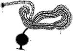
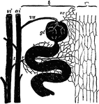
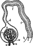
Leech Nephridium
"A nephridium of leech. F., Internal terminal funnel; C., glandular coil covered with blood vessels;…
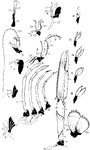
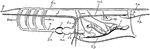
Norway Lobster Appendages
"Appendages of Norway lobster. Ex., Exopodite: En., endopodite; protopodite dark throughout; Ep., epipodite.…
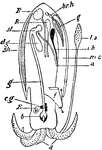
Cuttlefish Structure
"Diagram of the structure of Sepia. a., Eight short arms around mouth; l.a., one of the two long arms;…
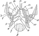
Cuttlefish Circulatory and Excretory Systems
"Diagram of circulatory and excretory systems in a Decapod-like Sepia. 1, Gill; 2, renal sac; 3, afferent…
Newt Anatomy
"Section through a young newt. c.t., Connective tissue; E., epidermis; D., dermis; S.C., spinal cord;…
Teleostean Circulation
"Diagram of Teleostean circulation. A., auricle; V., ventricle; v.a., ventral aorta; a.br., afferent…
Tadpole Dissection
"Dissection of tadpole. DL., Lower lip; H., ventricle of heart; DE., oesophagus; NA., head kidney; A.,…
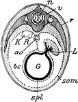
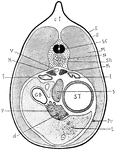
Bird Section
"Diagrammatic section of young bird. n., Spinal cord; v., vertebra; r., rib; L., liver; G., gut; som.…
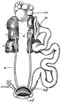
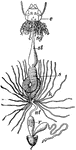
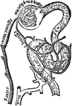
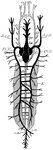
Female Pigeon Urogenital Organs
"Female urogenital organs of pigeon. K., Kidney with three lobes; u., ureter; cl., cloaca; ov., ovary;…
Fish Circulation Vessels
"Diagram of the principal vessels in the circulation of a Fish, lateral view. a, aorta; au., auricle;…
Female Bird Genital Organs
"Diagram of the female genital organs of a Bird. c, cloaca; i, intestine; k, kidney; o, ovary with ova…
Male Bird Genital Organs
"Diagram of the urino-genital organs of a male Bird. ad., adrenal body; c, cloaca; i, intestine; k,…
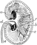
Frog Viscera
"General view of the viscera of a male frog, from the right side. a, stomach; b, urinary bladder; c,…
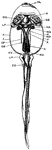
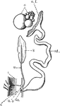
Male Rock Pigeon Genitalia
"Columba livia. Male urino-genital organs. adr, adrenal; cl. 2, urodaeum; cl. 3, proctodaeum; k, kidney;…
Female Rock Pigeon Genitalia
"Columba livia. Female urino-genital organs. cl. 2, urodaeum; cl. 3, proctodaeum; k, kidney; l. od,…
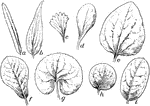
Leaf Shapes
"General outline of leaves. a, linear; b, lanceolate; c, wedge-shaped; d, spatulate; e, ovate; f, obovate;…
View of Organs from the Side
The chief organs of the body from the side. Labels: a, arch of the aorta or main artery of the trunk;…
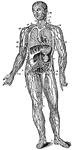
The Lymphatic Vessels and Glands
The lymphatic vessels and glands. Labels: 1, 2, 3, 4, 5, 6, Lymphatic vessels and glands of the lower…
The Respiratory System of a Small Mammal
The respiratory apparatus of other mammals is similar to humans in both structure and function. The…
Longitudinal Section of a Kidney
A longitudinal section of a kidney. Labels: 1, 2, 3, Parts of the Kidney. 4, Pelvis. 5, Ureter. 6, Renal…
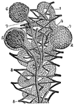
Diagram of the Structure of the Kidney
A diagram of the structure of the kidney. Labels: 1, Tubules or minute tubes. 2, Enlargement of a tubule…
Kidney
Plan of a longitudinal section through the pelvis and substance of the right kidney. Labels: a, the…
Cells from the Tubuli Uriniferi
The tubuli uriniferi are composed of a nearly homogeneous membrane, and are lined internally by epithelium.…
Blood Supply of the Kidney
Vascular supply of the kidney. Labels: a, part of arterial arch; b, interlobular artery; c, glomerulus;…
Malpighian Body
Relation of the Malpighian body to the uriniferous ducts and blood vessels. Labels: a, one of the interlobular…