The Comparative Anatomy ClipArt gallery offers 228 images of anatomy comparing numerous parts of humans and animals, showing similarities and differences among species and between different taxonomic orders.

The molar teeth of a human, horse and dog. The first image to the left in a molar tooth of a horse. Labels: 1, The enamel. 2, The ivory. 3, Canals for blood vessels. The center image is a molar tooth of a human. Label: 1, The enamel. 2, The ivory. 3, The cavity containing blood vessels. 4, 5, Artery and nerve. The image on the right is a molar tooth of a dog. Labels: 1, The enamel. 2, The ivory. 3, Cavity for blood-vessels.

Comparison of the Molar Teeth of a Human, Horse, and Dog

The molar teeth of a human, horse and dog. The first image to the left in a molar tooth of a horse.…
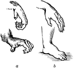
The power possessed by the hand of a human is chiefly depended upon the size and power of the thumb, which is more developed in humans than it is in the highest apes. The thumb of the human hand can be brought into exact opposition to the extremities of all the fingers, whether singly or in combination; while in those quandrumana which most nearly approach man, the thumb is so short, and the fingers are so weak that they can never be opposed to each other with any degree of force. The human foot is, in proportion to the side of the whole body, larger, broader, and stronger than that of any other mammal.

Comparison of the Hand and the Foot of a Monkey and Human

The power possessed by the hand of a human is chiefly depended upon the size and power of the thumb,…
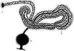
"A nephridium of leech. F., Internal terminal funnel; C., glandular coil covered with blood vessels; V., external terminal vesicle." -Thomson, 1916

Leech Nephridium

"A nephridium of leech. F., Internal terminal funnel; C., glandular coil covered with blood vessels;…
Nervous system of an adult ladybug. Ag, optic ganglion.

Nervous System of a Ladybug

Nervous system of an adult ladybug. Ag, optic ganglion.

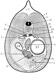
"Section through a young newt. c.t., Connective tissue; E., epidermis; D., dermis; S.C., spinal cord; M., muscle; N., notochord; Sh., mesodermic sheath of notochord; K., kidney; l., lung; S., spleen; ST., stomach; Pe., peritoneum; L., liver; d., duct of the pancreas (P); G.B., gall-bladder; V., dorsal aorta." -Thomson, 1916

Newt Anatomy

"Section through a young newt. c.t., Connective tissue; E., epidermis; D., dermis; S.C., spinal cord;…
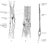
Olfactory and supporting cells in a frog and a human. A. Frog. B. Human. C. Human.

Olfactory and Supporting Cells

Olfactory and supporting cells in a frog and a human. A. Frog. B. Human. C. Human.
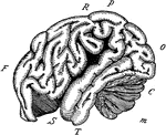
"The outline of the brain of an orang outang. Front portion F to O, cerebrum; C, cerebellum; M, medulla and spinal cord; F, the frontal lobe; P, the parietal lobe; O, the occipital lobe; T, the temporal lobe; R, the fissure of Rolando; S, the fissure of Silvius." -Galloway, 1915

Orangoutang Brain

"The outline of the brain of an orang outang. Front portion F to O, cerebrum; C, cerebellum; M, medulla…
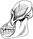
"Skull of adult orang outang." -Galloway, 1915

Orangoutang Skull

"Skull of adult orang outang." -Galloway, 1915

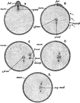
"Diagram illustrating the maturation and fertilization of the ovum. A, formation of first polar globule; B, beginning of fertilisation, sperms approaching the micropyle or aperture in the enclosing membrane of the ovum through which the sperm enters; C, formation of the male pronucleus; D, approximation of the male and female pronuclei; E, formation of segmentation-nucleus; female centrosome; male centrosome (the centrosomes are cell-structures not further referred to in this work); mem, egg-membrane; microp, micropyle; pol, polar bodies; female pronucleus; male pronucleus; seg. nucle, segmentation nucleus." -Parker, 1900

Ovum Fertilization

"Diagram illustrating the maturation and fertilization of the ovum. A, formation of first polar globule;…

"Heart and arterial system of pigeon. R.A., right auricle; R.V., right ventricle; L.V., left ventricle; L.A., left auricle; P.V., pulmonary veins; P., pectoral artery; Br., brachial artery; C., carotid artery; D.A., dorsal aorta; CL., coeliac; A.M., anterior mesenteric; R., renals; F., femoral; Sc., sciatic; IL., iliac; p.m., posterior mesenteric; C., caudal." -Thomson, 1916

Pigeon Arterial System

"Heart and arterial system of pigeon. R.A., right auricle; R.V., right ventricle; L.V., left ventricle;…
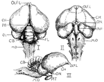
"Brain of pigeon (I. dorsal, II. ventral, III. lateral aspects). OLF.L., Olfactory lobes; C.H., cerebral hemispheres; PB., pineal body; OL., optic lobes; CB., cerebellum; FL., flocculus or lateral extension of cerebellum; M.O., medulla oblongata; PIT., pituitary body at end of infundibulum (INF.); O.N., optic nerves crossing in the chiasma." -Thomson, 1916

Pigeon Brain

"Brain of pigeon (I. dorsal, II. ventral, III. lateral aspects). OLF.L., Olfactory lobes; C.H., cerebral…
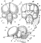
"Columba livia. The brain; A, from above; B, from below; C, from the left side. cb, cerebellum; c. h, cerebral hemispheres; f, flocculus; inf, infundibulum; m. o, medulla oblongata; o. l, optic lobes; o. t, optic tracts; pn, pineal body; II-XIII, cerebral nerves; sp. 1, first spinal nerve." -Parker, 1900

Rock Pigeon Brain

"Columba livia. The brain; A, from above; B, from below; C, from the left side. cb, cerebellum; c. h,…

"Columba livia. Part of left foot of an unhatched embryo. The cartilage is dotted. mtl. 2, second; mtl. 3, third; and mtl. 4, fourth metatarsal; ti, tibia; tl. 1, proximal tarsal cartilage; tl. 2, distal tarsal cartilage." -Parker, 1900

Rock Pigeon Embryo Foot

"Columba livia. Part of left foot of an unhatched embryo. The cartilage is dotted. mtl. 2, second; mtl.…
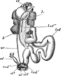
"Columba livia. Female urino-genital organs. cl. 2, urodaeum; cl. 3, proctodaeum; k, kidney; l. od, left oviduct; l. od', its cloacal aperture; l. od'', its coelomic funnel; l. od''', its coelomic aperture; ov, ovarty; r. od, right oviduct; r. od', its cloacal aperture; ur, ureter; ur', its cloacal aperture." -Parker, 1900

Female Rock Pigeon Genitalia

"Columba livia. Female urino-genital organs. cl. 2, urodaeum; cl. 3, proctodaeum; k, kidney; l. od,…

"Columba livia. Male urino-genital organs. adr, adrenal; cl. 2, urodaeum; cl. 3, proctodaeum; k, kidney; ts, tetis, that of the right side displaced; ur, ureter; ur', aperture of ureter; vd, vas deferens; vd', its cloacal aperture; v. s, vesicula seminalis." -Parker, 1900

Male Rock Pigeon Genitalia

"Columba livia. Male urino-genital organs. adr, adrenal; cl. 2, urodaeum; cl. 3, proctodaeum; k, kidney;…
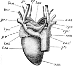
"A, heart of the pigeon, dorsal aspect. a. ao, arch of aorta; br. a, brachial artery; br. v, bachial vein; c. c, common carotid; ju, jugular; l. au, left auricle; l. p. a, left pulmonary artery; l. vn, left ventricle; pc. v, left pre-caval; ptc, post-caval; p. v, pulmonary veins; r. au, r. au', right auricle; r. p. a, right pulmonary artery; r. prc, right pre-caval; r.r vn, right ventricle." -Parker, 1900

Pigeon Heart

"A, heart of the pigeon, dorsal aspect. a. ao, arch of aorta; br. a, brachial artery; br. v, bachial…

"Columba livia. Left innominate of a nestling. The cartilage is dotted. ac, acetabulum; a. tr, anti-trochanter; il, pre-acetabular; and il', post-acetabular portion of ilium; is, ischium; i s. f, ischiatic foramen; ob. f, obturator notch; pu, pubis." -Parker, 1900

Rock Pigeon Innominate

"Columba livia. Left innominate of a nestling. The cartilage is dotted. ac, acetabulum; a. tr, anti-trochanter;…

"Columba livia. Left manus of a nestling. The cartilaginous parts are dotted. cp. 1, radiale; cp. 2, ulnare; mcp. 1, 2, 3, metacarpals; ph. 1, phalanx of first digit; ph. 2, ph. 2', phalanges of second digit; ph. 3, phalanx of third digit; ra, radius; ul, ulna." -Parker, 1900

Rock Pigeon Manus

"Columba livia. Left manus of a nestling. The cartilaginous parts are dotted. cp. 1, radiale; cp. 2,…
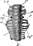
"Columba livia. Sacrum of a nestling (about fourteen days old), ventral aspect. c1, centrum of first sacral vertebra; c1, centrum of fifth caudal; c. r, first sacral rib; l1, centrum of first lumbar; l3, of third lumbar; s1, of fourth lumbar; s3, of sixth lumbar; tr. p, transverse process of first lumbar; tr. p', of fifth lumbar; tr. p'', of first sacral." -Parker, 1900

Rock Pigeon Sacrum

"Columba livia. Sacrum of a nestling (about fourteen days old), ventral aspect. c1, centrum of first…
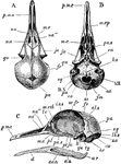
"Columba livia. Skull of young specimen. A, dorsal; B, ventral; C, left side. al. s, alisphenoid; au, angular; ar, articular; b. o, basi-occipital; d, dentary; e. o, ex-occipital; eu, aperture of Eustachian tube; f. m, foramen magnum; fr, frontal; i.o. s, inter-orbital septum; ju, jugal; lc, lachrymal; lb. s, lambdoidal suture; m. eth, mesethmoid; mx, maxilla; mx. p, maxillo-palatine process; na, na', na'', nasal; o. c, occipital condyle; or. fr, orbital plate of frontal; pa, parietal; pa. s, parasphenoid (rostrum); pl, palatine; p. mx, premaxilla; pt, pterygoid; qu, quadrate; s. an, supra-angular; s. o, supra-occipital; sq, squamosal; ty, tympanic cavity; IL-XII, foramina for cerebral nerves." -Parker, 1900

Rock Pigeon Skull

"Columba livia. Skull of young specimen. A, dorsal; B, ventral; C, left side. al. s, alisphenoid; au,…

"Female urogenital organs of pigeon. K., Kidney with three lobes; u., ureter; cl., cloaca; ov., ovary; od., oviduct; f.t., funnel at end of oviduct; r.r.od., rudimentary right oviduct." -Thomson, 1916

Female Pigeon Urogenital Organs

"Female urogenital organs of pigeon. K., Kidney with three lobes; u., ureter; cl., cloaca; ov., ovary;…

"Male urogenital organs of pigeon. T., testes; V., base of inferior vena cava; S.R., suprarenal bodies; K., kidneys with three lobes (1, 2, 3); u., ureter; v.d., vas deferens; vs., seminal vesicle; cl., cloaca." -Thomson, 1916

Male Pigeon Urogenital Organs

"Male urogenital organs of pigeon. T., testes; V., base of inferior vena cava; S.R., suprarenal bodies;…

"Heart and venous system of pigeon. R.A., Right auricle; R.V., right ventricle; L.V., left ventricle; L.A., left auricle; P.V., pulmonary veins; P.A., pulmonary arteries; J., jugular; Br., brachial; P., pectoral; H.V., hepatic; E.P., epigastric; I.V.C., inferior vena cava; C.M., coccygeo-mesenteric; I.V., iliac; F., femoral; R., renal; Sc., sciatic; Hyp., hypogastric or "renal-portal"; i.il., internal iliac; C., caudal." -Thomson, 1916

Pigeon Venous System

"Heart and venous system of pigeon. R.A., Right auricle; R.V., right ventricle; L.V., left ventricle;…

"Proneomenia. Nervous system. e.g., Cerebral ganglia; slg., sublingual; a.p.g., anterior pedal; p.p.g., posterior pedal; p.v.g., posterior viscerals; sl., sublingual connectives; cpc., cerebro-pedal connective; pe., longitudinal pedal nerves; la., longitudinal lateral nerves." -Thomson, 1916

Proneomenia

"Proneomenia. Nervous system. e.g., Cerebral ganglia; slg., sublingual; a.p.g., anterior pedal; p.p.g.,…
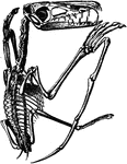
The skeleton of a pterodactyl, the prehistoric winged dinosaur.

Pterodactyl Skeleton

The skeleton of a pterodactyl, the prehistoric winged dinosaur.

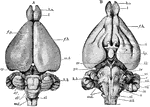
"Lepus cuniculus. Brain. A, dorsal view; B, ventral; b. o, olfactory lobe; cb', median lobe of cerebellum (vermis); cb'', lateral lobe of cerebellum; cr, cauro-cerebri; ep, epiphysis; f. b, parencephala; f. p, longitudinal fissure; h. b, hind-brain; hp, hypophysis; m. b, mid-brain (corpora quadrigemina); md, medulla oblongata; p.v, pons Varolii; i-xii, cranial nerves." -Parker, 1900

Rabbit Brain

"Lepus cuniculus. Brain. A, dorsal view; B, ventral; b. o, olfactory lobe; cb', median lobe of cerebellum…

"Lepus cuniculus. Longitudinal vertical sectioin of the brain. cb, cerebellum, showing arbor vitae; c. c, crus cerebri; ch1, parencephalon; ch2, temporal lobe; c. ma, corpus mammillare; f. m, foramen of Monro; inf, infundibulum; ly, lyra; m. o, medulla oblongata; o. ch, optic chiasma; olf, olfactory lobe; pty, pituitary body; vl. ip, velum interpositum; v. vn, valve of Vieussens; II, optic nerve." -Parker, 1900

Rabbit Brain

"Lepus cuniculus. Longitudinal vertical sectioin of the brain. cb, cerebellum, showing arbor vitae;…

"Dorsal view of rabbit's brain. olf.l., Olfactory lobes; c.h., cerebral hemispheres; o.l., optic lobes (corpora quadrigemina); cb., median part of cerrebellum; Fl., flocculus of cerebellum; 10, root of the tenth or vagus nerve; s.c., spinal cord." -Thomson, 1916

Dorsal View of Rabbit Brain

"Dorsal view of rabbit's brain. olf.l., Olfactory lobes; c.h., cerebral hemispheres; o.l., optic lobes…

"Under surface of rabbit's brain. olf.l., Olfactory lobes; o.t., olfactory tract; f.l., frontal lobe of cerebral hemisphere; ch., optic chiasma; l.c., infundibulum; c.m., corpus mammillare; 3, root of oculomotor; 4, root of pathetic; 5, root of trigeminal; 6, root of abducenss; 7-8, roots of facial and auditory; Fl., flocculus of cerebellum; 9, root of glosso-pharyngeal; 10, roots of vagus; H., 12th , or hypoglossal; p.v., pons Varolii." -Thomson, 1916

Under Surface of Rabbit Brain

"Under surface of rabbit's brain. olf.l., Olfactory lobes; o.t., olfactory tract; f.l., frontal lobe…
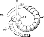
"Diagram of caecum in rabbit. s.i., Small intestine; s.r., sacculus rotundus; col., sacculated colon; c., caecum; v.a., vermiform appendix." -Thomson, 1916

Rabbit Caecum

"Diagram of caecum in rabbit. s.i., Small intestine; s.r., sacculus rotundus; col., sacculated colon;…

"Duodenum of rabbit. P., Pyloric end of stomach; g.b., gall-bladder with bile duct and hepatic ducts; p.d., pancreatic duct." -Thomson, 1916

Rabbit Duodenum

"Duodenum of rabbit. P., Pyloric end of stomach; g.b., gall-bladder with bile duct and hepatic ducts;…
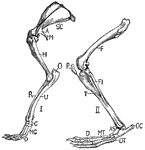
"Fore-limb and shoulder-girdle (I.) and hind-limb (II.) of rabbit. SC., Scapula; A., acromion; M., metacromion process; H., humerus; O., olecranon process; U., ulna; R., radius; C., carpals; MC., metacarpals; D., five digits; F., femur; P., patella; FI., fibula; T., tibia; OC., os calcis; AS., astragalus; DT., distal tarsals; MT., metatarsals; D., four digits." -Thomson, 1916

Rabbit Limbs

"Fore-limb and shoulder-girdle (I.) and hind-limb (II.) of rabbit. SC., Scapula; A., acromion; M., metacromion…
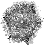
Lobule of rabbit's liver, vessels and bile ducts injected. Labels: a, central or intralobular vein; v, v, interlobular veins; c, interlobular bile duct.

Rabbit Liver Lobule

Lobule of rabbit's liver, vessels and bile ducts injected. Labels: a, central or intralobular vein;…
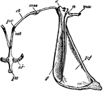
"Lepus cuniculus. Shoulder-girdle with anterior end of sternum of young specimen. a, acromion; af, pre-scapular fossa; c, coracoid; cl, ossified clavicle; ma, meta-cromion; mss, meso-scapular segment; ost, pre-sternum; pc, pre-coracoid; pf, post-scapular fossa; sr, sternal ribs." -Parker, 1900

Rabbit Shoulder Girdle

"Lepus cuniculus. Shoulder-girdle with anterior end of sternum of young specimen. a, acromion; af, pre-scapular…

"Side view of rabbit's skull. Pmx., Premaxilla; Na., nasal; Fr., frontal; Pa., parietal; Sq., squamosal; S.O., supraoccipital; Per., periotic; T., tympanic (the reference line points to the bony external auditory meatus, beneath it lies the inflated bulla); PO., paroccipital process." -Thomson, 1916

Rabbit Skull

"Side view of rabbit's skull. Pmx., Premaxilla; Na., nasal; Fr., frontal; Pa., parietal; Sq., squamosal;…
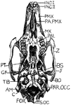
"Under surface of rabit's skull. Inc. I., First incisors; Inc. II., second incisors; PMX., premaxilla; PA.PMX., palatal process of premaxilla; MX., maxilla; PAL., palatine; Z., zygomatic arch; BS., basisphenoid; J., posterior part of jugal; BO., basioccipital; PAR.OCC., paroccipital process of exoccipital; SOC., supraoccipital; C., one of the condyles; AM., external auditory meatus; TB., tympanic bulla; GF., glenoid fossa; PT., pterygoid." -Thomson, 1916

Under Surface Rabbit Skull

"Under surface of rabit's skull. Inc. I., First incisors; Inc. II., second incisors; PMX., premaxilla;…

"Upper surface of rabbit's skull. N., Anterior nostril; PMX., premaxilla; NA., nasal; FR., anterior part of frontal; MX., posterior part of maxilla; j., anterior part of jugal; SO.F., supraorbital process of frontal; FRR., posterior part of frontal; JJ., posterior end of jugal protruding below zygomatic portion of squamosal (Z.SQ.); PA., parietal; AM., external auditory meatus; SO., supraoccipital; IP., interparietal; SQ., squamosal." -Thomson, 1916

Upper Surface Rabbit Skull

"Upper surface of rabbit's skull. N., Anterior nostril; PMX., premaxilla; NA., nasal; FR., anterior…
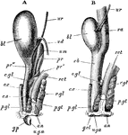
"Lepus cuniculus. The urogenital organs. A, of male; B, of female, from the left side. The kidneys and proximal ends of the ureters, and in B, the ovaries. Fallopian tubes and uteri are not shown. an, anus; bl, urinary bladder; c. c, corpus cavernosum; c. s, corpus spongiosum; c. gl, perineal gland; p. gl', aperture of its duct on the perineal space; pr, anterior, pr', posterior, and pr', lateral lobes of prostate; rct, rectum; r. gl, rectal gland; u. g. a, urino-genital aperture; u. m, uterus masculinus; ur, ureter; va, vagina; vb, vestibule; v. d, vas deferens." -Parker, 1900

Rabbit Urogenital Organs

"Lepus cuniculus. The urogenital organs. A, of male; B, of female, from the left side. The kidneys and…
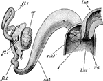
"Lepus cuniculus. The anterior end of the vagina, with the right uterus. Fallopian tube, and ovary. Part of the ventral wall of the vagina is removed, and the proximal end of the left uterus is shown in longitudinal section fl. t, Fallopian tube; fl. t', its peritoneal aperture; l. ut, left uterus; l. ut', left os uteri; r. ut, right uterus; r. ut', right os uteri; s, vaginal sepum; va, vagina." -Parker, 1900

Rabbit Vagina

"Lepus cuniculus. The anterior end of the vagina, with the right uterus. Fallopian tube, and ovary.…
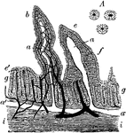
Vertical section of the intestinal mucous membrane of the rabbit. Two villi are represented, in one of which the dilated lacteal alone is shown, in the other the blood vessels and lacteal are both seen injected, the lacteal white, the blood vessels dark; a, the lacteal vessels of the villi; a', horizontal lacteal, which they join; b, capillary blood vessels in one of the villi; c, small artery; d, vein; e, the epithelium covering the villi; g, tubular glands or crypts of Lieberkuhn, some divided down the middle, others cut more irregularly; i, the submucous layer. A, cross-section of three tubular glands more highly magnified.

Rabbit's Intestinal Mucous Membrane

Vertical section of the intestinal mucous membrane of the rabbit. Two villi are represented, in one…
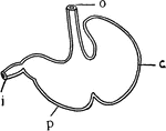
"Stomach of Rat (B)...c, cardiac portion; p, pyloric portion; o, esophagus; i, intestine." -Galloway, 1915

Rat Stomach

"Stomach of Rat (B)...c, cardiac portion; p, pyloric portion; o, esophagus; i, intestine." -Galloway,…
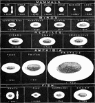
The illustration exhibits the typical characters of the red blood cells in the main divisions of Vertbrata. The fractions are those of an inch, and represent the average diameter. In the case of the oval cells, only the long diameter is here given. It is remarkable, that although the size of the red blood cells varies so much in the different classes of the vertebrate kingdom, that of the white corpuscles remains comparatively uniform, and thus are, in some animals, much greater, in others much less than the red corpuscles existing side by side with them.

Red Blood Cells in Vertebrata

The illustration exhibits the typical characters of the red blood cells in the main divisions of Vertbrata.…

"Diagram of the heart and branchial arches in a Reptile...a, aorta; au., auricle; c, carotid; c.v., cardinal veins (right and left); d.a., dorsal arteries; j, jugular veins; d.c., ductus Cuvieri; s.v., sinus venosus; v, ventricle." -Galloway, 1915

Reptile Heart

"Diagram of the heart and branchial arches in a Reptile...a, aorta; au., auricle; c, carotid; c.v.,…
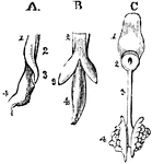
The development of the respiratory organs. A, is the esophagus of a chick on the fourth day of incubation, with the rudiments of the trachea on the lung of the left side, viewed laterally; 1, the inferior wall of the esophagus; 2, the upper portion of the same tube; 3, the rudimentary lung; 4, the stomach. B, is the same object seen from below, so that both lungs are visible. C, shows the tongue and respiratory organs of the embryo of a horse; 1, the tongue; 2, the larynx; 3, the trachea; 4, the lungs, viewed from the upper side.

Development of Respiratory Organs

The development of the respiratory organs. A, is the esophagus of a chick on the fourth day of incubation,…

The respiratory apparatus of other mammals is similar to humans in both structure and function. The lungs fill the cavity of the chest, and have the same general composition of lobes, lobules and air-cells. Labels: 1, Trachea. 2, Lungs. 3, Heart. 4, Diaphragm. 5, Liver. 6, Stomach. 7, Esophagus. 8, Kidney. 9, Intestines. 10, Bladder. 11, Cerebrum. 12, Cerebellum. 13, Medulla spinalis. 14, Vertebrae.

The Respiratory System of a Small Mammal

The respiratory apparatus of other mammals is similar to humans in both structure and function. The…
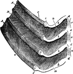
Portions of four ribs of a dog with the muscles between them. Labels: a, a, ventral ends of the ribs, joining at c the rib cartilages, b, which are fixed to cartilaginous portions, d, of the sternum. A, external intercostal muscle, ceasing between the rib cartilages, where the internal intercostal, B, is seen. Between the middle two ribs the external intercostal muscle has been dissected away, so as to display the internal which was covered by it.

Ribs of a Dog

Portions of four ribs of a dog with the muscles between them. Labels: a, a, ventral ends of the ribs,…
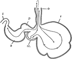
"Diagram of the stomach of a ruminant. o, esophagus; r, rumen or paunch; re., reticulum, or honeycomb; p, psalterium or manyplies; a, abomasum or rennet; i, intestine." -Galloway, 1915

Ruminant Stomach

"Diagram of the stomach of a ruminant. o, esophagus; r, rumen or paunch; re., reticulum, or honeycomb;…
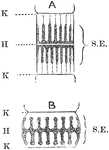
Diagram of a sarcomere in a moderately extended condition, A, and in a contracted condition, B. K, Krause's membranes; H, plane of Henson; S.E., poriferous sarcous element.

Sarcomere

Diagram of a sarcomere in a moderately extended condition, A, and in a contracted condition, B. K, Krause's…

Diagrams of sarcostyles of insects' wing muscles.  A, relaxed; B, contracted.

Sarcostyles of Insect Wings

Diagrams of sarcostyles of insects' wing muscles. A, relaxed; B, contracted.
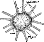
"Chromatophore of Sepia, magnified. nuc, nuclei in wall of sac; pigm, pigment; rad. mus, radiating strands of muscle." -Parker, 1900

Sepia Chromatophore

"Chromatophore of Sepia, magnified. nuc, nuclei in wall of sac; pigm, pigment; rad. mus, radiating strands…

Serous glands. Labels: a, rabbit's pancreas "loaded" (resting); c, "discharged" (active) (observed in the living animal). b, loaded; d, discharged, alveolus of parotis (fresh preparation).

Serous Glands of a Rabbit

Serous glands. Labels: a, rabbit's pancreas "loaded" (resting); c, "discharged" (active) (observed in…

"Side view of chief cranial nerves of Elasmobranchs." -Thomson, 1916

Shark Nerves

"Side view of chief cranial nerves of Elasmobranchs." -Thomson, 1916

"Side view of sheep's skull. PMX., Premaxilla; MX., maxilla; NA., nasal; J., Jugal; L., lachrymal; FR., frontal; PA., parietal; SQ., squamosal; CO., condyle; PP., paroccipital process." -Thomson, 1916

Sheep Skull

"Side view of sheep's skull. PMX., Premaxilla; MX., maxilla; NA., nasal; J., Jugal; L., lachrymal; FR.,…

"Stomach of sheep. a, OEsophagus; c, rumen or paunch; d, reticulum or honeycomb-bag; e, psalterium or many-plies; f, abomasum or reed; b, beginning of duodenum." -Thomson, 1916

Sheep Stomach

"Stomach of sheep. a, OEsophagus; c, rumen or paunch; d, reticulum or honeycomb-bag; e, psalterium or…
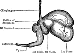
The four stomachs of the sheep, a grass-eating animal. The beginning of the intestines are also shown, which reach in their full length to 28 times the size of the sheep's body.

Sheep, Stomachs of

The four stomachs of the sheep, a grass-eating animal. The beginning of the intestines are also shown,…
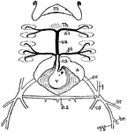
"Heart and adjacent vessels of skate. v., Ventricle; c.a., conus arteriosus; p.i., posterior innominate; v.a., ventral aorta; a.i., anterior innominate; Th., thyroid; m., mouth; a., auricle; s.v., sinus venosus; s.c., precaval sinus or sinus of Cuvier; h.s., hepatic sinus; j., jugular; br., brachials; cd., cardinal; epg., epigastric." -Thomson, 1916

Skate Heart

"Heart and adjacent vessels of skate. v., Ventricle; c.a., conus arteriosus; p.i., posterior innominate;…
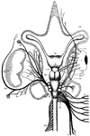
"Dissection of nerves of skate. CH., Cerebral hemispheres; O.TH., optic thalami; OL., optic lobes; M., medulla; 4v., posterior part of cerebellum, covering fourth ventricle; OB., olfactory bulb; OC., olfactory capsule; SO., superior oblique muscle; E., eye; SR., superior rectus; ER., external rectus; SO.VII., superficial ophthalmic branch of VII.; SO.V., superficial ophthalmic branch of V.; OP., ophthalmicus profundus; A.C., auditory capsule; B.Pl., brachial plexus; R.F., recurrent facial; C.T., chorda tympani; F.P., facial proper; Hy., hyoidean; Hyomn., hyomandibular; E.M., external mandibular; M.M., mandibular muscle; Sp., spiracle; P.sp., prespiracular; Pl., palatine; O.B., outer buccal; Mn., mandibular; Mx., maxillary; I.B., inner buccal; L., lateral branch of X.; Py., pyloric branch; C., cardiac branch." -Thomson, 1916

Skate Nerves

"Dissection of nerves of skate. CH., Cerebral hemispheres; O.TH., optic thalami; OL., optic lobes; M.,…
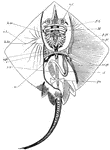
Skeleton of skate. nc, nasal capsules; pq, palato-pterygo-quadrate cartilage; M, Meckel's cartilage; hm, hyo-mandibular; h.br., fifth hypobranchial; v.pl., vertebral plate; st, sensory tubes; sc, scapular region; c, coracoid region; p.pt., anterior basal cartilage or pro-pterygium; m.pt., meso pterygium; mt.pt., meta-pterygium; pu., pubic bar; st, stomach; sv, spiral valve of intestine. -Thomson, 1916

Skate Skeleton

Skeleton of skate. nc, nasal capsules; pq, palato-pterygo-quadrate cartilage; M, Meckel's cartilage;…

"Under surface of skull and arches of skate. l.1, First labial cartilage; R., rostrum; tr., trabecular region; n.c., nasal capsule; a.o., antorbital cartilage; p.pt.q., palato-pterygo-quadrae; M.c., Meckel's cartilage; h.m., hyo-mandibular; h.br.1-5, hypobranchials; c.br.5, fifth ceratobranchial; c.h., cerato-hyal; l.2-4, labial cartilages." -Thomson, 1916

Skate Skull

"Under surface of skull and arches of skate. l.1, First labial cartilage; R., rostrum; tr., trabecular…

"Side view of skate's skull. l1., First labial cartilage; n.c., nasal capsule; a.o., antorbital; p.pt.q., palato-pterygo-quadrate; M.c., Meckel's cartilage; h.m., hyo-mandibular; e.h., epi-hyal; c.h., cerato-hyal; h.h., hypo-hyal; h.br.1-5, hypobranchials; c.br., ceratobranchial; e.br., epibranchial; p.br.1., first prebranchial; i.h., inter-hyal; m.pt., meta-pterygoid; 2, 5, 7, foramina of exit of the corresponding nerves." -Thomson, 1916

Skate Skull

"Side view of skate's skull. l1., First labial cartilage; n.c., nasal capsule; a.o., antorbital; p.pt.q.,…