Abdomen
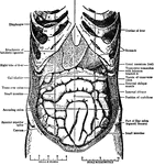
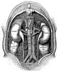
"The Abdominal Viscera in situ, as seen when the abdomin is laid open and the great omentum…
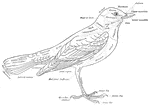
Principal Organs of the Thorax and Abdomen
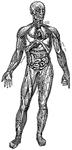
"The principal muscles are seen on the left, and superficial veins on the right." — Blaisedell, 1904
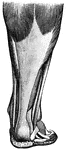
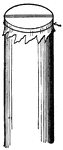
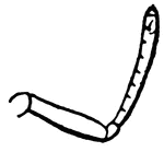
Achillles tendon
"Tendons are white, glistening cords, or straps, which connect the muscles with the bones." —…
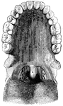
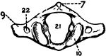
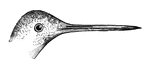
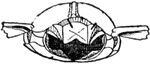
Back view of the adult mouth
"The head is represented as having been thrown back, and the tongue drawn forward. A, B…
Ailmentary Canal of Bluebird
An illustration of the "ailmentary canal of Bluebird, reduced; after Audobon. a,b, gullet or oesophagus;…
Air Passage
"To illustrate roughly the passage of air through the glottis, force air through such a tube by blowing…
Capillaries of the Air Sac
"Diagram showing the capillary network of the air sacs and origin of the pulmonary veins.. A,…
Diagrammatic view of an air sac
"A, epithelial lining wall; B, partition between two adjacent sacs, in which run capillaries;…
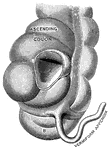
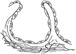
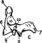
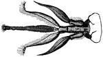
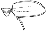
Vermiform appendix
"A, a portion of the colon laid open to show the valve between the large and small intestine;…
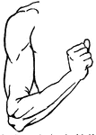
Muscles of the Arm
"Muscles on the front of the arm. Note the while cords, the tendons at the wrist." —Davison, 1910
Arteries
"The Right Axillary and Branchial Arteries, with Some of their Main Branches." — Blaisedell, 1904
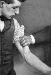
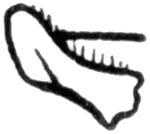
Branchial artery
"Showing how firm pressure may be made with the fingers to compress the branchial artery of the left…
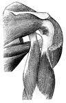
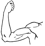
Muscles of the Back and Shoulder
"Some of the Larger Muscles on the Back of the Shoulder and the Arm." — Blaisedell, 1904
Vertical Section of the Back
"The spinal column below the twelfth dorsal vertebra at A has been removed, as well as the…
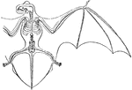
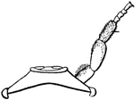
Skeleton and Wing Membranes of the Noctule Bat
The bat genus Nyctalus (Noctule bats) are Evening bats. They are distributed in the temperate and subtropical…
Leg of Bear
This illustration shows the plantigrade leg of a bear. Plantigrade means that the animal walks flat…
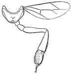
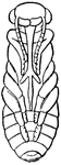
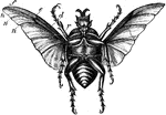
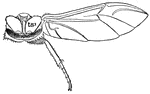
Mouth and Tongue of a Bee
"The structure of the mouth in insects exhibits very remarkable modifications, and these are of the…
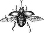
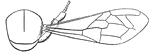
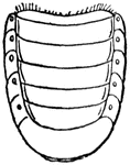
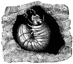
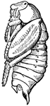
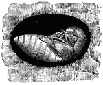
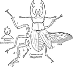
Large Beetle
"Under surface of large beetle, with deeply concave and comparatively small wings, shows that the nervures…
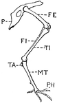
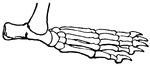
Leg of Bird
This illustration shows the leg of a bird. P. Pelvis, FE. Femur, TI. Tibia, FI. Fibula, TA. Tarsus,…
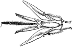
Wing of Bird
"Shows how the bones of the arm (a), forearm (b), and hand (c), are twisted, and form a conical screw."—Pettigrew,…
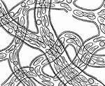
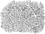
Circulation of blood
"Showing how the circulation of blood in the web of a frog's foot looks as seen under the microscope."…
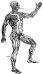
Superficial Muscles of the Body
"A single muscle rarely or never contracts alone, but always in harmony with a number of other muscles.…
Bowfin
The bowfin, a primitive freshwater fish, also known as the freshwater dogfish is a voracious fish.