This human anatomy ClipArt gallery offers 116 illustrations of human systemic circulation of the cardiovascular system. This includes veins, arteries, and capillaries involved in systemic circulation of blood throughout the body. General views of the circulatory system are also included here.

Veins of the upper extremity . Labels: 1, axillary artery; 2, axillary veins; 3, 4, basilic; 5, point where median basilic joins basilic; 6, posterior basilic vein; 8, anterior basilic vein; 9, point where cephalic enters axillary; 10, portion same vein; 11, point where median cephalic enters cephalic; 12, lower portion cephalic vein; 13, median cephalic vein; 14, median vein; 15, anastromosing branch; 16, cephalica-pollicis veins; 17, veins of fingers; 18, palmar veins.

Veins of the Arm and Shoulder

Veins of the upper extremity . Labels: 1, axillary artery; 2, axillary veins; 3, 4, basilic; 5, point…

Arteries of the arm. Labels: 1, axillary artery; 2, thoracica acromialis; 3, superior thoracic; 4, subcapular; 5, inferior scapular; 6, 7, branches to the teres and subscapularis; 8, anterior circumflex; 9, brachial; 10, profunda; 11, posterior circumflex; 12, profunda; 13, muscular branches; 14, branches to brachialis interus; 15, recurrent ulnar.

Arteries of the Arm

Arteries of the arm. Labels: 1, axillary artery; 2, thoracica acromialis; 3, superior thoracic; 4, subcapular;…

Superficial veins of the upper extremity (arm). Labels: 1, axillary artery; 2, axillary veins; 3, basilic vein; 5, point where the median basilic joins the basilic vein; 9, point where the cephalic enters the axillary vein; 11, point where the median cephalic centers the cephalic vein; 12, lower portion of the cephalic vein; 14, median vein; 15, anastomosing branch; 16, cephalicapellicis vein; 17, subcutaneous veins of the fingers; 18, subcutaneous palmar veins.

Veins of the Arm

Superficial veins of the upper extremity (arm). Labels: 1, axillary artery; 2, axillary veins; 3, basilic…

The arterial system.

The Arterial System

The arterial system.

"The Right Axillary and Branchial Arteries, with Some of their Main Branches." — Blaisedell, 1904

Arteries

"The Right Axillary and Branchial Arteries, with Some of their Main Branches." — Blaisedell, 1904
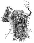
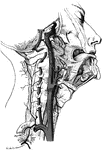
Arteries of the neck. 1, occipital artery; 2. facial vein; 3, spinal accessory nerve; 4. facial artery; 5, internal jugular vein; 6, hypoglossal nerve; 7. communicaus noni nerve; 8, lingual artery; 9, pneumogastric; 10, superior larnygeal nerve; 11, phrenio; 12, superior thyroid artery; 13, sterno-mastoid; 14, common carotid; 15, clavicle; 16, sterno-hyoid; 17, subclavian; 18, omo-hyoid; 19, subclavian; 20, sympathetic ganglia; 21, apex lung.

Arteries

Arteries of the neck. 1, occipital artery; 2. facial vein; 3, spinal accessory nerve; 4. facial artery;…

The main arteries of the body. Labels: Crd, and Crs, right and left coronary arteries of the heart, cut short near their origin; Aa, and aA, aortic arch; At, thoracic aorta; Aab, abdominal aorta; K, renal artery; Sd, right, and Ssi, left subclavian; Cd, right, and Cs, left carotid; Ax, axillary artery; B, brachial artery; U, ulner artery; R, radial artery; Ai, common iliac artery; I, external iliac artery; C, femoral artery; Po, popliteal artery; Ta, anterior, and Tp, posterior tibial artery; Pe, peroneal artery.

The Main Arteries of the Body

The main arteries of the body. Labels: Crd, and Crs, right and left coronary arteries of the heart,…
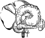
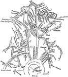
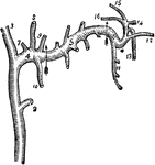
Diagram showing the branching, anastomoses, and confluence of arteries.

Arteries

Diagram showing the branching, anastomoses, and confluence of arteries.

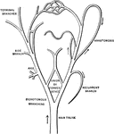
Arteries of the abdominal organs. Labels: 1, the liver; 2, the stomach; 3, upper gut; 4, pancreas; 6, great mesenteric artery; 7, gastric branch; 8, spleen; 9, pyloric branch; 10, pancreatic branch;    11, hepatic artery; 12, duodinal branch; 13, cystic artery; 14, branches to the stomach; 15, 16, spleenic arteries; 17, gastro-epiploic artery; 18, descending aorta; 19, great mesenteric artery.

Arteries of the Abdominal Organs

Arteries of the abdominal organs. Labels: 1, the liver; 2, the stomach; 3, upper gut; 4, pancreas; 6,…

Arteries of the palm of the head and front of the forearm. Labels: 3, deep part of the raised pronator of the radius; 4, long supinator muscle; 5, long flexor of the thumb; 6, square pronator; 7, deep flexor of the fingers; 8, cubital flexor of the wrist; 9, annular ligament; 10, the brachial artery; 12, radial artery; 13, recurring radial artery joining the end of the upper deep one; 4, superficial veins; 15, cubital artery; 16, superficial palmary arch; 17, magna artery of the thumb and radial artery of the index; 18, back cubital recurring artery; 19, front interosseous artery; 20, back interosseous artery.

Arteries of the Hand and Forearm

Arteries of the palm of the head and front of the forearm. Labels: 3, deep part of the raised pronator…
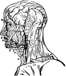
The arteries of the head and neck.

Arteries of the Head and Neck

The arteries of the head and neck.

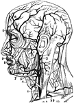
Arteries of the head and neck. Labels: 1, primitive carotid artery; 2, occipital branch to the back part of the skull; 4, upper hyoides artery; 5, lower pharyngeal artery; 6, masseter artery; 7, submental artery; 8, lower coronary artery; 9, upper coronary artery; 10, deep branch; 11, back cervical artery; 12, continuation and fold of the occipital; 13, descending branch for muscles of the neck; 14, posterior auricular; 15, temporal artery; 16, parietal branches; 17, frontal branches; 19, orbitary branches; 20, subclavian artery.

Arteries of the Head and Neck

Arteries of the head and neck. Labels: 1, primitive carotid artery; 2, occipital branch to the back…
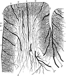
Arteries of the lower extremity (thigh/upper leg).

Arteries of the Lower Extremity

Arteries of the lower extremity (thigh/upper leg).

Arteries of the lower extremity (thigh/upper leg).

Arteries of the Lower Extremity

Arteries of the lower extremity (thigh/upper leg).

Diagram of the arteries of the pelvis and thigh.

Arteries of the Pelvis and Thigh

Diagram of the arteries of the pelvis and thigh.

Arteries of the trunk.

Arteries of the Trunk

Arteries of the trunk.

Diagram of the arteries of the upper extremity (arm/thoracic area).

Arteries of the Upper Extremity

Diagram of the arteries of the upper extremity (arm/thoracic area).

Distribution of the cortical arteries. Labels" 1, Medullary arteries; 1', Group of medullary arteries in the sulcus between tow adjacent convolutions; 1", Arteries situated among the short association fibers. 2, Cortical arteries; a, Capillary network with fairly wide meshed, situated beneath the pia; b, Network with more compact, polygonal meshes, situated in the cortex; c, Transitional network, with wider meshes; d, Capillary network in the white substance.

Cortical Arteries

Distribution of the cortical arteries. Labels" 1, Medullary arteries; 1', Group of medullary arteries…

Major arteries of the body. The kidneys and spleen are also shown with their respective arteries. Labels: 1, cheek arteries; 2, coeliac axis; 3, gastric artery; 4, hepatic artery; 5, spleenic artery; 6, supra-renal artery on right side; 7, right renal artery; 8, lumbar arteries; 9, upper mesenteric artery; 10, the two spermatic arteries; 11, the lower mesenteric; 12, the middle sacras; 13, the common iliac; 14, internal iliac of the right side; 15, external iliac; 16, episgastric artery; 17, circumflex iliac artery; 18, femoral artery.

Major Arteries

Major arteries of the body. The kidneys and spleen are also shown with their respective arteries. Labels:…

A portion of an artery.

Artery

A portion of an artery.

Transverse section through a small artery and vein. Labels: A, artery; V, vein; e, epithelial lining; m, middle muscular and elastic coat, thick in the artery, much thinner in the vein; a, outer coat of areolar tissue, magnified 350 diameters.

Diagram of an Artery and a Vein

Transverse section through a small artery and vein. Labels: A, artery; V, vein; e, epithelial lining;…

Transverse section through the wall of a large artery. Labels: A, tunica intima; B, tunica media; C, tunica externa.

Section Through Artery Wall

Transverse section through the wall of a large artery. Labels: A, tunica intima; B, tunica media; C,…

"The dotted line shows the course of the branchial artery." — Blaisedell, 1904

Brancial artery

"The dotted line shows the course of the branchial artery." — Blaisedell, 1904

"The dotted line shows the course of the right femoral artery." — Blaisedell, 1904

Femoral artery

"The dotted line shows the course of the right femoral artery." — Blaisedell, 1904

"The Femoral Artery of Right Leg." — Blaisedell, 1904

Leg artery

"The Femoral Artery of Right Leg." — Blaisedell, 1904

"Artery in the Front of the Right Leg." — Blaisedell, 1904

Leg artery

"Artery in the Front of the Right Leg." — Blaisedell, 1904

Portion of an artery, showing the several coats of which it is composed, separated from each other. Labels: 1. the internal or serious coat. 2. the middle or fibrous coat. 3. the external or cellular coat.

Portion of an Artery

Portion of an artery, showing the several coats of which it is composed, separated from each other.…
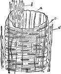
Structure of an artery. Labels: A, internal coat, with b, its inner layer of pavement epithelium (endothelium); c, middle coat, with transverse fibers; d, outer coat, with longitudinal fibers.

Structure of an Artery

Structure of an artery. Labels: A, internal coat, with b, its inner layer of pavement epithelium (endothelium);…

Surface view of an artery from the mesentery of a frog, ensheathed in a perivascular lymphatic vessel. Labels: a, The artery, with its circular muscular coat (media) indicated by broad transverse markings, with an indication of the adventitia outside. 1. Lymphatic vessel: its wall is a simple endothelial membrane.

Surface View of an Artery

Surface view of an artery from the mesentery of a frog, ensheathed in a perivascular lymphatic vessel.…
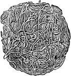
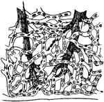
Relations of the artery, vein, and capillaries.

Relations of Artery, Vein, and Capillaries

Relations of the artery, vein, and capillaries.

A portion of fibrin, showing its fibrous structure and netlike arrangement of its fibers. In a short time after the blood is taken from the body it separates into two portions, by a process called coagulation or clot; the white substance which forms the upper part of the clot is called fibrin, and the red mass under it, the red particles. The fibrin is the material from which all solids of the body are formed.

Blood Clotting Fibers

A portion of fibrin, showing its fibrous structure and netlike arrangement of its fibers. In a short…
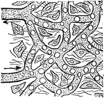
Blood plasma passing out of capillary to feed the body cells.

The Course of Blood

Blood plasma passing out of capillary to feed the body cells.

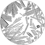
By pricking the end of the finger with a needle, we can obtain a drop of blood for examination. Place it on the slide, cover with a glass, and put it under a microscope. The red disks will be seen to group themselves in rows, while the white disk will seem to draw apart, and to be constantly changing their form. After gradual evaporation, the crystals may be seen.

Blood Crystals

By pricking the end of the finger with a needle, we can obtain a drop of blood for examination. Place…

Diagram of the course of the blood. Labels: RA, right auricle; RV. right ventricles; LV and LA, left auricle and left ventricle; VCI and VCS, vena cava inferior and superior; Ao aorta; Lg, lungs; Al, alimentary canal; Th D, thoracic duct; Lct,lacteals; HA and HV, hepatic artery and vein.

Blood Flow Diagram

Diagram of the course of the blood. Labels: RA, right auricle; RV. right ventricles; LV and LA, left…

A particle of human blood as it appears when transparent and floating. 2. the same, seen as illuminated. 3. the same, one half illuminated. 4. a particle of frog's blood floating. 5. the same, seen edgewise. All these objects are magnified 500 diameters.

Blood Particle

A particle of human blood as it appears when transparent and floating. 2. the same, seen as illuminated.…

Diagram of the blood vascular system, showing that it forms a single closed circuit with two pumps in it, consisting of the right and left halves of the heart, which are represented separate in the diagram. Labels: ra and rv, right auricle and ventricle; la and lv, left auricle and ventricle; ao, aorta; sc, systemic capillaries; vc, venae cavae; pa, pulmonary artery; pc, pulmonary capillaries; pv, pulmonary veins.

Blood Vascular System

Diagram of the blood vascular system, showing that it forms a single closed circuit with two pumps in…
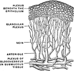
Termination of the blood vessels in the mucous membrane of the stomach.

Blood Vessels in the Mucous Membrane of the Stomach

Termination of the blood vessels in the mucous membrane of the stomach.
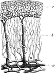
The blood vessels of the skin.

Blood Vessels of the Skin

The blood vessels of the skin.

Plan of the blood vessels of the stomach, as they would be seen in a vertical section. Labels: a, arteries, passing up from the vessels of submucous coat; b, capillaries branching between and around the tubes; c, superficial plexus of capillaries occupying the ridges of the mucous membrane; d, vein formed by the union of veins which, having collected the blood of the superficial capillary plexus, are seen passing down between the tubes.

Blood Vessels of the Stomach

Plan of the blood vessels of the stomach, as they would be seen in a vertical section. Labels: a, arteries,…
"Showing how the circulation of blood in the web of a frog's foot looks as seen under the microscope." — Blaisedell, 1904

Circulation of blood

"Showing how the circulation of blood in the web of a frog's foot looks as seen under the microscope."…
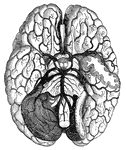
"Arteries and their Branches at the Base of the Brain." — Blaisedell, 1904

Blood vessels of the brain

"Arteries and their Branches at the Base of the Brain." — Blaisedell, 1904

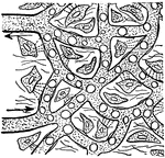
"Blood plasma passing out of capillary to feed the body cells." —Davison, 1910

Capillaries

"Blood plasma passing out of capillary to feed the body cells." —Davison, 1910

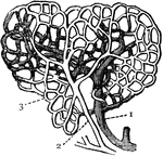
Capillaries. 1: Vein; 2: Artery; 3: Capillary.

Capillaries

Capillaries. 1: Vein; 2: Artery; 3: Capillary.

A large capillary from the frog's mesentery eight hours after irritation had been set up, showing emigration of leucocytes. Labels: a, Cells in the act of traversing the capillary wall; b, some already escaped.

Capillaries Showing Emigration of Leucocytes

A large capillary from the frog's mesentery eight hours after irritation had been set up, showing emigration…

Diagrammatic representation of a capillary seen from above and in section. Labels: a, the wall of the capillary with, b, the nuclei; c, nuclei belonging to the connective tissue in which the capillary is supposed to be lying; d, the canal of the capillary.

Diagram of a Capillary

Diagrammatic representation of a capillary seen from above and in section. Labels: a, the wall of the…

Isolated capillary network formed by the junction of several hallowed-out cells, and containing colored blood-corpuscles in a clear fluid. Labels: p, p, pointed cell-processes extending in different directions for union with neighboring capillaries.

Capillary Network

Isolated capillary network formed by the junction of several hallowed-out cells, and containing colored…
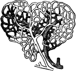
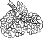
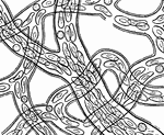
A network of capillaries.

Capillary Network

A network of capillaries.

Injected capillary vessel of muscle.

Capillary Vessels of Muscle

Injected capillary vessel of muscle.

The internal carotid and vertebral arteries.

Carotid Artery

The internal carotid and vertebral arteries.

The external carotid artery.

The Carotid Artery

The external carotid artery.

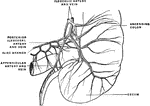
Arteries and veins of the cecum and vermiform appendix seen from behind.

Arteries and Veins of the Cecum and Appendix

Arteries and veins of the cecum and vermiform appendix seen from behind.

An ideal view of the circulation in the lungs and system.

Circulation

An ideal view of the circulation in the lungs and system.

Circulation of blood. 1: Capillaries of lungs; 2: Capillaries of system.

Circulation

Circulation of blood. 1: Capillaries of lungs; 2: Capillaries of system.

"Diagram of circulation. r.a., Right auricle receiving superior vena cava (s.v.c.) and inferior vena cava (i.v.c.); r.v., right ventricle; p.a., pulmonary artery to lungs (L.); p.v., right pulmonary vein; l.a., left auricle; l.v., left ventricle; ao., aortic arch; d.ao., dorsal aorta giving off arteries to liver (ii.), to gut (g.), to body (B.); po.v., portal veins; h.v., hepatic vein." -Thomson, 1916

Circulation

"Diagram of circulation. r.a., Right auricle receiving superior vena cava (s.v.c.) and inferior vena…

Diagram of circulation. Labels: L, left side of heart; R, right side of heart; a,a,a arterial system; b,b, capillaries; c,c,c veins; Alim., alimentary canal; Liv., liver; p, portal vein; H,hepatic vein; Lymph., lymphatic duct and tributaries; Pulm., lungs; Pa, pulmonary artery; Pu, pulmonary vein.

Diagram of Circulation

Diagram of circulation. Labels: L, left side of heart; R, right side of heart; a,a,a arterial system;…

"<em>R.A.</em>, right auricle; <em>L.A.</em>, left auricle; <em>R.V.</em>, right ventricle; <em>L.V.</em>, left ventricle; <em>P.A.</i., pulmonary artery; <em>A</em>, pulmonary artery and vein of right lung; <em>B</em>, pulmonary artery and vein of left lung; <em>C</em>, carotid artery to head, showing branch of left subclavian artery; <em>D</em>, portal vein; <em>F</em>, hepatic artery; <em>G</em>, superior vena cava, bringing blood from head and upper limbs to right auricle." &mdash; Blaisedell, 1904

Diagram of the circulation of the blood

"R.A., right auricle; L.A., left auricle; R.V., right ventricle; L.V.,…
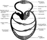
The blood is made to circulate within the system of closed tubes in which it is contained by means of the alternate contraction and relaxation of the heart.

Blood Circulation

The blood is made to circulate within the system of closed tubes in which it is contained by means of…

Diagram of circulation.

Diagram of Circulation

Diagram of circulation.

Diagram of circulation.

Diagram of Circulation

Diagram of circulation.

1: Left ventricle of the heart. 2 and 3: Aorta. 5: Arteries that extend to the lower extremities. 6: Arteries of the neck. 7: Arteries of the arms.

Circulatory

1: Left ventricle of the heart. 2 and 3: Aorta. 5: Arteries that extend to the lower extremities. 6:…