This human anatomy ClipArt gallery offers 116 illustrations of human systemic circulation of the cardiovascular system. This includes veins, arteries, and capillaries involved in systemic circulation of blood throughout the body. General views of the circulatory system are also included here.
The Circulatory Organs
The circulatory organs. Labels: 1, The left auricle. 2, The right auricle. 3, The left ventricle. 4,…
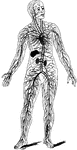
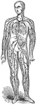
Diagram of the Circulatory System
Diagram of the circulatory system, showing that it forms a single closed circuit with two pumps in it,…
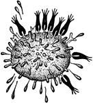
Cytolysis, Stage 1
"Ehrlich's diagrams illustrating the mechanism of immunity and cytolysis. The figures in black indicate…

Cytolysis, Stage 2
"Ehrlich's diagrams illustrating the mechanism of immunity and cytolysis. The figures in black indicate…
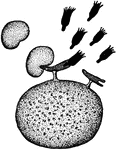
Cytolysis, Stage 3
"Ehrlich's diagrams illustrating the mechanism of immunity and cytolysis. The figures in black…
Cytolysis, Stage 4
"Ehrlich's diagrams illustrating the mechanism of immunity and cytolysis. The figures in black…
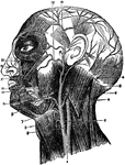
Arteries of the Face and Head
The arteries of the face and head. Labels: 1, common carotid; 2, internal carotid; 3, external carotid;…
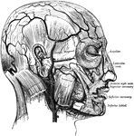
Facial Arteries
The arteries of the face and scalp. The muscle tissue of the lips must be supposed to have been cut…
Facial Arteries
"1, primitive carotid artery dividing itself into carotid external and carotid internal; 3, occipital…
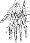
Hand Arteries
"The arteries of the hand, showing the communications or anastomoses of different arteries and the fine…
Arteries of the Head
Arteries of the head. Labels: 1, common carotid; 2, internal carotid; 3, external carotid; 4, occipital;…
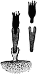
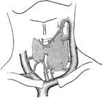
Vascular Supply of Kidney
Vascular supply of kidney. Labels: a, partof arterial arch; b, arterial branch passing upwards through…
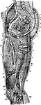
Arteries of the Leg
Arteries of the leg. Labels: 1, extensor propius pollicis; 2, articular arteries; 3, anterior tibia;…
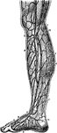
Veins of the Leg
Veins of the leg. Labels: 1, saphenous; 2, collateral branch; 3, anastomosis; 4, internal saphenous;…
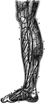
Veins of the Legs
Superficial veins of the legs. Labels: 1, saphena major; 2, collateral branch; 3, anastomosis of veins;…
Section of Liver Showing Intralobular Capillary Network
Section of liver injected from hepatic vein, showing intralobular capillary network.
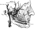
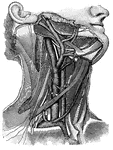
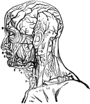
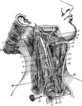
Blood vessels of the neck
"Showing the carotid artery and jugular vein on the right side, with some of their main branches; also…
Nerves
"a, axis cylinder; b, inner border of white substance; c, c, outer border of same; d, d, tubular membrane;…
Arteries of the Pelvis and Thigh
Arteries of the pelvis and thigh. Labels: 1, inferior extremity of abdominal aorta; 2, right primitive…
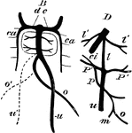
Communication of the Portal and Systemic Circulation
To show the sites at which communications occur between the portal and systemic circulations.
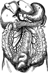
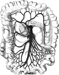
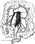
The Portal Vein and its Branches
The portal vein and its branches. Labels: l, liver, under surface; gb, gall bladder; st, stomach; sp,…
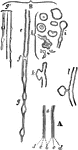
Reticulum of Fibrin
The clotting of the blood is due to the development in it of a substance called fibrin which appears…
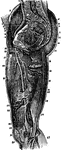
Arteries of the Thigh
Arteries of the thigh. Labels: 1, aorta; 2, common iliac; 3, external iliac; 4, epigastric; 5, circumflex…
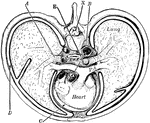
Thorax
"Cross-section of thorax. A, bronchus, entering the lung; B, the aorta cut at its origin and again at…
Anterior Tibial Artery
Anterior tibial artery. Labels: 1, extensor proprius pollicis pedis muscle and tendon; 2, 2, articular…
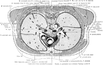
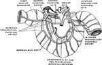
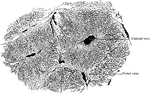
Cross Section of the Trunk at the Level of Junction of the Manubrium
Section at the level of the junction of the manubrium and corpus sterni, exposing the great vessels…

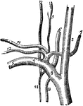
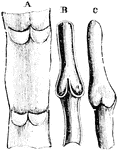
Valves of a Vein
Valves of a vein. In the lower part of the figure are seen the parietal valve; the upper part shows…

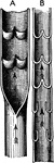
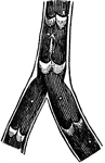
Valves of Veins
Diagram showing valves of veins. A, part of a vein laid open and spread out with two pairs of valves.…
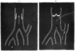
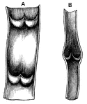
Valves of Veins
Valves of veins. A shows a vein cut open between the segments of two valves. B shows appearance of valves…
Blood Flow in the Valves of Veins
A, vein with valves open. B, vein with valves closed: stream of blood passing off by lateral channel.
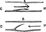
Action of the Valves of the Veins
Diagram to illustrate the mood of action of the valves of the veins. Labels: C, the capillary; H, the…

Action of Valves
Diagram to illustrate the mode of action of the valves of the veins. Labels: C, the capillary end, and…
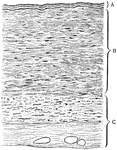
Section Through Vein Wall
Transverse section through the wall of a vein. Labels: A, tunica intima; B, tunica media; C, tunica…

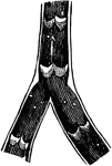
Structure of a Vein
Structure of a vein with valves. A, part of a vein, laid open, with two pairs of valves; B, longitudinal…

Veins
The portal system of veins. a: Portal vein. b: Splenic vein. c: Right gastro-epiploic vein. d: Inferior…
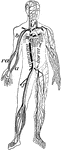
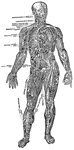
Veins and arteries
"Chief veins and arteries of the body. a, place of the heart; the veins are in black. On the right side…
Development of Veins in the Liver
Diagram illustrating the development of veins about the liver. B, dc, ducts of Cuvier, right and left;…

Formation of Large Veins
Diagram showing the formation of large veins by convergence of small, and the branching of veins.
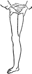
Superficial Veins of Leg
Superficial veins of lower extremity. Labels: 1, veins of the foot; 2, internal saphenous vein; 3, superficial…
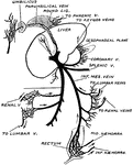
Principal Venous Trunks
Sketch of the principal venous trunks. Labels: 1, superior vena cava; , inferior vena cava; 3, right…