Clipart tagged: ‘arteries’
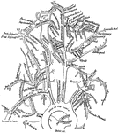
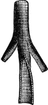
Arteries of the Arm
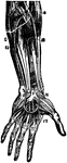
Arteries of the arm. Labels: 1, axillary artery; 2, thoracica acromialis; 3, superior thoracic; 4, subcapular;…
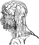
Arteries
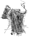
Arteries of the neck. 1, occipital artery; 2. facial vein; 3, spinal accessory nerve; 4. facial artery;…
The Main Arteries of the Body
The main arteries of the body. Labels: Crd, and Crs, right and left coronary arteries of the heart,…
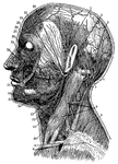
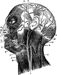
Arteries and Nerves
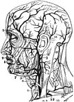
"Superficial arteries and nerves of the face and neck. 1, Temporal artery; 2, artery behind the ear;…
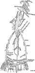
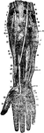
Arteries of the Hand and Forearm
Arteries of the palm of the head and front of the forearm. Labels: 3, deep part of the raised pronator…
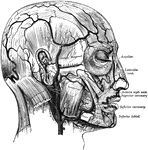
Arteries of the Head and Neck
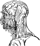
Arteries of the head and neck. Labels: 1, primitive carotid artery; 2, occipital branch to the back…
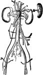
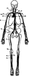
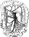
Major Arteries
Major arteries of the body. The kidneys and spleen are also shown with their respective arteries. Labels:…
Bird Arteries
Arteries of the trunk of a bird. 1: The aorta. 2: The vena cava. 3: A cerebral artery. The small lines…
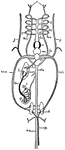
Blood Circulation
This illustration shows a representation of the circulation of the blood, in its essential features.…
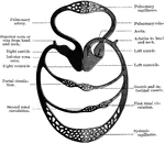
Blood Vascular System
Diagram of the blood vascular system, showing that it forms a single closed circuit with two pumps in…
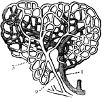
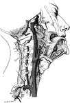
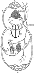
Diagram of Circulation
Diagram of circulation. Labels: L, left side of heart; R, right side of heart; a,a,a arterial system;…
Blood Circulation
The blood is made to circulate within the system of closed tubes in which it is contained by means of…
Arteries of the Face and Head
The arteries of the face and head. Labels: 1, common carotid; 2, internal carotid; 3, external carotid;…
Facial Arteries
The arteries of the face and scalp. The muscle tissue of the lips must be supposed to have been cut…
Facial Arteries
"1, primitive carotid artery dividing itself into carotid external and carotid internal; 3, occipital…
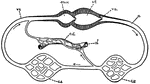
Fish Circulation
"Diagram of the principal vessels in the circulation of a fish, ventral view. a, aorta; au., auricle;…
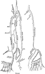
Dissection of Forearm
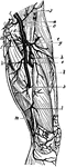
Deep dissection of front of the forearm. Labels: 1, supinator longus; 2, ulnar nerve; 3, brachialis;…
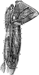
Dissection of the Forearm
Deep dissection of the front of the forearm. Labels: 1, supinator longus; 2, ulnar nerve; 3, brachialis…
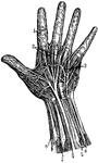
Hand Arteries
"The arteries of the hand, showing the communications or anastomoses of different arteries and the fine…
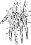
Hand Nerves
"Nerves of the had. 1, Nerves of the skin; 2, tendons; 3, arteries of the palm of the hand; 4, elbow…
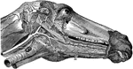
Head of a Horse Showing Arteries
Arteries of the head- the left maxillary ramus being remove. Labels: 1, occipital; 2, internal carotid;…
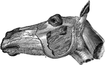
Head of a Horse Showing Arteries
Facial arteries of the left side. Labels: a, maxillo-muscular; a', posterior masseter; b, c, posterior…
Arteries of the Head
Arteries of the head. Labels: 1, common carotid; 2, internal carotid; 3, external carotid; 4, occipital;…
Heart
The heart is the organ that propels the blood and causes it to circulate through the arteries, veins,…
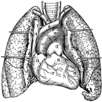
Heart and Lungs
1, The trachea or windpipe; 2 and 3, right and left common carotid arteries; 4 and 5, right and left…
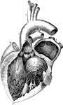
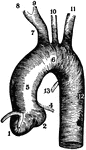
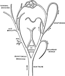
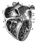
A Diagram of the Heart
The left auricle and ventricle opened and a part of their anterior and left walls removed. The pulmonary…
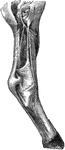
Leg of a Horse Showing Arteries
Arteries of the right posterior limb- external view. Labels: 1, popiteal; 2, posterior tibial; 3, anterior…
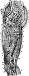
Arteries of the Leg
Arteries of the leg. Labels: 1, extensor propius pollicis; 2, articular arteries; 3, anterior tibia;…
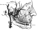
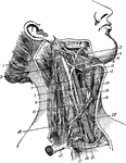
Neck of a Horse Showing Arteries
Arteries of the neck exposed on the left side. Labels: a, anterior aorta; a', left brachial; a",…
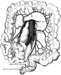
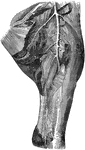
Arteries of the Pelvis and Thigh
Arteries of the pelvis and thigh. Labels: 1, inferior extremity of abdominal aorta; 2, right primitive…
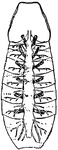
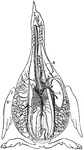
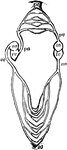
Scorpion
"View from below of a scorpion (Buthus occitanus) opened and dissected so as to show the pericardium…
Thigh of a Horse Showing Arteries
Internal view of left thigh-showing the arteries. Labels: 1, femoral; a, profunda femoris; b, superficialis…
Arteries of the Thigh
Arteries of the thigh. Labels: 1, aorta; 2, common iliac; 3, external iliac; 4, epigastric; 5, circumflex…
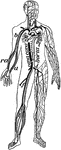
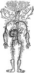
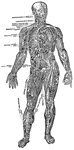
Veins and Arteries of the Body
Chief veins and arteries of the body. Labels: a, place of the heart; the veins are in the back. On the…