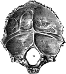
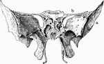
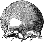
Occipital Bone of the Human Skull
Occipital bone of the human skull, inner surface. It is situated at the back and base of the skull.…
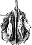
Parietal Bone of the Human Skull
Parietal bone of the human skull, inner surface. The parietal bones form the greater part of the sides…
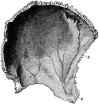
Frontal Bone of the Human Skull
Frontal bone of the human skull, outer surface. The frontal bone forms the forehead, roof of the orbital…
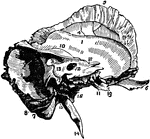
Temporal Bone of the Human Skull
Temporal bone of the human skull. The temporal bones are situated at the sides and base of the skull.…
Sphenoid Bone of the Human Skull
Sphenoid bone, situated the anterior part of the base of the skull, articulating with all the other…
Ethmoid Bone of the Human Skull
Ethmoid bone, posterior surface. The ethmoid bone is an exceedingly light, spongy bone, placed between…
Human Skull
The skull. Labels: a, nasal bone; b, superior maxillary; c, inferior maxillary; d, occipital; e, temporal;…
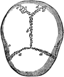
Human Skull at Birth
The skull at birth, superior suerface. The cranial bones of the infant at birth are not fullyformed…
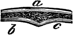
Human Joint, Dentated Suture
A toothed, or dentated suture. This is one type of immovable articulation. It is found in the union…
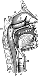
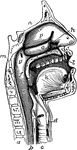
The Mouth, Nose, and Pharynx
The mouth, nose, and pharynx, with the larynx and commencement of gullet (esophagus), seen in section.…
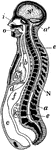
Longitudinal Section of Body
Diagrammatic longitudinal section of the body. Labels: a, the neural tube, with its upper enlargement…
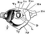
The Atlas (1st Cervical Vertebra)
The atlas, which is the first cervical vertebra. Labels: Aa, body of atlas, D, odontoid process of axis;…
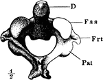
The Axis (2nd Cervical Vertebra)
The axis, which is the second cervical vertebra. Labels: D, odontoid process of axis; Fas, facet on…
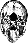
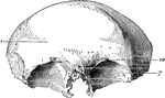
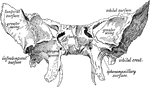
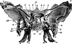
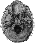
Base of the Skull
The base of the skull. "The lower jaw has been removed. At the lower part of the figure is the hard…
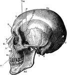
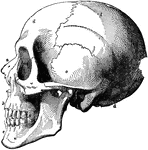
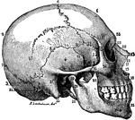
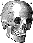
The Skull
A side view of the skull. Labels: O, occipital bone; T, temporal bone; Pr, parietal bone; F, frontal…
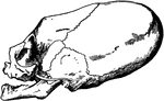
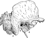
Distorted Skull of Child
Skull of a child of the tribe of Chinook Indians (inhabiting the neighborhood of the Columbia River),…
The Mouth, Nose, and Pharynx
The mouth, nose, and pharynx, with the commencement of the gullet (esophagus) and larynx, as exposed…
The Squamous Portion of the Temporal Bone
The squamous portion of the temporal bone of the skull (ossa temporalia).
The Mastoid and Petrous Portions of the Temporal Bone
The mastoid and petrous portions of the temporal bone of the skull (ossa temporalia).
Cranial Sutures
The bones of the top of the head are fastened together by what are called sutures which are locked together…
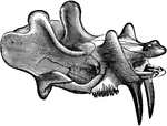
Upper Two Vertebrae
The upper two vertebrae of the spinal column, involved in head movement. In performing the rotary motion…
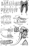
Teeth of Man and Several Animal Species
1. Dentition (teeth) of man. 2. Dentition of hyena. 3. Dentition of pig. 4. Dentition of Patagonian…
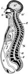
Longitudinal Section of the Body
Diagrammatic longitudinal section of the Body. Labels: a, the neural tube, with its upper enlargement…
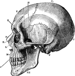
Side View of the Skull
A side view of the skull. Labels: O, occipital bone; T, temporal; Pr, parietal; F, frontal; S, sphenoid;…
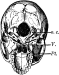
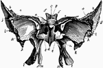
Base of the Skull
The base of the skull. The lower jaw has been removed. At the lower part of the figure is the hard palate…
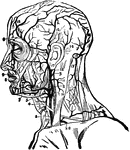
Arteries of the Head and Neck
Arteries of the head and neck. Labels: 1, primitive carotid artery; 2, occipital branch to the back…
Bone Structure
If we divide any of the long bones longitudinally, we find two kinds of structure, the hard or compact,…
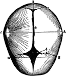
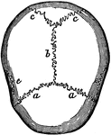
Skull Sutures
Sutures of the skull. Labels: a,a, the coronal suture, from the Latin corona, crown, so called from…
Frontal Bone
A front view of the frontal bone; a,a, frontal sinuses; b, the temporal arch, beneath which lies the…
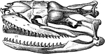
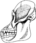
Dinoceras Mirabile
The skull and upper jaw of an early rhinoceros-like mammal from the Cenozoic time.
Boa Constrictor Skull
"Skull of Boa constrictor. a, quadrate bone; b, b, halves of lower jaw." -Cooper, 1887
Skate Skull
"Under surface of skull and arches of skate. l.1, First labial cartilage; R., rostrum; tr., trabecular…
Skate Skull
"Side view of skate's skull. l1., First labial cartilage; n.c., nasal capsule; a.o., antorbital; p.pt.q.,…
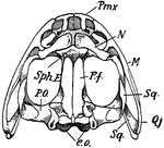
Upper Surface of Frog Skull
"Skull of frog. Upper surface-- Pmx., premaxilla; N., nasal; M., maxilla; Sq., squamosal; Q.j., quadrato-jugal;…
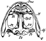
Lower Surface of Frog Skull
"Skull of frog. Lower surface-- Pmx., premaxilla; M., maxilla; Q.j., quadrato-jugal; Q., quadrate; Pt.,…
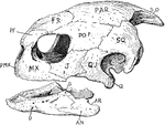
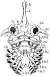
Turtle Skull
"Skull of turtle. "S.O., supra-occipital; PAR., parietal; FR., frontal; P.F., pre-frontal; PO.F., post-frontal;…
Lizard Skull
"Side view of skull of Lacerta. px., Premaxilla; mx., maxilla; l., lachrymal; j., jugal; t.pa., transpalatine;…
Bird Skull Disarticulation
"Disarticulation of bird's skull. Membrane bones shaded. B.Oc., basioccipital; E.Oc., exoccipital; S.Oc.,…
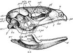
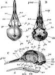
Rabbit Skull
"Side view of rabbit's skull. Pmx., Premaxilla; Na., nasal; Fr., frontal; Pa., parietal; Sq., squamosal;…
Upper Surface Rabbit Skull
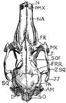
"Upper surface of rabbit's skull. N., Anterior nostril; PMX., premaxilla; NA., nasal; FR., anterior…
Under Surface Rabbit Skull
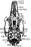
"Under surface of rabit's skull. Inc. I., First incisors; Inc. II., second incisors; PMX., premaxilla;…
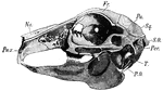
Sheep Skull
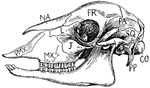
"Side view of sheep's skull. PMX., Premaxilla; MX., maxilla; NA., nasal; J., Jugal; L., lachrymal; FR.,…
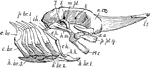
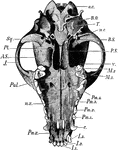
Horse Skull
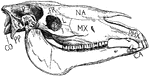
"Side view of horse's skull. P., Parietal; FR., frontal; NA., nasal; PMX., premaxilla; MX., maxilla;…
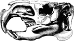
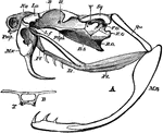
Dog Skull
"Lower surface of dog's skull. o.c., Occipital condyle; B.O., basioccipital; T., tympanic bulla; m.c.,…
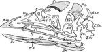
Fish Skull
"Salmo fario, the entire skull, from the left side. art, articular; branchiost, branchiostegal rays;…
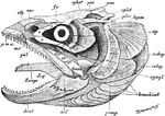
Rattlesnake Skull
"A, lateral view of skull of rattlesnake (Crotalus). B. O, basi-occipital; B. S, basi-sphenoid; E. O,…
Rock Pigeon Skull
"Columba livia. Skull of young specimen. A, dorsal; B, ventral; C, left side. al. s, alisphenoid; au,…
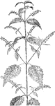
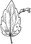
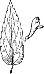
Common Skullcap
Of the Mint family (Labiatae), the leaf of the common skullcap (Scutellaria galericulata).