Clipart tagged: ‘vein’
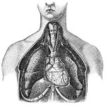
Principal Organs of the Thorax and Abdomen
"The principal muscles are seen on the left, and superficial veins on the right." — Blaisedell, 1904
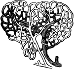
Capillaries of the Air Sac
"Diagram showing the capillary network of the air sacs and origin of the pulmonary veins.. A,…
The Arteries and Veins of a Section of the Skin
The arteries and veins of a section of the skin. A, arterial branches. B, capillary, or hair-like vessels,…
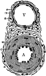
Diagram of an Artery and a Vein
Transverse section through a small artery and vein. Labels: A, artery; V, vein; e, epithelial lining;…
Muscular fibers of the auricle
L.A., left auricle; R.A., right auricle; A, opening of the inferior vena…
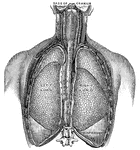
Vertical Section of the Back
"The spinal column below the twelfth dorsal vertebra at A has been removed, as well as the…
Blood Flow Diagram
Diagram of the course of the blood. Labels: RA, right auricle; RV. right ventricles; LV and LA, left…
Circulation of blood
"Showing how the circulation of blood in the web of a frog's foot looks as seen under the microscope."…
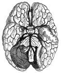
Blood vessels of the brain
"Arteries and their Branches at the Base of the Brain." — Blaisedell, 1904
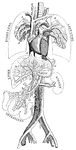
Diagram of the circulation of the blood
"R.A., right auricle; L.A., left auricle; R.V., right ventricle; L.V.,…
The Circulatory Organs
The circulatory organs. Labels: 1, The left auricle. 2, The right auricle. 3, The left ventricle. 4,…
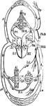
Cuttlefish Organs
"Central organs of the circulaion, gills, and renal organs of Sepia officinalis. a, aorta; v, vena cava;…
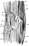
Elbow-Joint
"The superficial veins in front of the elbow-joint. B', tendon of biceps muscle; Bi, brachialis internus…
Gland
"Diagram to show the working parts of a gland. v and a are blood tubes with thin-walled branches around…
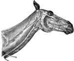
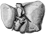
Head and Neck of a Horse Showing Veins
Veins of the face and neck. Labels: 1, glosso-facial; A, its facial portion; 2, jugular; 3, occipial;…
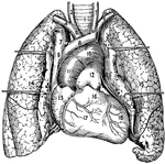
Heart and Lungs
1, The trachea or windpipe; 2 and 3, right and left common carotid arteries; 4 and 5, right and left…

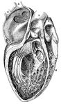
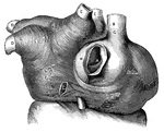
Cavities of the heart
"A, B, right pulmonary veins, S, openings of the left pulmonary veins; E, D, C,…
Intestinal absorption
"A, a fold of peritoneum; B, lacteals and lymphatic glands; C, veins of intestines;…
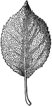
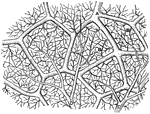
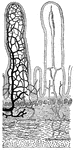
Reticulated Vein Leaf
"Reticulated leaves are those the veins of which branch and ramify in all directions, forming a complete…

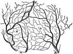
Parallel-Veined Leaves
"Parallel-veined leaves are those in which the veins proceed from their origin to their termination…
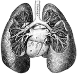
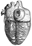
Lungs
"Relative Postion of the Lungs, the Heart, and Some of the Great Vessels belonging to the latter. A,…
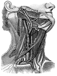
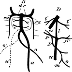
Blood vessels of the neck
"Showing the carotid artery and jugular vein on the right side, with some of their main branches; also…
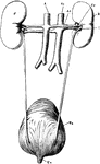
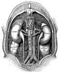
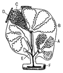
Renal Organs
"The renal organs, viewed from behind. R, right kidney; A, aorta; Ar, right renal artery; Vc, inferior…
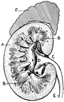
Section of Human Kidney
This illustration shows a section of a human kidney (A, Cortical substance; B, Pyramids; C, Hilum; D,…
Transverse section of the small intestine
"In the figure on the left are seen the artery and vein of a villus. In the right figure are represented…
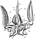
Thoracic Duct
"Human Thoracic Duct and Azygous Veins. a, receptacle of the chyle; b, trunk of the thoracic duct, opening…

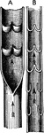
Valves of Veins
Diagram showing valves of veins. A, part of a vein laid open and spread out with two pairs of valves.…
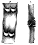
Valves of Veins
Valves of veins. A shows a vein cut open between the segments of two valves. B shows appearance of valves…
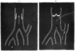
Blood Flow in the Valves of Veins
A, vein with valves open. B, vein with valves closed: stream of blood passing off by lateral channel.
Section Through Vein Wall
Transverse section through the wall of a vein. Labels: A, tunica intima; B, tunica media; C, tunica…

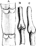
Structure of a Vein
Structure of a vein with valves. A, part of a vein, laid open, with two pairs of valves; B, longitudinal…
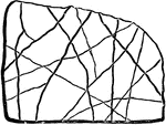
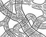
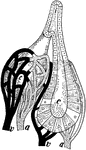
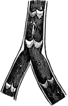
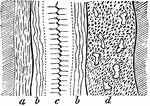
Vein-form Condition
This illustration shows two very irregular veins. a represents regular veins, whereas b shows an intersecting…
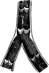
Vein-form Condition
This illustration shows two large veins, of irregular character that cross one another.
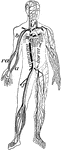
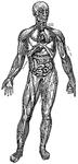
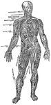
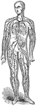
Veins and arteries
"Chief veins and arteries of the body. a, place of the heart; the veins are in black. On the right side…
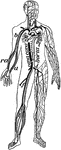
Veins and Arteries of the Body
Chief veins and arteries of the body. Labels: a, place of the heart; the veins are in the back. On the…
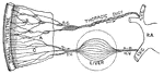
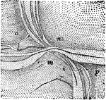
Development of Veins in the Liver
Diagram illustrating the development of veins about the liver. B, dc, ducts of Cuvier, right and left;…
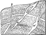
Development of Great Veins
Diagram illustrating the development of the great veins. dc, ducts of Cuvier; j, jugular veins; h, hepatic…

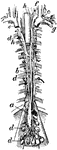
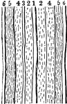
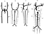
Formation of Large Veins
Diagram showing the formation of large veins by convergence of small, and the branching of veins.