The Mammal Anatomy: Internal Organs ClipArt gallery offers 192 views of the internal organs and other soft tissue of various species of mammals.
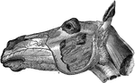
Head of a Horse Showing Arteries
Facial arteries of the left side. Labels: a, maxillo-muscular; a', posterior masseter; b, c, posterior…
Head of a Horse Showing Nerves
Nerves of the right side of the head- the maxillary ramus and cheek being removed. Labels: a, superior…
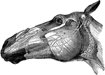
Head of a Horse Showing Nerves
Left side of the face- showing the distribution of the facial portions of the fifth and the seventh…
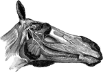
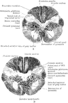
Head of a Horse Showing Nerves
Ninth, tenth, eleventh, and twelfth cranial, first cervical, and part of the sympathetic nerves- the…
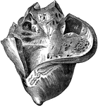
Heart of a Horse
Right side of the heart-laid open. Labels: 1, right ventricle; a, its external wall; b, carneae columnae;…
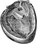
Heart of a Horse
Left side of the heart-laid open. Labels: 1, left ventricle; a, its external wall; b, carneae columnae;…
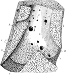
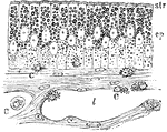
Hepatic Vein in the Liver of a Pig
Section of a portion of liver passing longitudinally through a considerable hepatic vein, from the pig.…
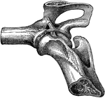
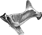
Hip Joint Ligaments
Ligaments of the hip joint- infero-internal view. Labels: a, cotyloid ligament; b, round ligament; c,…
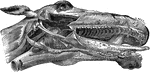
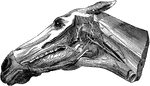
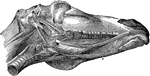
Horse Head
Right infero-lateral view of the head; the maxillary ramus, cheek, parotid gland, and upper lip being…
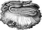
Intestines of an Ox
Mesentery and intestines of an ox. Labels: 1, duodenum; 2, small intestines; 3, caecum; 4, colon; 5,…
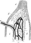
Longitudinal Section of Kidney of a Dog
Longitudinal section of injected kidney of dog, showing general arrangement of blood vessels of cortex…
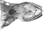
Kidney of a Hog
Horizontal section of the kidney of a hog. Labels: a, cortical substance; b, medullary substance; c,…
Kidney of a Horse
Horizontal section of the right kidney. Labels: a, fibrous capsule detached; b, cortical layer; c, medullary…
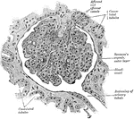
Section Through an Ape's Kidney
A section through the cortex of an ape's kidney. A Malpighian corpuscle, together with the beginning…
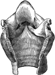
Larynx Cavity of a Horse
Cavity of the larynx- opened posteriorly. Labels: a, lateral ventricles of the larynx; b, middle ventricle;…
Larynx Muscles of a Horse
Muscles of the larynx-left lateral view. Labels: a, hyoepiglottideus; b, hyothyroideus; c, cricothyroideus.
Larynx Muscles of a Horse
Muscles of the larynx-left lateral view-the thyroid wing being removed. Labels: a, arytenoideus; b,…
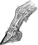
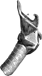
Section Through the Horse Leg and Hoof
Longitudinal section through the digit of a horse. Labels: 1, skin; 2, extensor pedis tendon; 3, synovial…
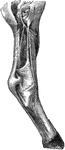
Leg of a Horse Showing Arteries
Arteries of the right posterior limb- external view. Labels: 1, popiteal; 2, posterior tibial; 3, anterior…
Leg of a Horse Showing Nerves
Carpal and metacarpal nerves-internal aspect. q, external branch of median; r, internal branch of median,…
Lens of a Rabbit
Meridional section through the lens of a rabbit. Labels: 1, Lens capsule; 2, epithelium of lens; 3,…
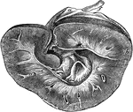
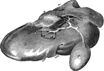
Liver and Diaphragm of a Horse
Posterior view of the liver and diaphragm in situ. Labels: a, left lobe; b, right lobe; c, quadrate…
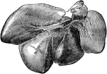
Liver of a Hog
Liver of a hog-posterior view. Labels: a, right external lobe; c, left external lobe; d, left internal…
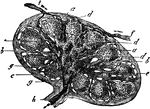
Liver of an Ox
Posterior view of the liver of an ox. Labels: a, left lobe; b, right lobe; c, spigelian lobe; d, quadrate…
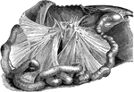
Lymphatic Gland of a Horse
Section of a lymphatic gland. Labels: a, capsule; b, trabeculae of cortical portion; c, trabeculae of…
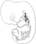
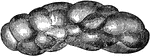
Lymphatics in a Rabbit Embryo
Developing lymphatics in rabbit embryo of 14 days. Lymphatic vessels are heavily shaded; veins are light.…
Lymphatics of Rabbit's Diaphragm
Lymphatics of central tendon of rabbit's diaphragm, stained with silver nitrate. The ground substance…
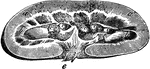
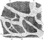
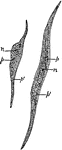
Intestinal Tract of Macropus Bennetti
S, cut end of duodenum; R, cut end of rectum; C, caecum; C2, accessory caecum; C.L., colic loop of hind-gut.
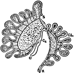
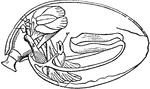
Mammal Ovum
"Mammalian ovum. ov., Ovum; f., follicular capsule; f2., follicle cells; f.c., follicle cells forming…
Mammal Vertebrae
This is a diagram of a trunk vertebra in a mammal. c, centrum; ch., position originally occupied by…
Section Through the Junction of the Medulla and Cord of the Orang
Two section through the junction between the cord and medulla of the Orang. A is at a slightly lower…
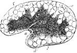
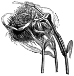
Medullary Substance from a Mesenteric Gland of an Ox
A small portion of medullary substance from a mesenteric gland of an ox, d, trabeculae; a, part of a…
Medullary Substance of an Inguinal Gland
Section of medullary substance of an inguinal gland of an ox. Labels: a, glandular substance or pulp…
Mesenteric Gland
Section of a mesenteric gland from the ox. Labels: a, Hilus; b, medullary substance; c, cortical substance…
Mesenteries of a Horse
The two mesenteries; the great colon being removed. Labels: a, anterior mesentery; b, mesenteric glands;…
Mucous Membrane
Section of mucous membrane of the small intestine. One the left a villus is seen in seen in section.…
Mucous Membrane
Section of mucous membrane of the small intestine. One the left a villus is seen in seen in section.…
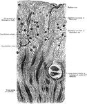
Mucous Membrane of Frog's Intestine
Mucous membrane of a frog's intestine during fat absorption. Labels: ep, epithelium; str, striated borer;…
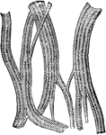
Muscle Fibres
"Plain muscle fibres. n, nucleus of muscle cell; p, undifferentiated cell protoplasm; p', the differentiated…
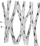
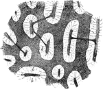
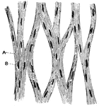
Unstriped Muscle of a Guinea Pig
Unstriped muscle, or plain muscle, forms the proper muscular coats. Shown is a plexus of bundles of…
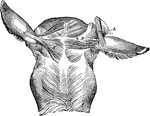
Muscles from Gluteal Region of a Horse
Deep-seated muscles of the gluteal region. Labels: a, deep anterior portion of gluteus maximus; b, gluteus…
Muscles From the Femoral Region of the Horse
Muscles of the sublumbar and internal deep femoral regions-seen from below. Labels: a, quadratus lumborum;…
Muscles From the Femoral Region of the Horse
The diaphragm, and superficial muscles of the internal femoral region-viewed from below. A, the diaphragm;…
Neck Muscles of a Horse
The neck muscles of a horse-lateral view. Labels: a, oblique capitis posticus; b, obliquus capitis anticus;…
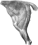
Thigh and Haunch Muscles of a Horse
The muscles of the thigh and haunch- left side; the external fascia being removed. Labels: a, tensor…
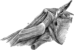
Muscles of the External Ear of a Horse
Muscles of the external ear- posterior view. Labels: a, inferior and b, superior layer of the scuto-auricularis…
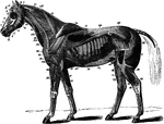
Neck Muscles of the Horse
The deep muscles of the horse. Labels: 1, temporalis; 1', stylo-maxillaris; 2, rectus capitis anticus…
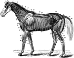
Superficial Muscles of the Horse
The superficial muscles of the horse with the panniculus and tunica abdominalis removed. Labels: 1,…
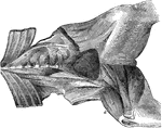
Muscles of the Maxillary Space of a Horse
Right infero-lateral view of the muscles of the maxillary space, the ramus and hyoid cornu are cut away.…
Horse Leg Muscles
Muscles of the anterior limb-external view. Labels: a, antea-spinatus; b, postea-spinatus; c, teres…
Horse Leg Muscles
Muscles of the anterior limb-internal view. Labels: a, subscapularis; b, teres internus; c, coracohumeralis;…
Horse Leg Muscles
External view of the muscles of the anterior limb-showing the deeper ones of the upper region. Labels:…
Horse Leg Muscles
Internal view of the deep muscles of the anterior limb. Labels: a, caput parvum of triceps extensor…
Horse Leg Muscles
Anterior tibial group of muscles of the right limb, seen from before and the outside. Labels: a, flexor…
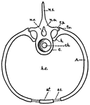
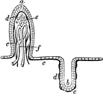
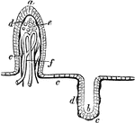
Muscular System
"Diagram showing the muscular system. M, ventral, N, dorsal valve; l, loop; V, mouth; Z, extremity of…