Different forms of ganglion cells. A, a, round ball-shaped unipolar cell from the human Gasserian ganglion. Two cells only show the process f; b, spindle shaped; c, multipolar ganglion cell from he spinal cord of the ox. d, D, Purkinjee ganglion cells from human cerebellum; ax, axis cylinder process; p, protoplasmic process; h, h, two cells surrounded with a nucleated sheath.

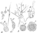
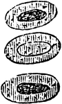
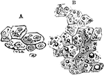
Different Forms of Ganglion Cells

Different forms of ganglion cells. A, a, round ball-shaped unipolar cell from the human Gasserian ganglion.…

Touch corpuscles are found in the papillae of the skin of the fingers and toes, or among its epithelium; they may be simple or compound; when simple, they are large and slightly flattened transparent nucleated ganglion cells, enclosed in a capsule; when compound the capsule contains several smalls cells. Shown are the papillae from the skin of the hand, freed from the cuticle and exhibiting tactile corpuscles. A. Simple papilla with four nerve fibers; a, tactile corpuscles; b, nerves with cells and fine elastic filaments; b, tactile corpuscle with transverse nuclei; c, entering nerve with neurilemma or perineurium; d and e, nerve fibers winding round the corpuscle.

Touch Corpuscles

Touch corpuscles are found in the papillae of the skin of the fingers and toes, or among its epithelium;…

The corpuscles of Grandy have been noticed in the beaks and tongues of birds. They consist of corpuscles oval or spherical, contained within a delicate nucleated sheath, and containing several cells, tow or more compressed vertically. The cells are granular and transparent, with a nucleus. The nerve enter on one side and, laying aside its medullary sheath, terminated in or between the cells. Show is a corpuscle of Grandy, from the tongue of a duck.

Corpuscle of Grandy

The corpuscles of Grandy have been noticed in the beaks and tongues of birds. They consist of corpuscles…

Touch corpuscles are found in the papillae of the skin of the fingers and toes, or among its epithelium; they may be simple or compound; when simple, they are large and slightly flattened transparent nucleated ganglion cells, enclosed in a capsule; when compound the capsule contains several smalls cells. Show is a touch corpuscle of Meissner, from the skin of the human hand.

Touch Corpuscle of Meissner

Touch corpuscles are found in the papillae of the skin of the fingers and toes, or among its epithelium;…

The illustration exhibits the typical characters of the red blood cells in the main divisions of Vertbrata. The fractions are those of an inch, and represent the average diameter. In the case of the oval cells, only the long diameter is here given. It is remarkable, that although the size of the red blood cells varies so much in the different classes of the vertebrate kingdom, that of the white corpuscles remains comparatively uniform, and thus are, in some animals, much greater, in others much less than the red corpuscles existing side by side with them.

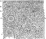
Red Blood Cells in Vertebrata

The illustration exhibits the typical characters of the red blood cells in the main divisions of Vertbrata.…

Acetic acid (dilute) causes the nucleus of the red blood cells in the frog to become more clearly defined; if the action is prolonged, the nucleus becomes strongly granulated, and all the coloring matter seems to be concentrated in it, the surrounding cell substance and outline of the cell becoming almost invisible; after a time the cells lose their color altogether. The cells shown represent the successive stages of change. A similar loss of color occurs in the red cells of human blood, which , however, from the absence of nuclei, seem to disappear entirely.

Effect of Acetic Acid on Red Blood Cells

Acetic acid (dilute) causes the nucleus of the red blood cells in the frog to become more clearly defined;…

When a 2 percent fresh solution of tannic acid is applied to frog's blood it causes the appearance of a sharply defined little knob, projecting from the free surface (Robert's macula): the coloring matter becomes at the same concentrated in the nucleus, which grows more distinct. A somewhat similar effect is produced on the human blood corpuscle.

Effect of Tannin on Red Blood Cells

When a 2 percent fresh solution of tannic acid is applied to frog's blood it causes the appearance of…

A 2 percent solution of boric acid applied to nucleated red blood cells of a frog will cause the concentration of all he coloring matter in the nucleus; the colored body thus formed gradually quits its central position , and comes to be partly, sometime entirely, protruded from the surface of the now colorless cell. When applied to non-nucleated mammalian corpuscle its effect merely resembles that of other dilute acids.

Effect of Boric Acid on Red Blood Cells

A 2 percent solution of boric acid applied to nucleated red blood cells of a frog will cause the concentration…

If the red blood cells of a frog be first exposed to the action of water vapor (which renders their outer pellicle more readily permeable to gases), and then acted on by carbonic acid, the nuclei immediately become clearly defined and strong granulated, when air or oxygen is admitted the original appearance is at once restored. The upper and lower cells in this figure show the effect of carbonic acid; the middle one the effect of the readmission of air. The effects can be reproduced five or six times in succession. If, however, the action of the carbonic acid be much prolonged, the granulated of the nucleus become permanent; it appears to depend on a coagulation of the paraglobulin.

Effect of Gases on Red Blood Cells

If the red blood cells of a frog be first exposed to the action of water vapor (which renders their…

The effect of heat on red blood cells up to 50-60 degrees C. (120-140 degrees F.) is to cause the formation of a number of bud-like processes.

Effect of Heat on Red Blood Cells

The effect of heat on red blood cells up to 50-60 degrees C. (120-140 degrees F.) is to cause the formation…

A, Three colored blood corpuscles. B, Three colorless blood corpuscles acted on by acetic acid; the nuclei are very clearly visible.

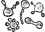
Colorless and Colored Blood Corpuscles

A, Three colored blood corpuscles. B, Three colorless blood corpuscles acted on by acetic acid; the…

The walls of capillaries are composed of a single layer of elongated or radiate, flattened and nucleated cells, so joined and dovetailed together as to form a continuous transparent membrane.

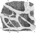
Capillaries Showing Nucleated Endothelial Membrane

The walls of capillaries are composed of a single layer of elongated or radiate, flattened and nucleated…

The form of the capillary network presents considerable variety in the different textures of the body: the varieties consists principally of modifications of two chief kind of mesh, the rounded and the elongated. The kind in which the meshes or interspaces have a roundish form is the most common, and prevails in those parts which the capillary network is most dense, such as the lungs. Shown is the network of capillary vessels of the air cells of the horse's lung magnified. Labels: a, capillaries proceeding from b, terminal branched of the pulmonary artery.

Capillary Vessels of Air Cells

The form of the capillary network presents considerable variety in the different textures of the body:…

A large capillary from the frog's mesentery eight hours after irritation had been set up, showing emigration of leucocytes. Labels: a, Cells in the act of traversing the capillary wall; b, some already escaped.

Capillaries Showing Emigration of Leucocytes

A large capillary from the frog's mesentery eight hours after irritation had been set up, showing emigration…
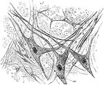
The nerve cells that compose the ganglia are generally unipolar, and seldom bipolar; sometimes two cells are said to exist in the same envelope, constituting the twin cells of Dogiel. The cells are large and have very large nucleoli. Shown are isolated nerve cells from the frog's heart. Labels: I, Usual form. II, Twin cell. C, capsule; N,nucleus; N', nucleolus; P, process.

Nerve Cells

The nerve cells that compose the ganglia are generally unipolar, and seldom bipolar; sometimes two cells…
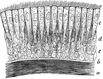
Ciliary epithelium of the human trachea. Labels: a, layer of longitudinally arranged elastic fibers; b, basement membrane; c, deepest cells, circular in form; d, intermediate elongated cells; e, outermost layer of cells fully developed and bearing cilia.

Ciliary Epithelium

Ciliary epithelium of the human trachea. Labels: a, layer of longitudinally arranged elastic fibers;…

Terminal branch of a bronchial tube, with its infundibula and air cells from the margin of the lung of a monkey, injected with quicksilver. Labels: a, terminal bronchial twig; b, infundibula and air cells.

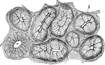
Air Cells of a Monkey

Terminal branch of a bronchial tube, with its infundibula and air cells from the margin of the lung…
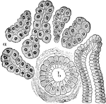
Two small infundibula or groups of air cells, a, with air cells, b, and the ultimate bronchial tubes, c, with which the air cells communicates. From a newborn child.

Infundibula

Two small infundibula or groups of air cells, a, with air cells, b, and the ultimate bronchial tubes,…

From a section of the lung of a cat, stained with silver nitrate. Labels: A. D., alveolar duct or intercellular passage; S, alveolar septa; N, alveoli or air cells, lined with large flat, nucleated cells, with some smaller polyhedral nucleated cells; M, Unstriped muscular fibers. Circular muscular fibers are seem surrounding the interior of the alveolar duct, and at one part is seen a group of small polyhedral cells continued from the bronchus.

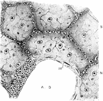
Air Cells from a Cat's Lung

From a section of the lung of a cat, stained with silver nitrate. Labels: A. D., alveolar duct or intercellular…

A section of the submaxillary gland of a dog. Showing gland cells, b, and a duct, a, in section.

Gland Cells of a Dog

A section of the submaxillary gland of a dog. Showing gland cells, b, and a duct, a, in section.

From a section through a true salivary gland. Labels: a, the gland alveoli, lined with albuminous "salivary cells;" b, intralobular duct cut transversely.

True Salivary Gland

From a section through a true salivary gland. Labels: a, the gland alveoli, lined with albuminous "salivary…

From the section through a mucous gland in a quiescent state. The alveoli are lined with transparent mucous cells, and outside these are the semilunes.

Section of a Mucous Gland

From the section through a mucous gland in a quiescent state. The alveoli are lined with transparent…

From the section through a mucous gland after prolonged electrical stimulation. The alveoli are lined with small granular cells.

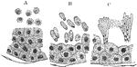
Section of a Mucous Gland After Stimulation

From the section through a mucous gland after prolonged electrical stimulation. The alveoli are lined…

Alveoli of true salivary gland. A, at rest; B, in the first stage of secretion; C, after prolonged secretion.

Changes in Gland Cells

Alveoli of true salivary gland. A, at rest; B, in the first stage of secretion; C, after prolonged secretion.

Section of a mucous gland from the tongue. Labels: A, opening of the duct on the free surface; C, basement membrane with nuclei; B, flattened epithelial cells lining duct. The duct divides into several branches, which are convoluted and end blindly, being lined throughout by columnar epithelium. D, lumen of one of the tubuli of the gland.

Mucous Gland from Tongue

Section of a mucous gland from the tongue. Labels: A, opening of the duct on the free surface; C, basement…

Taste goblet from dog's epiglottis (laryngeal surface near the base) , precisely similar in structure to those found in the tongue. Labels: a, depression in epithelium over goblet; below the letter are seen the fine hair-like processes in which the cells terminate; c, two nuclei of the axial (gustatory) cells. The more superficial nuclei belong to the superficial (encasing) cells; the converging lines indicate the fusiform shape of the encasing cells.

Taste Goblet from Dog's Epiglottis

Taste goblet from dog's epiglottis (laryngeal surface near the base) , precisely similar in structure…

Auerbach's nerve plexus in the small intestine. The plexus consist of fibrillated substance, and is made up of trabeculae of various thicknesses. Nucleus-like elements and ganglion cells are imbedded in the plexus, the whole of which is enclosed in a nucleated sheath.

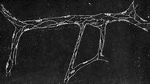
Auerbach's Nerve Plexus

Auerbach's nerve plexus in the small intestine. The plexus consist of fibrillated substance, and is…

Portion of a lobule of liver. Labels: a, bile capillaries between liver cells, the network which is well seen; b, blood capillaries.

Portion of a Lobule of Liver

Portion of a lobule of liver. Labels: a, bile capillaries between liver cells, the network which is…

Hepatic cells and bile capillaries, from the liver of a child three months old. Both figures represent fragments of a section carried through the periphery of a lobule. The red corpuscles of the blood are recognized by their circular contour; vp, corresponds to an interlobular vein in immediate proximity with which are the epithelial cells of the biliary ducts, to which , at the lower part of the figures, the much larger hepatic cells suddenly succeed.

Hepatic Cells and Bile Capilaries

Hepatic cells and bile capillaries, from the liver of a child three months old. Both figures represent…

Lymphatics of central tendon of rabbit's diaphragm, stained with silver nitrate. The ground substance has been shaded diagrammatically to bring out the lymphatic clearly. Lymphatics, l, lined by long narrow endothelial cells, and showing valves, v, at frequent intervals.

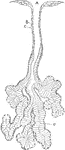
Lymphatics of Rabbit's Diaphragm

Lymphatics of central tendon of rabbit's diaphragm, stained with silver nitrate. The ground substance…

A, Liver cells. B, Liver cells, containing various sized particles of fat.

Liver Cells

A, Liver cells. B, Liver cells, containing various sized particles of fat.

Section of the villus of a rat killed during fat absorption. Labels: ep, epithelium; str, striated border; c, lymph cells; c', lymph cells in the epithelium; l, central lacteal containing disintegrating lymph corpuscles

Villus of a Rat

Section of the villus of a rat killed during fat absorption. Labels: ep, epithelium; str, striated border;…

The tubuli uriniferi are composed of a nearly homogeneous membrane, and are lined internally by epithelium. A, Portion of a secreting tubule from the cortical substance of the kidney. B, The epithelial or gland cells.

Cells from the Tubuli Uriniferi

The tubuli uriniferi are composed of a nearly homogeneous membrane, and are lined internally by epithelium.…

Transverse section of a developing Malpighian capsule and tuft from a fetus at about the fourth month. Labels: a, flattened cells growing to form the capsule; b, more rounded cells; continuos with the above, reflected around c, and finally enveloping it; c, mass of embryonic cells which later become developed into blood vessels.

Development of Malpighian Capsule

Transverse section of a developing Malpighian capsule and tuft from a fetus at about the fourth month.…

Epithelial elements of a Malpighian capsule and tuft, with the commencement of a urinary tubule showing the afferent and efferent vessel. Labels: a, layer of flat epithelium forming the capsule; b, similar, but rather larger epithelial cells, places in the walls of the tube; c, cells, covering the vessels of the capillary tuft; d, commencement of the tubule, somewhat narrower than the rest of it.

Epithelial Elements of a Malpighian Capsule

Epithelial elements of a Malpighian capsule and tuft, with the commencement of a urinary tubule showing…

Epithelium of the bladder. Labels: a, one of the cells of the first row; b, a cell of the second row; c, cells in situ, of first, second, and deepest layers.

Epithelium of the Bladder

Epithelium of the bladder. Labels: a, one of the cells of the first row; b, a cell of the second row;…

Vertical section of epidermis of the prepuce. Labels: a, stratum corneum, of very few layers, the stratum lucidum and stratum granulosum not being distinctly represented; b, c, d, and e, the layers f the stratum Malpighi, a certain number of the cells in layers d and e showing signs of segmentation; layers c consists chiefly of prickle or ridge and furrow cells; f, basement membrane; g, cells in cutis vera.

Epidermis

Vertical section of epidermis of the prepuce. Labels: a, stratum corneum, of very few layers, the stratum…

Section of gray matter of anterior cornu of a calf's spinal cord; a, nerve fibers of white matter in transverse section, showing axis cylinder in the center of each; r, large stellate nerve cells with nuclei and three prolongations.

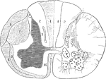
Gray Matter of Spinal Cord

Section of gray matter of anterior cornu of a calf's spinal cord; a, nerve fibers of white matter in…

Section of a spinal cord, one half of which shows the tracts of the white matter, and the other half (right) shows the position of the nerve cells in the gray matter. 7, 10, 9, and 3 are tracts of descending degeneration, 1, 4, 6, and 8, of the ascending degeneration.

Section of the Spinal Cord

Section of a spinal cord, one half of which shows the tracts of the white matter, and the other half…

The five layers of the cortical gray matter of the cerebrum. 1, Superficial layer with abundance of neuroglia and few small multipolar ganglion cells. 2, A thin layer of a large number of closely packed small ganglion cells of pyramidal shape. 3, The most important layer, and the thickest of all: it contains many large pyramidal ganglion cells, each with a process running off from the apex vertically towards the free surface, and lateral processes at the base which are always branched. 4, Numerous ganglion cells come are large and others small, forming the granular formation of Meynert. 5, Spindle-shaped and branched ganglion cells of moderate size arranged chiefly parallel to the free surface.

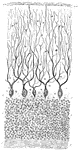
Cortical Gray Matter of the Cerebrum

The five layers of the cortical gray matter of the cerebrum. 1, Superficial layer with abundance of…

Vertical section of dog's cerebellum. Labels: p m, pia mater; p, corpuscles of Purkinje, which are branched nerve cells lying in a single layer and sending single processed downwards and more numerous ones upwards, which branch continuously and extend through the deep "molecular layer" towards the free surface; g, dense layer of ganglionic corpuscles., closely resembling nuclear layers of retina; f, layer of nerve fibers, with a few scattered ganglionic corpuscles. This last layer (f) constitutes part of the white matter of the cerebellum, while the layer between it and the free surface are gray matter.

Cerebellum of Dog's Brain

Vertical section of dog's cerebellum. Labels: p m, pia mater; p, corpuscles of Purkinje, which are branched…

Section through the olfactory mucous membrane of the newborn child. Labels: a, non-nuclear; and b, nucleated portions of the epithelium; c, nerves; dd, glands.

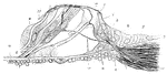
Cells of the Olfactory Mucous Membrane

Section through the olfactory mucous membrane of the newborn child. Labels: a, non-nuclear; and b, nucleated…

Cells from the olfactory region of the rabbit. Labels: st, supporting cells; r, r', varieties of rod-cells; f, ciliated cell; s, cilia-like process; b, cells from Bowman's gland.

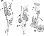
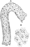
Cells from the Olfactory Region of a Rabbit

Cells from the olfactory region of the rabbit. Labels: st, supporting cells; r, r', varieties of rod-cells;…

Vertical section of the organ of Corti from the dog. Labels: 1 to 2, homogeneous layer of the so-called membrana basilaris; u, vestibular layer; v, tympana layer, with nuclei and protoplasm; a, prolongation of tympanal periosteum of lamina spiralis ossea; c, thickened commencement of the membrana basilaris near the point of perforation of the nerves h; d, blood-vessel ( vas spirale) ; e, blood-vessel; f, nerves; g, the epithelium of the sulcus spiralis internus; i, internal or tufted cell, with basil process k, surrounded with nuclei and protoplasm (of the glanular layer), into which the nerve-fibers radiate; l, hairs of the internal hair-cell ; n, base or foot of inner pillar of organ of Corti, whose under half is missing, while the next pillar beyond, o, presents both middle portion and base; r s d, three external hair-cells; t, bases of two neighboring hair or tufted cells; x, so-called supporting cell of Hensen; W, nerve-fiber terminating in the first of the external hair cells.

Corti from the Dog

Vertical section of the organ of Corti from the dog. Labels: 1 to 2, homogeneous layer of the so-called…

Vertical section of rabbit's cornea. Labels: anterior epithelium, showing the different shapes of the cells at various depths from the free surface; b, portion of the substance of cornea.

Magnified Rabbit's Cornea

Vertical section of rabbit's cornea. Labels: anterior epithelium, showing the different shapes of the…

Vertical section of rabbit's cornea, stained with gold chloride. Labels: e, Laminated anterior epithelium. Immediately beneath this is the anterior elastic lamina of Bowman. n, Nerves forming a delicate sub-epithelial plexus, and sending up fine twigs between the epithelial cells to end in a second plexus on the free surface; d, Descemet's membrane, consisting of a fine elastic layer, and a single layer of epithelial cells. the substance of the cornea, f, is seen to be fibrillated, and contains many layers of branched corpuscles, arranged parallel to the free surface, and here seen edgewise.

Section of Rabbit's Cornea

Vertical section of rabbit's cornea, stained with gold chloride. Labels: e, Laminated anterior epithelium.…
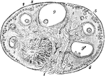
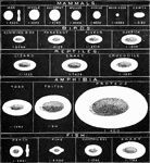
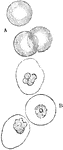
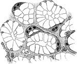
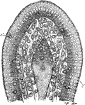
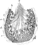
View of a section of the ovary of the cat. Labels: 1, outer covering and free border of the ovary; 1', attached border; 2, the ovarian stroma, presenting a fibrous and vascular structure; 3, granular substance lying external to the fibrous stroma; 4, blood-vessels; 5, ovigerms in their earliest stages occupying part of the granular layer near the surface; 6, ovigerms which have begun to enlarge and to pass more deeply into the ovary; 7, ovigerms round which the Graafian follicles and tunica granulosa are now formed and which have passed somewhat deeper into the ovary and are surrounded by the fibrous stroma; 8, more advanced Graafian follicle with the ovum imbedded in the layer of cells constituting the proligerous disc; 9, the most advanced follicle containing the ovum; 9', a follicle from which the ovum has accidentally escaped; IO, corpus luteum.

Ovary of the Cat

View of a section of the ovary of the cat. Labels: 1, outer covering and free border of the ovary; 1',…

Section of the ovary of a cat. Labels: A, germinal epithelium; B, immature Graafian follicle; C, stroma of ovary; D, vitelline membrane containing the ovum; E, Graafian follicle showing lining cells; F, follicle from which the ovum has fallen out.

Ovary of the Cat

Section of the ovary of a cat. Labels: A, germinal epithelium; B, immature Graafian follicle; C, stroma…

From a section of the testis of a dog, showing portions of seminal tubes. A, seminal epithelial cells, and numerous small cells loosely arranged; B, the small cells or spermatoblasts converted into spermatozoa; groups of these is a further stage of development.

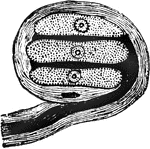
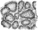
Testis of a Dog, Section of the

From a section of the testis of a dog, showing portions of seminal tubes. A, seminal epithelial cells,…

Section of a tubule of the testicle of a rat, to show the formation of the spermatozoa. Labels: a, spermatozoa; b, seminal cells; c, spermatoblasts, to which the spermatozoa are sill adherent; d, membrana propria; e, fibro-plastic elements of the connective tissue.

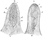
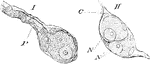
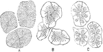
Section of a Tubule of the Testicle of a Rat

Section of a tubule of the testicle of a rat, to show the formation of the spermatozoa. Labels: a, spermatozoa;…

Vertical section of area pellucida and area opaca (left extremity of figure) of blastoderm of a fresh laid egg (unincubated). Labels: S, superficial layer corresponding to epiblast; D, deeper layer, corresponding to hypoblast; and probably in part to mesoblast; M, large "formative cells," filled with yolk granules, and lying on the floor of the segmentation cavity; A, the white yolk immediately underlying the segmentation cavity.

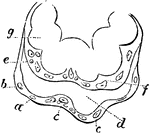
Blastoderm of an Egg

Vertical section of area pellucida and area opaca (left extremity of figure) of blastoderm of a fresh…

Vertical section of blastoderm of chick (1st day of incubation). S, epiblast consisting of short columnar cells; D, hypoblast, consisting of a single layer of flattened cells; M, formative cells." They are seen on the right of the figure, passing in between the epiblast and hypoblast to form the mesoblast; A, white yolk granules. Many of the large "formative cells" are seen containing these granules.

Blastoderm of a Chick

Vertical section of blastoderm of chick (1st day of incubation). S, epiblast consisting of short columnar…

Extremities of a placental villus. Labels: a, lining membrane of the vascular system of the mother; b, cells immediately lining a; d, space between the maternal and fetal portion of the villus; e, internal membrane of the chorion; f, internal cells of the villus, or cells of the chorion; g, loop of umbilical vessels.

Placental Villus

Extremities of a placental villus. Labels: a, lining membrane of the vascular system of the mother;…

Diagram of part of digestive tract of a chick (4th day). The black line represents hypoblast , the outer shading mesoblast; lg, lung diverticulum with expanded end forming primary lung vesicles; St, stomach; l, two hepatic diverticulum, with their terminations united by solid rows of hypoblast cells; p, diverticulum of the pancreas with the vesicular diverticula coming from it.

Digestive Tract of a Chick

Diagram of part of digestive tract of a chick (4th day). The black line represents hypoblast , the outer…

The Antheridium is the male organ of plants. Within it are produced the sperms or their equivalents, which fertilize the female cells or eggs.

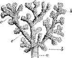
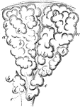
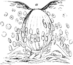
Section through Antheridium of Liverwort

The Antheridium is the male organ of plants. Within it are produced the sperms or their equivalents,…
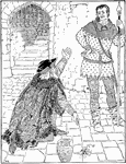
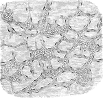
An illustration of the story, Picciola by X. B. Saintine. Count Charney was imprisoned by the Emperor of France for allegedly planning to assassinate him. In this image, he is begging the guard to send a letter the the emperor to save a little flower that is growing in the stone. A girl delivers this letter and the emperor realizes that this man could not have him killed and releases him.

Picciola

An illustration of the story, Picciola by X. B. Saintine. Count Charney was imprisoned by the Emperor…

Fall cells. Labels: a, filled with oil; b, exhausted of oil, the cell wall shriveled.

Fat Cells

Fall cells. Labels: a, filled with oil; b, exhausted of oil, the cell wall shriveled.

American linden, showing wire guard.

American linden, showing wire guard.

American linden, showing wire guard.

"Each cell forms a small, hexagonal cup, closed on one side only by a pyramidal base. These bees always accommodate themselves to the nature of the hive."

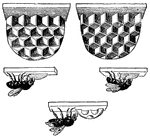
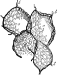
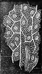
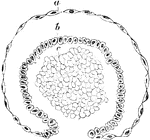
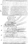
Cells Constructed by Bees

"Each cell forms a small, hexagonal cup, closed on one side only by a pyramidal base. These bees always…

A. large cell intended for the larvae of the <em>queens</em>B. middling-sized cells intended for the larvae of the <em>male</em>C. small cells intended for he larvae of the <em>workers</em>

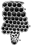
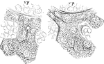
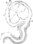
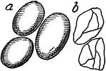
The Cells of a Beehive

A. large cell intended for the larvae of the queens B. middling-sized cells intended for the…