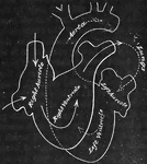
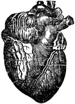
This human anatomy ClipArt gallery offers 96 illustrations of human coronary and pulmonary circulation of the cardiovascular system, which includes the organs and vessels involved in the flow of blood through the heart and lungs, where oxygen-depleted blood is sent to the lungs and oxygenated blood returns to the heart. Detailed views of the heart and views showing the relationship between the heart and lungs are also included in this category.
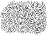
Capillaries of the Air Sac
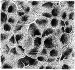
"Diagram showing the capillary network of the air sacs and origin of the pulmonary veins.. A,…

Aorta
"Aorta: a, ascending arch of aorta; ss, coronary arteries; b', innominata artery; b, right subelavian;…
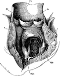
Aorta
The left ventricle and the commencement of the aorta laid open. Mpm, Mpl, the papillary muscles. From…

The Aorta
The aorta. A, from in front; B, from behind, with the origin of its principal branches. Labels: 1,2,…

Thoracic Aorta
The thoracic aorta. The three branches from left to right are the unnamed ones. The primitive left carotid…
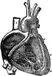
Right Atrium and Ventricle of the Heart
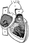
The right auricle (atrium) and ventricle of the heart opened, and a part of their right and anterior…
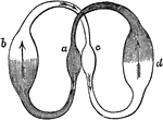
Blood Circulation in the Heart
Diagram showing the circulation of blood in the heart. Let a represent the right side of the…

The Course of Blood in the Heart
Diagram of the rush of blood when the heart beats. The valves (v) open above are closed below while…
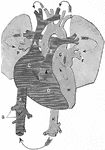
Circulation of Blood
A diagram illustrating the circulation of the blood. Labels: A, vena cava descending (superior); Z,…

Respiration Diagram
A diagram of how respiration works. A balloon is attached to the end of a small oil lamp chimney. The…
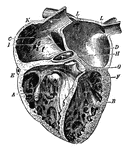
Heart
The heart is the organ that propels the blood and causes it to circulate through the arteries, veins,…

Heart
"The heart and blood-vessels diagrammatically represented. L, lung; M, intestine; P, liver; dotted lines…
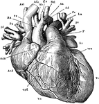
Heart
"The heart and the great blood-vessel attached to it, seen from the side towards the sternum. The left…
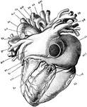
Heart
"The heart vied from its dorsal aspect. ci, inverior vena cava; Vc, coronary vein; Atd, right auricle;…
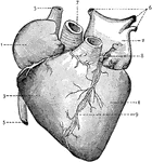
Heart
External view of the Heart. 1: Right Auricle; 2: Left Auricle; 3: Right Ventricle; 4: Left Ventricle;…

Heart
Front view of the heart and great vessels. The pulmonary artery has been cut short close to its origin.…

Heart
"Heart and blood vessels: A, B, Superior and inferior venae cavae; C, right auricle; D, right ventricle;…
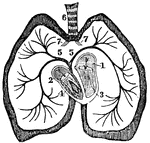
The Heart
A diagram of the heart. Labels: 1. Left auricle. 2, Right auricle. 3, Left ventricle. 4, Right ventricle.…
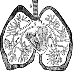
The Heart
A diagram of the heart. Labels: 1. Right auricle. 2, Left auricle. 3, Right ventricle. 4, Left ventricle.…
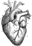
Heart
The heart. Labels: A, the right ventricle; B, the left ventricle; C, the right auricle; D, the left…

Heart and Blood Vessels
The heart and blood vessels diagrammatically represented, showing the direction of blood flow.
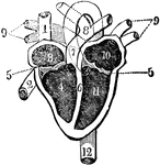
Heart and its Chambers
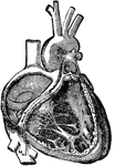
View of the heart with its several chambers exposed and the vessels in connection with them. Labels:…
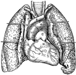
Heart and Lungs
1, The trachea or windpipe; 2 and 3, right and left common carotid arteries; 4 and 5, right and left…
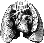
Heart and Lungs
The heart and lungs. 1, right ventricle; 3, right auricle (atrium); 6, 7, pulmonary artery; 9, aorta;…
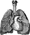
Relative Position of the Heart and Lungs
A view of the bronchia and blood vessels of the lungs, as shown by dissection, as well as the relative…
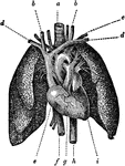
Heart and Lungs
The heart, showing its relative position to the lungs. The heart is almost wholly covered up by the…

Heart Divided into Left and Right Halves
Diagram of the heart completely divided into right and left halves, and of a double (systematic and…
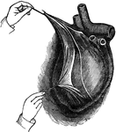
Heart in the Pericardium
View of the heart enclosed in its bag, or pericardium, which is a serious membrane. It is here laid…
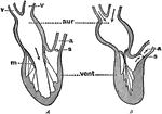
Diagram Showing the Pumping of the Heart
Diagram to illustrate the action (pumping) of the heart. Labels: aur., auricle; vent., ventricle; v,…
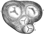
Heart Valves
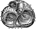
A diagram showing the peculiar fibrous structure of the heart and the shape of the valves. A, triscupid…
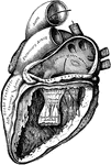
Heart with Left Auricle and Ventricle Laid Open
The left auricle and ventricle laid open, the posterior walls of both being removed.
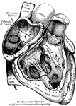
Heart with Right Auricle and Ventricle Laid Open
The right auricle and ventricle laid open, the anterior walls of both being removed.
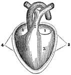
A Diagram of the Heart
A diagram of the heart. Labels: 1, Right and left auricle. 2, Right and left ventricle. 3, 4, The pericardium.…
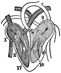
A Diagram of the Heart
A diagram of the heart. Labels: 1, Right auricle. 2, Right ventricle. 9, Left auricle. 10, Left ventricle.…
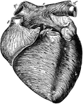
Anterior View of the Heart
Anterior view of the heart, dissected, after long boiling to show the superficial muscular fibers. The…
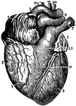
Anterior View of the Heart
An anterior view of the heart in a vertical position with its vessels injected.
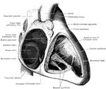
Auricle and Ventricle of the Heart
The cavities of the right auricle and right ventricle of the heart.

Beating heart
"Diagram of the rush of blood when the heart beats. The valves v open above are closed below while the…

Cavities of the heart
"A, B, right pulmonary veins, S, openings of the left pulmonary veins; E, D, C,…
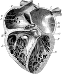
Chambers of the Heart
The chambers of the heart. Labels: A, right ventricle; B, left ventricle; C, right auricle; D, left…
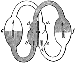
Heart, Compartments of
Diagram showing the compartments of the heart. The smaller compartments are the auricles (also known…
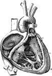
A Diagram of the Heart
The right auricle and ventricle opened, and a part of their right and anterior walls removed, so as…

A Diagram of the Heart
The left auricle and ventricle opened and a part of their anterior and left walls removed. The pulmonary…
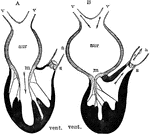
Diagram of the Action of the Heart
Diagram to illustrate the action of the heart. Labels: aur, auricle (atrium); vent. ventricle; v, veins;…
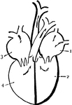
Diagram of the heart
"Diagram of the passages of the heart. 1. left auricle. 2. left ventricle, 3. Right auricle. 4. Right…

Heart, Front View of
A representation of the heart as it really appears showing the front view. At a is the right…
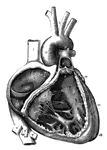
Human Heart
The interior of the heart is divided longitudinal into the right and left sections. Each right and left…


